. 2017 Mar 14;8(11):17700-17711.
doi: 10.18632/oncotarget.10775.

Capsazepine inhibits JAK/STAT3 signaling, tumor growth, and cell survival in prostate cancer

Affiliations

Capsazepine inhibits JAK/STAT3 signaling, tumor growth, and cell survival in prostate cancer

Jong Hyun Lee et al. Oncotarget. .

Abstract

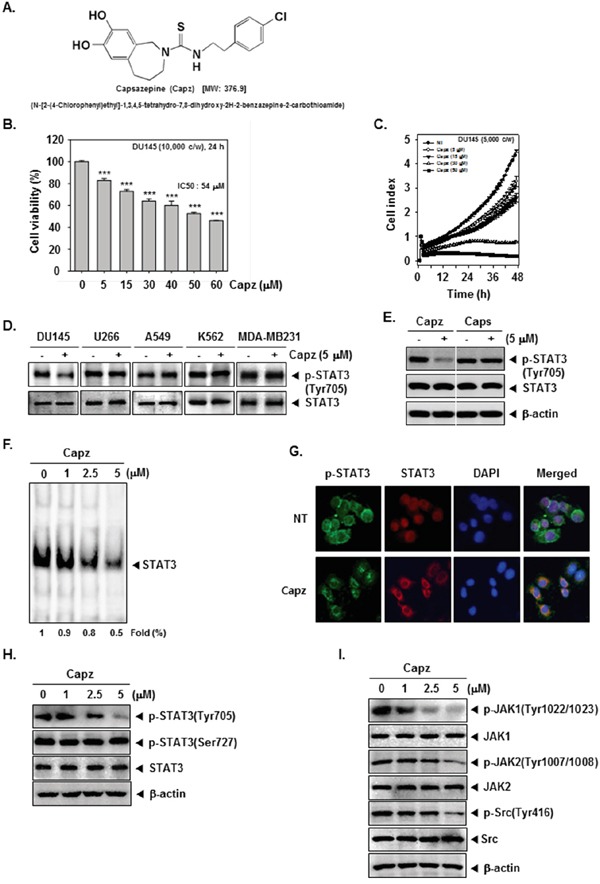
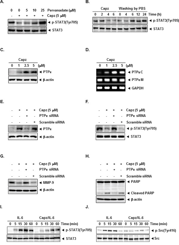
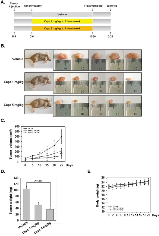
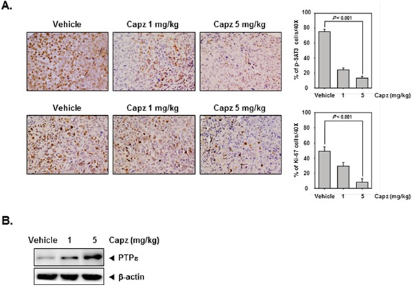
Persistent STAT3 activation is seen in many tumor cells and promotes malignant transformation. Here, we investigated whether capsazepine (Capz), a synthetic analogue of capsaicin, exerts anticancer effects by inhibiting STAT3 activation in prostate cancer cells. Capz inhibited both constitutive and induced STAT3 activation in human prostate carcinoma cells. Capz also inhibited activation of the upstream kinases JAK1/2 and c-Src. The phosphatase inhibitor pervanadate reversed Capz-induced STAT3 inhibition, indicating that the effect of Capz depends on a protein tyrosine phosphatase. Capz treatment increased PTPε protein and mRNA levels. Moreover, siRNA-mediated knockdown of PTPε reversed the Capz-induced induction of PTPε and inhibition of STAT3 activation, indicating that PTPε is crucial for Capz-dependent STAT3 dephosphorylation. Capz also decreased levels of the protein products of various oncogenes, which in turn inhibited proliferation and invasion and induced apoptosis. Finally, intraperitoneal Capz administration decreased tumor growth in a xenograft mouse prostate cancer model and reduced p-STAT3 and Ki-67 expression. These data suggest that Capz is a novel pharmacological inhibitor of STAT3 activation with several anticancer effects in prostate cancer cells.

Keywords: PTPe; STAT3; apoptosis; capsazepine; prostate cancer.


Conflict of interest statement

CONFLICTS OF INTEREST

The authors declare no competing financial interests.

Figures

Figure 1
Figure 1. Capz inhibits the STAT3 signaling pathway by inhibiting constitutive JAK1/2 and Src activation
A. The chemical structure of Capsazepine (Capz). B. DU145 cells (1 × 10⁴ cells/well) were treated with the indicated concentrations of Capz for 24 h and cell viability was determined by MTT assay. C. The cell proliferation assay was performed using the Roche xCELLigence Real-Time Cell Analyzer (RTCA) DP instrument (Roche Diagnostics GmbH, Germany). DU145 cells (5 × 10³ cells/well) were seeded onto 16-well E-plates and continuously monitored using impedance technology. D. DU145, U266, A549, K562, and MDA-MB231 cells (1 × 10⁶ cells/well) were treated with Capz (5 μM) for 6 h. Whole-cell extracts were prepared and immunoblotted with antibodies for p-STAT3(Tyr705) and STAT3. E. DU145 cells (1 × 10⁶ cells/well) were treated with Capz or Caps (5 μM) for 6 h. Whole-cell extracts were prepared and immunoblotted with antibodies for p-STAT3(Tyr705) and STAT3. F. DU145 cells (1 × 10⁶ cells/well) were treated with the indicated concentrations of Capz for 6 h and nuclear STAT3 levels were measured using EMSA. G. Capz inhibited phosphorylation and translocation of STAT3 to the nucleus. DU145 cells (4 × 10⁴ cells/well) were incubated with or without 5 μM Capz for 6 h and intracellular p-STAT3 and STAT3 distributions were analyzed by immunocytochemistry. H. DU145 cells (1 × 10⁶ cells/well) were treated with 0, 1, 2.5, or 5 μM Capz for 6 h. Whole-cell extracts were prepared and immunoblotted with antibodies for p-STAT3(Tyr705) and p-STAT3(Ser727). The same blots were stripped and reprobed with STAT3 antibody to verify equal protein loading. I. Equal amounts of lysates were analyzed by Western blot using antibodies against p-JAK1(Tyr1022/1023), p-JAK2(Tyr1007/1008), and p-Src(Tyr416). The same blots were stripped and reprobed with JAK1, JAK2, and Src antibodies to verify equal protein loading.
Figure 2
Figure 2. Capz increases protein tyrosine phosphatase epsilon (PTPε) levels in prostate cancer cells
A. DU145 cells (1 × 10⁶ cells/well) were treated with the indicated concentrations of pervanadate and 5 μM Capz for 6 h. Whole-cell extracts were prepared and immunoblotted with antibody for p-STAT3(Tyr705). The same blots were stripped and reprobed with STAT3 antibody to verify equal protein loading. The results shown are representative of two independent experiments. B. DU145 cells (1 × 10⁶ cells/well) were treated with 5 μM Capz for the indicated durations (left) or treated for 6 h and washed with PBS twice to remove Capz before resuspension in fresh medium (right). Cells were removed at indicated times and lysed to prepare whole-cell extracts. C. DU145 cells (1 × 10⁶ cells/well) were treated with the indicated concentrations of Capz for 6 h. Whole-cell extracts were prepared and immunoblotted with antibody for PTPε. The same blots were stripped and reprobed with β-actin antibody to verify equal protein loading. The results shown are representative of two independent experiments. D. DU145 cells (1 × 10⁶ cells/well) were treated with the indicated concentrations of Capz for 6 h. Total RNA was extracted and PTPε C and PTPε M expression were examined by Reverse Transcription-Polymerase Chain Reaction (RT-PCR). Glyceraldehyde-3-phosphate dehydrogenase (GAPDH) was used as an internal control to verify equal RNA loading. E. DU145 cells (1 × 10⁶ cells/well) were transfected with either scrambled or PTPε-specific siRNA (50 nM). After 48 h, cells were treated with 5 μM Capz for 6 h. Whole-cell extracts were prepared and immunoblotted with antibody for PTPε. The same blots were stripped and reprobed with β-actin antibody to verify equal protein loading. The results shown are representative of three independent experiments. F. Equal amounts of lysates were analyzed by Western blot using antibody against p-STAT3(Tyr705). The same blots were stripped and reprobed with STAT3 antibody to verify equal protein loading. G and H. DU145 cells (1 × 10⁶ cells/well) were transfected with either scrambled or PTPε-specific siRNA (50 nM). After 48 h, cells were treated with 5 μM Capz for 24 h. Whole-cell extracts were prepared and immunoblotted with antibodies for MMP-9 and PARP. The same blots were stripped and reprobed with β-actin antibody to verify equal protein loading. I. LNCaP cells (1 × 10⁶ cells/well) were treated with 5 μM Capz for 6 h and then stimulated with IL-6 (25 ng/mL) for the indicated times. Whole-cell extracts were prepared and immunoblotted with antibodies for p-STAT3(Tyr705) and STAT3. J. Equal amounts of lysates were analyzed by Western blot using antibodies against p-Src(Tyr416) and Src.
Figure 3
Figure 3. Capz inhibits invasion and promotes apoptosis in prostate cancer cells
A. An invasion assay was performed using the Roche xCELLigence Real-Time Cell Analyzer (RTCA) DP instrument (Roche Diagnostics GmbH, Germany). DU145 cell invasion activity (4 × 10⁴ cells/well) was measured in a matrigel-coated CIM (cellular invasion/migration)-Plate 16 with the indicated concentrations of Capz. B. DU145 cells were seeded in a matrigel invasion chamber overnight in the absence of a serum, incubated with the indicated concentrations of Capz for 24 h, and then subjected to the invasion assay. Representative photographs of stained cells on the lower side of matrigel membrane from one of the three experiments are shown. C. DU145 cells (1 × 10⁶ cells/well) were treated with 5 μM of Capz for 24 h. The cells were incubated with a FITC-conjugated Annexin V antibody and then analyzed by flow cytometry. D. DU145 cells (1 × 10⁶ cells/well) were seeded into 6 well plates and treated with 5 μM of Capz for 24 h. The cells were fixed and incubated with TUNEL reaction solution and then analyzed by flow cytometry. E. DU145 cells (1 × 10⁶ cells/well) were treated with the indicated concentrations of Capz for 24 h. Whole-cell extracts were prepared; 20 μg portions of those extracts were resolved via 10% SDS-PAGE and probed with pro-caspase-3, cleaved caspase-3, and PARP antibodies. The same blots were stripped and reprobed with β-actin antibody to verify equal protein loading. F. Equal amounts of lysates were analyzed by Western blot using antibodies against Bcl-2, Bcl-xl, and Survivin. The same blots were stripped and reprobed with β-actin antibody to verify equal protein loading. G. Equal amounts of lysates were analyzed by Western blot using antibodies against Cyclin D1 and COX-2. The same blots were stripped and reprobed with β-actin antibody to verify equal protein loading. H. Equal amounts of lysates were analyzed by Western blot using antibodies against VEGF and MMP-9. The same blots were stripped and reprobed with β-actin antibody to verify equal protein loading. The results shown are representative of two independent experiments.
Figure 4
Figure 4. Antitumor effects of Capz in human prostate cancer xenograft mouse model
A. Aschematic representation of the experimental protocol. DU145 cells (1× 10⁷ cells/mice) were injected subcutaneously into the right flanks of the mice. The animals were randomized into three groups 1 week after tumor cell injection based on tumor volume. Group I (control) was treated with PBS (100 μL i.p. 3 times/week), group II with Capz (1 mg/kg i.p. 3 times/week), and group III with Capz (5 mg/kg i.p. 3 times/week) (n = 8). B. Necropsy photographs of mice bearing subcutaneously implanted prostate tumors. C. Tumor diameters were measured every 5 days with Digimatic calipers, and tumor volumes were calculated using the formula V = 4/3 πr3 (n = 8). D. Tumor volumes (mean ± SE) based on tumor diameters measured on the last day of the experiment at autopsy using Digimatic calipers. E. Capz did not alter body weights, indicating the doses used were not toxic.
Figure 5
Figure 5. Capz reduces levels of oncogenic biomarkers in prostate tissues
A. Immunohistochemical analysis indicated that Capz inhibited p-STAT3 expression compared to control group (Top panels). Percentages with positive staining for the given biomarkers are shown. The photographs were taken at 40× magnification. Immunohistochemical analysis of the proliferation marker Ki-67 indicated that Capz treatment inhibited prostate cancer cell proliferation in mice (bottom panels). B. Western blot analysis showed that Capz treatment reduced PTPε levels in whole cell extracts from mouse tissues. Western samples from three mice in each group were analyzed and representative data are shown.

Similar articles

Cited by

References

    1. Siveen KS, Sikka S, Surana R, Dai X, Zhang J, Kumar AP, Tan BK, Sethi G, Bishayee A. Targeting the STAT3 signaling pathway in cancer: role of synthetic and natural inhibitors. Biochim Biophys Acta. 2014;1845:136–154. - PubMed
    1. Masciocchi D, Gelain A, Villa S, Meneghetti F, Barlocco D. Signal transducer and activator of transcription 3 (STAT3): a promising target for anticancer therapy. Future medicinal chemistry. 2011;3:567–597. - PubMed
    1. Chai EZ, Shanmugam MK, Arfuso F, Dharmarajan A, Wang C, Kumar AP, Samy RP, Lim LH, Wang L, Goh BC, Ahn KS, Hui KM, Sethi G. Targeting transcription factor STAT3 for cancer prevention and therapy. Pharmacol Ther. 2015 - PubMed
    1. Kim C, Cho SK, Kapoor S, Kumar A, Vali S, Abbasi T, Kim SH, Sethi G, Ahn KS. beta-Caryophyllene oxide inhibits constitutive and inducible STAT3 signaling pathway through induction of the SHP-1 protein tyrosine phosphatase. Molecular carcinogenesis. 2014;53:793–806. - PubMed
    1. Aggarwal BB, Sethi G, Ahn KS, Sandur SK, Pandey MK, Kunnumakkara AB, Sung B, Ichikawa H. Targeting signal-transducer-and-activator-of-transcription-3 for prevention and therapy of cancer: modern target but ancient solution. Ann N Y Acad Sci. 2006;1091:151–169. - PubMed

MeSH terms