. 2016 Jul 25:7:12282.
doi: 10.1038/ncomms12282.

Plexins function in epithelial repair in both Drosophila and zebrafish

Affiliations

Plexins function in epithelial repair in both Drosophila and zebrafish

Sa Kan Yoo et al. Nat Commun. .

Abstract

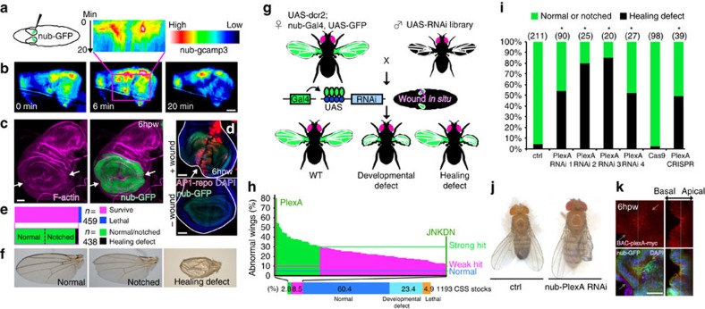
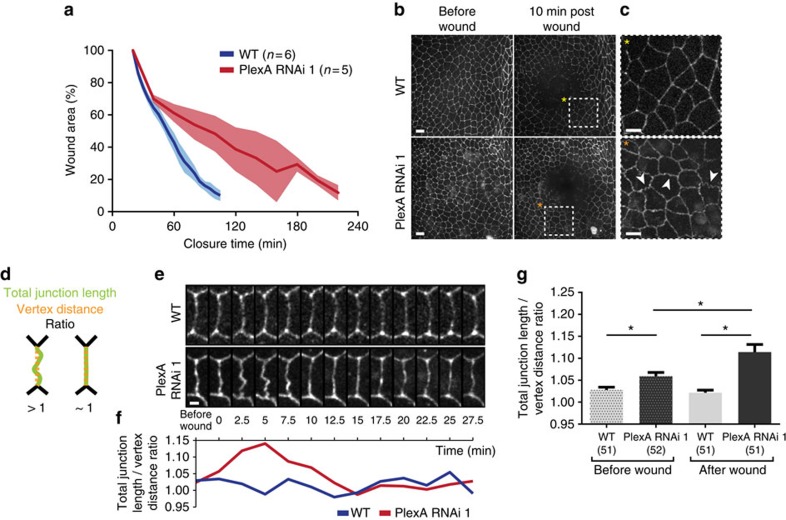
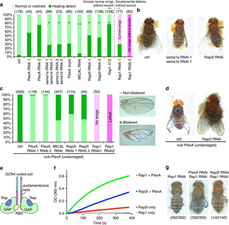
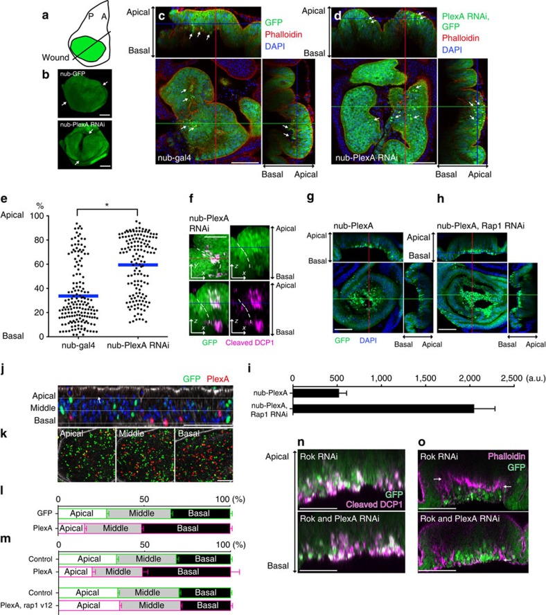
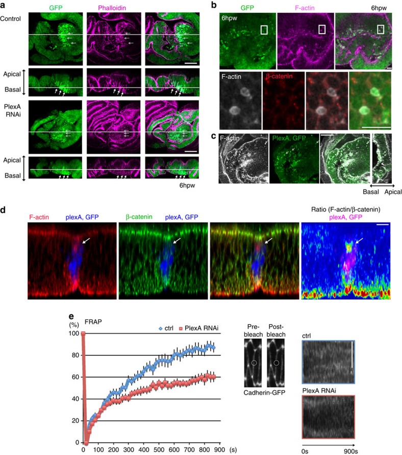
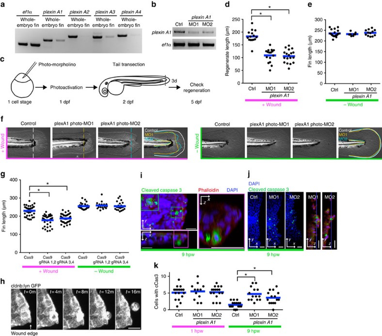
In most multicellular organisms, homeostasis is contingent upon maintaining epithelial integrity. When unanticipated insults breach epithelial barriers, dormant programmes of tissue repair are immediately activated. However, many of the mechanisms that repair damaged epithelia remain poorly characterized. Here we describe a role for Plexin A (PlexA), a protein with particularly well-characterized roles in axonal pathfinding, in the healing of damaged epithelia in Drosophila. Semaphorins, which are PlexA ligands, also regulate tissue repair. We show that Drosophila PlexA has GAP activity for the Rap1 GTPase, which is known to regulate the stability of adherens junctions. Our observations suggest that the inhibition of Rap1 activity by PlexA in damaged Drosophila epithelia allows epithelial remodelling, thus facilitating wound repair. We also demonstrate a role for Plexin A1, a zebrafish orthologue of Drosophila PlexA, in epithelial repair in zebrafish tail fins. Thus, plexins function in epithelial wound healing in diverse taxa.


Figures

Figure 1
Figure 1. An RNAi screen identifies a role for PlexA in wing disc repair.
(a) Diagram of in situ wounding of wing discs (Supplementary Movie 1). (b) Damage induces a transient calcium flash in wing discs (Supplementary Movie 2). The top inset is a kymograph derived from the region shown in the rectangular box. (c,d) F-actin accumulates and the AP-1 reporter is activated at the wound edges (white arrows), which have fused by 6 h post wounding (h.p.w.). (e,f) Adult wing phenotypes after wounding wing discs of L3 larvae. (g,h) Scheme of the wing disc-specific RNAi screen and classes of phenotypes obtained. Overall, 1,193 CSS RNAi lines were used. (i,j) Four independent PlexA RNAi transgenes and the wing disc-specific PlexA knockout with CRISPR induce healing defects. *P<0.05, two-tailed χ2-test with the Bonferroni correction (RNAi), two-tailed χ2-test (CRISPR). (k) PlexA expression in wounded wing discs of PlexA-myc BAC transgenic flies. Arrows indicate the wound location. Scale bars, 50 μm.
Figure 2
Figure 2. PlexA regulates wound closure in the epithelium of the notum.
(a) PlexA RNAi slows down wound closure upon laser wounding in the pupal notum at 13 APF. Shadowed areas represent s.d. (b,c) Expression of E-Cad-GFP in notum epithelia. Cells in the PlexA RNAi epithelium exhibit wiggly cell boundaries around the wound (indicated with white arrowheads) when compared with controls. Scale bars, (b) 10 μm and (c) 5 μm. (d) Schematic diagram of the ratio used to quantify the membrane defects. (e,f) Quantification of the junction length and vertex distance ratio over time in controls and PlexA RNAi. The membrane morphology change reaches its maximum at 5 min after wounding. Scale bar, 2 μm. (g) Junction length and vertex distance ratio for controls and PlexA RNAi both before and after wound for five independent pupae for each condition. Error bars represent s.e.m. *P<0.05; Kruskal–Wallis test with Dunn's post-test.
Figure 3
Figure 3. PlexA signaling regulates wound repair.
(a,b) Double knockdown of sema1a and sema1b, expression of PlexA Δcyto, knockdown of Rap2l or expression of constitutively active Rap1 V12 perturbs wound healing but MICAL knockdown does not. Note that only right wings are wounded. The flies were held in a hole of a sponge, which looks brown beneath flies, for image acquisition. *P<0.05, two-tailed χ2-test with the Bonferroni correction. (c,d) Ectopic expression of PlexA in the wing pouch induces blisters in adult wings. This effect is prevented by Rap2l or MICAL knockdown and enhanced by Rap1 knockdown. (e) Schematic diagram of the coiled-coil PlexAcyto fusion protein to enable constitutive dimerization. (f) The Rap GAP activity of Coiled-coil PlexAcyto was measured using the photometric assay. The slope of the linear portion of the reaction curves reflects the initial rate of GTP hydrolysis (see Methods for details). The rate of GTP hydrolysis of Rap1 catalysed by PlexA (0.166 O.D. per min) is approximately ninefold higher than that of Rap2l (0.019 O.D. per min). (g) Rap1 knockdown-induced loss of adult wings is suppressed by inhibition of PlexA or Rap2l.
Figure 4
Figure 4. PlexA regulates delamination of epithelial cells.
(a) Diagram of a bisected wing disc (A, anterior; P, posterior). (b) 3D reconstruction of wounded wing discs at 18 h.p.w. (Supplementary Movie 4). White arrows indicate the wound sites. (c,d) Control discs have many basally extruded cells at18 h.p.w. In PlexA RNAi discs, unextruded round cells with intense GFP signals are observed in the middle of the epithelial layer at 18 h.p.w. White arrows indicate examples of round cells with intense GFP fluorescence. (e) z axis location of the round cells with intense GFP fluorescence in damaged wing discs at 18 h.p.w. *P<0.05; two-tailed Mann–Whitney U-test. (f) The small cells with intense GFP signals in the middle of the epithelial layer contain the activated effector caspase DCP-1. Dotted lines indicate the wound edge. (g,h) Ectopic expression of PlexA induces basal cell extrusion, which is enhanced by concomitant inhibition of Rap1. (i) Quantification of basally extruded cells. Error bars represent s.e.m. (nub-PlexA: n=6, nub-PlexA, Rap1 RNAi: n=6). (jl) PlexA expression in single-cell clones. Cells expressing PlexA also express RFP. Control cells express GFP alone. PlexA expression displaces cells to the basal side cell autonomously. Error bars represent s.e.m. (n=17 discs). (m) Basal displacement of cells by PlexA expression in single-cell clones is suppressed by Rap1 activation. Error bars represent s.e.m. (top: n=5, bottom: n=6). (n) Rok RNAi-mediated apoptosis is not affected by PlexA inhibition. (o) Rok RNAi induces cell delamination, with F-actin separating the epithelium from the basally extruded cells. PlexA inhibition perturbs Rok RNAi-induced cell delamination. White arrows indicate the plane of F-actin accumulation. Scale bars, 50 μm.
Figure 5
Figure 5. PlexA mediates uncoupling of F-actin and adherens junctions.
(a) In control discs the round cells with intense GFP signals have pronounced accumulation of F-actin at 6 h.p.w. In PlexA RNAi discs, these cells have less accumulation of F-actin. White arrows indicate examples of round cells with intense GFP fluorescence. Data are representative of more than 10 images. (b) The round cells with intense GFP signals have actin rings around them at 6 h.p.w. (c) Extruded cells by ectopic expression of PlexA have actin rings around them (indicated with white arrows). Data are representative of more than five images. (d) Ratiometric analysis shows that cells with elevated PlexA have more F-actin uncoupled with adherens junctions compared with the surrounding cells. (e) FRAP analysis of cadherin–GFP in the pupal notum at 13 APF. PlexA RNAi inhibits fluorescence recovery of cadherin–GFP after photobleaching compared with control. Error bars represent s.e.m. (ctrl: n=20, PlexA RNAi: n=18). Right pictures show the process of photobleaching and kymographic analyses. Scale bars, 50 μm (a,c), 10 μm (b,d) and 5 μm (e).
Figure 6
Figure 6. Plexin A1 regulates wound repair in zebrafish fins.
(a) RT–PCR of plexins. plexin A1 and A3 are relatively enriched in tail fins at 2 d.p.f. (b) RT–PCR of plexin A1 following knockdown. Two independent splice morpholinos were used. (c) Diagram of photo-morpholino injection, photoactivation and tail transection. (d) Regenerated fin length at 3 days post wounding (d.p.w.), i.e., 5 d.p.f. *P<0.05; one-way ANOVA with Dunnett's post-test. (e) Fin length at 5 d.p.f. in the absence of wounding. (f) Images of 5 d.p.f.-larval tail fins with/without wounding. Note that wounding affects both the length and width of the wounded tail fins in plexin A1 morphants. (g) CRISPR-mediated plexin A1 inhibition impairs fin regeneration at 3 d.p.w. (5 d.p.f.) but not the fin length without wounding. *P<0.05; one-way ANOVA with Dunnett's post-test. (h) Epithelial cell extrusion around a wound of Tg(cldnb:lynGFP). On average, 2.75 (n=4, s.e.m.=0.48) cells were observed to be extruded for 5 h imaging, but note that all the extrusion events were not captured because imaging was done only in a part of the tail fin. (i) An extruded cell is undergoing apoptosis. (j) Representative images of transected tail fins at 9 h.p.w. (k) Knockdown of Plexin A1 increases apoptotic cells at wounds at 9 h.p.w. but not 1 h.p.w. *P<0.05; one-way ANOVA with Dunnett's post-test.

Similar articles

Cited by

References

    1. Niethammer P., Grabher C., Look A. T. & Mitchison T. J. A tissue-scale gradient of hydrogen peroxide mediates rapid wound detection in zebrafish. Nature 459, 996–999 (2009). - PMC - PubMed
    1. Yoo S. K., Freisinger C. M., LeBert D. C. & Huttenlocher A. Early redox, Src family kinase, and calcium signaling integrate wound responses and tissue regeneration in zebrafish. J. Cell Biol. 199, 225–234 (2012). - PMC - PubMed
    1. Poss K. D. Advances in understanding tissue regenerative capacity and mechanisms in animals. Nat. Rev. Genet. 11, 710–722 (2010). - PMC - PubMed
    1. Campos I., Geiger J. A., Santos A. C., Carlos V. & Jacinto A. Genetic screen in Drosophila melanogaster uncovers a novel set of genes required for embryonic epithelial repair. Genetics 184, 129–140 (2010). - PMC - PubMed
    1. Lesch C., Jo J., Wu Y., Fish G. S. & Galko M. J. A targeted UAS-RNAi screen in Drosophila larvae identifies wound closure genes regulating distinct cellular processes. Genetics 186, 943–957 (2010). - PMC - PubMed

Publication types