. 2016 Oct 6;19(4):462-475.
doi: 10.1016/j.stem.2016.06.018. Epub 2016 Jul 21.

Tissue Mechanics Orchestrate Wnt-Dependent Human Embryonic Stem Cell Differentiation

Affiliations

Tissue Mechanics Orchestrate Wnt-Dependent Human Embryonic Stem Cell Differentiation

Laralynne Przybyla et al. Cell Stem Cell. .

Abstract

Regenerative medicine is predicated on understanding the mechanisms regulating development and applying these conditions to direct stem cell fate. Embryogenesis is guided by cell-cell and cell-matrix interactions, but it is unclear how these physical cues influence stem cells in culture. We used human embryonic stem cells (hESCs) to examine whether mechanical features of the extracellular microenvironment could differentially modulate mesoderm specification. We found that, on a hydrogel-based compliant matrix, hESCs accumulate β-catenin at cell-cell adhesions and show enhanced Wnt-dependent mesoderm differentiation. Mechanistically, Src-driven ubiquitination of E-cadherin by Cbl-like ubiquitin ligase releases P120-catenin to facilitate transcriptional activity of β-catenin, which initiates and reinforces mesoderm differentiation. By contrast, on a stiff hydrogel matrix, hESCs show elevated integrin-dependent GSK3 and Src activity that promotes β-catenin degradation and inhibits differentiation. Thus, we found that mechanical features of the microenvironmental matrix influence tissue-specific differentiation of hESCs by altering the cellular response to morphogens.


Figures

Figure 1
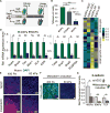
Figure 1. Substrate Stiffness Does Not Affect hESC Self-Renewal
(A) Actin staining in hESCs on soft versus stiff substrates is shown. (B) Percentage of cells positive for phosphorylated Histone H3 in hESCs on soft or stiff substrates is shown. (C) mRNA expression of self-renewal markers in hESCs grown on soft or stiff substrates for 5 days is shown. (D) Immunofluorescent staining in hESCs grown in the indicated conditions for 5 days is shown. (E) mRNA expression of self-renewal markers in hESCs replated from soft or stiff gels onto Matrigel-coated tissue culture plates cultured for two passages is shown. (F) Panel analysis data of mRNA expression levels of 77 genes involved in self-renewal and lineage specification compared in hESCs grown on soft or stiff substrates for 5 days. Linear regression analysis yields an R2 value of 0.9788. Data are represented as mean of at least three independent experiments ± SD. All scale bars, 20 μm. See also Figure S1.
Figure 2
Figure 2. The Induction of Mesoderm Differentiation in hESCs Is Enhanced on Compliant Matrices
(A) Time course of differentiation for hESCs on soft and stiff hydrogels is shown. (B) Percentage of NCAM-high cells as analyzed by flow cytometry in cells differentiated on gels of the indicated stiffness. Light gray bars indicate percentages in uninduced cells on the corresponding stiffness. Statistics refer to the bars depicting differentiated cells. (C) Heatmap showing a subset of genes from RNA-seq analysis that are significantly upregulated and correspond to gene ontology designations for self-renewal (top) or mesoderm differentiation (bottom). Table S1 includes associated reads per kilobase per million mapped reads (RPKM) values. (D) mRNA expression of genes involved in mesoderm differentiation (left) and EMT (right) in hESCs differentiated on soft or stiff gels is shown. (E) Immunofluorescent staining in cells grown in the indicated conditions is shown. (F) Immunofluorescent staining in cells grown in the indicated conditions is shown. (G) Relative E-cadherin protein expression levels as analyzed by flow cytometry in cells grown in the indicated conditions. Data are represented as mean of at least three independent experiments ± SD (**p < 0.001 and *p < 0.05). TCP, tissue culture plastic. All scale bars, 20 μm. See also Figure S2, Table S1, and Movie S1.
Figure 3
Figure 3. Cell-Secreted Wnts Are Critically Involved in Mechanically Mediated Mesoderm Differentiation
(A) Diagram depicting the mechanistic question addressed in this figure. All figure panels are from data obtained on day 3.5 at the end of mesoderm induction. (B) mRNA expression of Wnt genes in cells differentiated on soft or stiff gels is shown. (C) Immunofluorescent staining in cells differentiated on soft or stiff gels is shown. (D) Relative average fluorescent intensity in 7×TCF-GFP Wnt activity reporter hESCs differentiated in the indicated conditions is shown. (E) mRNA expression of mesoderm and EMT markers in cells differentiated in the indicated conditions. IWP2 is a Wnt inhibitor. (F) Immunofluorescent staining for NCAM for cells differentiated in the indicated conditions is shown. (G) mRNA expression in cells differentiated in the indicated conditions. All bars are set relative to expression levels in cells differentiated on 400-Pa gels (= 1). CM, conditioned media. (H) mRNA expression in cells differentiated in the indicated conditions is shown. CM, conditioned media. (I) mRNA expression of SFRP family proteins in cells differentiated on soft or stiff gels is shown. (J) Immunofluorescent staining in cells differentiated in the indicated conditions is shown. (K) mRNA expression of Sfrp1 in cells differentiated in the indicated conditions. Data are represented as mean of at least three independent experiments ± SD (**p < 0.001 and *p < 0.05). All scale bars, 20 μm. See also Figure S3.
Figure 4
Figure 4. E-Cadherin-Based AJs Are Required for Compliance-Driven Mesoderm Induction
(A) mRNA expression of Wnt3a in hESCs and mesoderm-induced cells on soft or stiff gels is shown. (B) Immunofluorescent quantification of nuclear divided by junctional β-catenin levels in uninduced hESCs on soft gels and in cells induced for 1 day on soft gels is shown. (C) The proposed pathway involved in enhancing mesoderm differentiation on soft gels is shown. (D) Western blot analysis of protein levels of β-catenin and E-cadherin with β-actin as a loading control in hESCs on soft or stiff gels is shown. (E) Immunofluorescent staining in hESCs on soft or stiff gels is shown. (F) Immunofluorescent staining shows localization of E-cadherin, β-catenin, and P120-catenin in hESCs on soft gels with and without induction of an shRNA against E-cadherin. (G) hESCs on soft gels have reciprocal junctional stabilization of E-cadherin, β-catenin, and P120-catenin. (H) mRNA expression in cells differentiated in the indicated conditions. Differentiation was performed on soft gels in each case. Data are represented as mean of at least three independent experiments ± SD (*p < 0.05). All scale bars, 20 μm. See also Figure S4.
Figure 5
Figure 5. Elaboration of Cell-Cell versus Cell-ECM Contacts Varies across Substrate Stiffness
(A) The proposed relationship between cell coordination in hESCs on soft versus stiff gels and the ability of cells to become primed for mesoderm differentiation are shown. (B) Immunofluorescent staining in hESCs on soft and stiff gels is shown. (C) Immunofluorescent staining in hESCs on soft gels with and without induction of an shRNA against E-cadherin is shown. (D) Immunofluorescent staining in hESCs on stiff gels with or without an integrin β1-blocking antibody (AIIB2) is shown. (E) Western blot for β-catenin with β-actin as a loading control in hESCs on stiff gels with or without an integrin β1-blocking antibody is shown. (F) mRNA expression in cells differentiated in the indicated conditions. Data are represented as mean of at least three independent experiments ± SD (*p < 0.05). All scale bars, 20 μm.
Figure 6
Figure 6. β-Catenin Is Actively Degraded in hESCs on Stiff Gels
(A) Diagram depicts the question addressed in this figure. (B) mRNA expression of β-catenin in hESCs on soft or stiff gels is shown. (C) Immunofluorescent staining in hESCs on stiff gels with or without proteasome inhibitor MG132 is shown. (D) mRNA expression in hESCs cultured on stiff gels with or without proteasome inhibitor MG132 and then induced to differentiate is shown. (E) Immunofluorescent staining in hESCs on stiff gels with or without the GSK3β inhibitor CHIR99021 is shown. (F) mRNA expression levels in cells differentiated in the indicated conditions are shown. (G) Immunofluorescent staining in hESCs on stiff gels with or without induction of an shRNA against CBLL1 is shown. (H) mRNA expression levels in cells differentiated in the indicated conditions are shown. (I) Immunofluorescent staining in hESCs on stiff gels with or without an integrin β1-blocking antibody is shown. (J) Immunofluorescent staining in hESCs on stiff gels with or without addition of the SFK inhibitor PP1 is shown. (K) Depiction of the proteins activated in hESCs on stiff gels that are responsible for enhancing the degradation of β-catenin. Data are represented as mean of at least three independent experiments ± SD (**p < 0.001 and *p < 0.05). All scale bars, 20 μm. See also Figure S5.
Figure 7
Figure 7. SFKs Induce E-Cadherin Internalization to Initiate the Signaling Cascade Associated with EMT and Mesoderm Differentiation
(A) Immunofluorescent staining of non-phosphorylated (left) and phosphorylated (right) SFKs in cells on soft gels that were never induced for differentiation (top) or that were induced for 2 hr (bottom) is shown. (B) Diagram depicts corresponding cell types to those differentiating to mesoderm on a soft gel in an appropriately staged chicken embryo. (C) Immunofluorescent staining of cells in a gastrulating chick embryo (stage HH4). Top layer of cells (i) has high levels of unphosphorylated SFKs, while cells that have ingressed below the epithelial sheet (ii, white arrow) lose expression (left panels). This loss is anticorrelated to the upregulation of phosphorylated SFKs in cells that have ingressed (right panels). (D) Immunofluorescent staining in cells induced for differentiation on soft gels for 1 day with or without induction of an shRNA against CBLL1. Top and bottom panels are from the same image. (E) Immunofluorescent staining in cells differentiated on soft or stiff gels is shown. (F) Immunofluorescent quantification of nuclear Kaiso levels in cells differentiated on stiff gels with or without CHIR99021 or on soft gels is shown. (G) Model figure depicts how cells are primed to undergo differentiation on soft gels and the relationship to mesoderm differentiation in vivo. (H) Detailed signaling pathways that act upon differentiation of hESCs cultured on soft gels. Data are represented as mean of at least three independent experiments ± SD (*p < 0.05). All scale bars, 20 μm. See also Figure S6.

Comment in

Similar articles

Cited by

References

    1. Bauwens CL, Peerani R, Niebruegge S, Woodhouse KA, Kumacheva E, Husain M, Zandstra PW. Control of human embryonic stem cell colony and aggregate size heterogeneity influences differentiation trajectories. Stem Cells. 2008;26:2300–2310. - PubMed
    1. Burdsal CA, Damsky CH, Pedersen RA. The role of E-cadherin and integrins in mesoderm differentiation and migration at the mammalian primitive streak. Development. 1993;118:829–844. - PubMed
    1. Buxboim A, Rajagopal K, Brown AEX, Discher DE. How deeply cells feel: methods for thin gels. J Phys Condens Matter. 2010;22:194116. - PMC - PubMed
    1. Chapman SC, Collignon J, Schoenwolf GC, Lumsden A. Improved method for chick whole-embryo culture using a filter paper carrier. Dev Dyn. 2001;220:284–289. - PubMed
    1. Chapman SC, Brown R, Lees L, Schoenwolf GC, Lumsden A. Expression analysis of chick Wnt and frizzled genes and selected inhibitors in early chick patterning. Dev Dyn. 2004;229:668–676. - PubMed

Publication types