. 2016 Sep 16;291(38):20149-62.
doi: 10.1074/jbc.M116.734079. Epub 2016 Jul 21.

Identification of a Distinct Substrate-binding Domain in the Bacterial Cysteine Methyltransferase Effectors NleE and OspZ

Affiliations

Identification of a Distinct Substrate-binding Domain in the Bacterial Cysteine Methyltransferase Effectors NleE and OspZ

Ying Zhang et al. J Biol Chem. .

Abstract

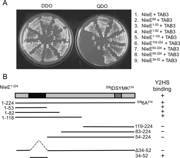
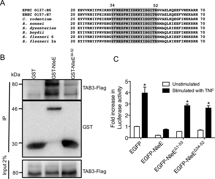
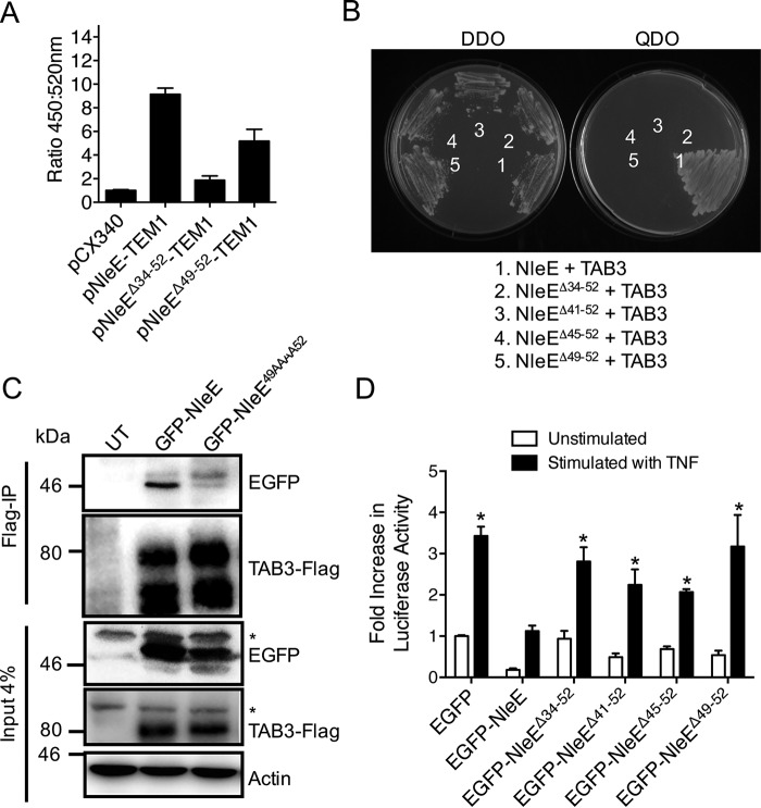
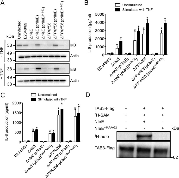
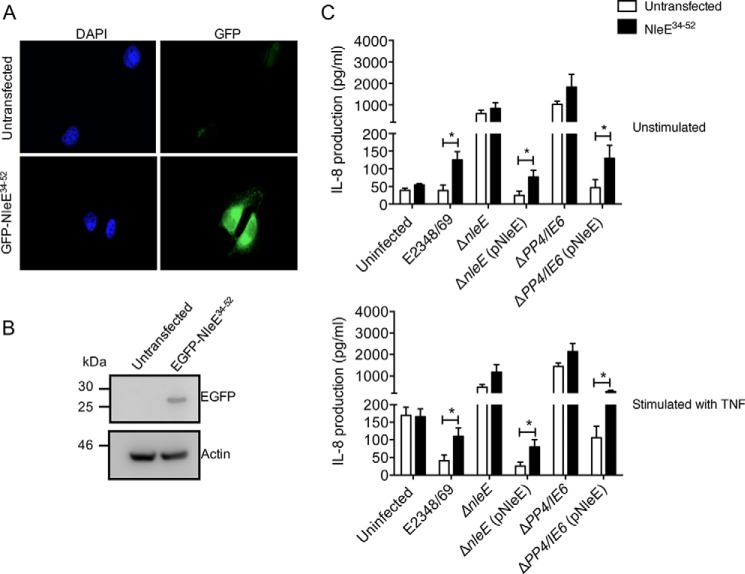
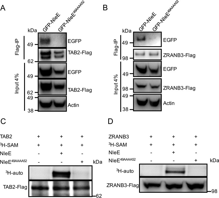
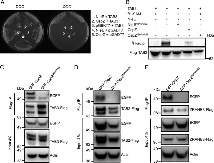
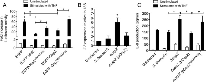
The type III secretion system effector protein NleE from enteropathogenic Escherichia coli plays a key role in the inhibition of NF-κB activation during infection. NleE inactivates the ubiquitin chain binding activity of host proteins TAK1-binding proteins 2 and 3 (TAB2 and TAB3) by modifying the Npl4 zinc finger domain through S-adenosyl methionine-dependent cysteine methylation. Using yeast two-hybrid protein interaction studies, we found that a conserved region between amino acids 34 and 52 of NleE, in particular the motif (49)GITR(52), was critical for TAB2 and TAB3 binding. NleE mutants lacking (49)GITR(52) were unable to methylate TAB3, and wild type NleE but not NleE(49AAAA52) where each of GITR was replaced with alanine restored the ability of an nleE mutant to inhibit IL-8 production during infection. Another NleE target, ZRANB3, also associated with NleE through the (49)GITR(52) motif. Ectopic expression of an N-terminal fragment of NleE (NleE(34-52)) in HeLa cells showed competitive inhibition of wild type NleE in the suppression of IL-8 secretion during enteropathogenic E. coli infection. Similar results were observed for the NleE homologue OspZ from Shigella flexneri 6 that also bound TAB3 through the (49)GITR(52) motif and decreased IL-8 transcription through modification of TAB3. In summary, we have identified a unique substrate-binding motif in NleE and OspZ that is required for the ability to inhibit the host inflammatory response.

Keywords: Escherichia coli (E. coli); NF-κB (NF-κB); infection; inflammation; substrate specificity.


Figures

FIGURE 1.
FIGURE 1.
Mapping of NleE-TAB3 interaction domains. A, growth of S. cerevisiae AH109 on medium lacking histidine, adenine, leucine, and tryptophan (QDO) to select for protein-protein interactions (right panel) or medium lacking leucine and tryptophan (DDO) to select for plasmid maintenance only (left panel). Yeast are co-expressing NleE and TAB3 (1), NleE6A and TAB3 (2), NleE1–53 and TAB3 (3), NleE1–82 and TAB3 (4), NleE1–118 and TAB3 (5), NleE119–224 and TAB3 (6), NleE83–224 and TAB3 (7), NleE54–224 and TAB3 (8), and NleE34–52 and TAB3 (9). B, schematic representation of NleE and various deletion profiles with TAB3 binding capacity indicated in the right panel. Y2HS, yeast two-hybrid system.
FIGURE 2.
FIGURE 2.
Functional analysis of the TAB3-binding domain of NleE. A, alignment of the N-terminal regions of NleE and OspZ from A/E pathogens and Shigella. Amino acids 34–52 are shaded. B, pulldown of TAB3-FLAG by immobilized GST, GST-NleE, and GST-NleE34–52. C, -fold increase in NF-κB-dependent luciferase activity in HeLa cells expressing EGFP, EGFP-NleE, EGFP-NleEΔ1–53, or EGFP-NleEΔ34–52 and left unstimulated or stimulated with TNF for 8 h where indicated. Results are the mean ± S.E. (error bars) of three independent experiments carried out in duplicate. *, significantly different from unstimulated HeLa cells expressing EGFP only (p < 0.0001, unpaired two-tailed t test). EHEC, enterohemorrhagic E. coli; IP, immunoprecipitation.
FIGURE 3.
FIGURE 3.
Translocation and function of NleE mutants lacking the TAB3-binding domain. A, translocation of NleE-TEM1, NleEΔ34–52-TEM1, and NleEΔ49–52-TEM1 fusions in HeLa cells. The ratio of blue fluorescence at 450 nm and green fluorescence at 520 nm is presented. HeLa cells were infected with EPEC E2348/69 (pCX340), EPEC E2348/69 (pNleE-TEM1), EPEC E2348/69 (pNleEΔ34–52-TEM1), or EPEC E2348/69 (pNleEΔ49–52-TEM1). Results are the mean ± S.E. (error bars) of three independent experiments carried out in triplicate. *, significantly different from EPEC E2348/69 carrying pCX340 only (p < 0.0001, unpaired two-tailed t test). B, growth of S. cerevisiae AH109 on medium lacking histidine, adenine, leucine, and tryptophan (QDO) to select for protein-protein interactions (right panel) or medium lacking leucine and tryptophan (DDO) to select for plasmid maintenance only (left panel). Yeast are co-expressing NleE and TAB3 (1), NleEΔ34–52 and TAB3 (2), NleEΔ41–52 and TAB3 (3), NleEΔ45–52 and TAB3 (4), and NleEΔ49–52 and TAB3 (5). C, immunoprecipitation (IP) of TAB3-FLAG and detection of EGFP-NleE and EGFP-NleE49AAAA52 binding in HEK293T cells. Actin is a loading control. * indicates nonspecific bands. D, -fold increase in NF-κB-dependent luciferase activity in HeLa cells expressing EGFP, EGFP-NleE, EGFP-NleEΔ34–52, EGFP-NleEΔ41–52, EGFP-NleEΔ45–52, or EGFP-NleEΔ49–52 and stimulated with or without TNF for 8 h where indicated. Results are the mean ± S.E. (error bars) of three independent experiments carried out in duplicate. *, significantly different from unstimulated HeLa cells expressing EGFP only (p < 0.02, unpaired two-tailed t test). UT, untransfected.
FIGURE 4.
FIGURE 4.
Importance of the TAB3-binding domain to NleE function during infection. A, immunoblot of IκB degradation in HeLa cells infected with derivatives of EPEC E2348/69 for 2 h and left unstimulated or stimulated with TNF for 30 min. B and C, IL-8 production from HeLa cells infected with derivatives of EPEC E2348/69 as indicated for 2 h and left unstimulated or stimulated with TNF for 8 h. Results are the mean ± S.E. (error bars) of three independent experiments carried out in duplicate. *, significantly different from unstimulated HeLa cells or HeLa cells stimulated with TNF and infected with EPEC E2348/69 (p < 0.02, unpaired two-tailed t test). D, [3H]AdoMet (SAM) labeling of full-length TAB3 immunopurified from HEK293T cells and incubated with either purified GST-NleE or GST-NleE49AAAA52 in the presence of [3H]AdoMet. [3H]AdoMet labeling was determined by 3H autoradiography (auto).
FIGURE 5.
FIGURE 5.
Inhibition of NleE activity during infection by overexpression of NleE34–52. A, representative immunofluorescence fields showing HeLa cells or HeLa cells expressing the GFP-NleE34–52 fusion protein (green). Cell nuclei were counterstained with DAPI (blue). B, immunoblot of untransfected HeLa cells or HeLa cells stably expressing the EGFP-NleE34–52 fusion protein using antibodies to GFP. Actin is a loading control. C, IL-8 production from HeLa cells or HeLa cells stably expressing EGFP-NleE34–52 infected with derivatives of EPEC E2348/69 for 2 h and left unstimulated (top panel) or stimulated with TNF (bottom panel) for 8 h. Results are the mean ± S.E. (error bars) of three independent experiments carried out in duplicate. *, EGFP-NleE34–52-expressing HeLa cells significantly different from HeLa cells when infected with E2348/69 or mutants complemented with pNleE (p < 0.05, unpaired two-tailed t test).
FIGURE 6.
FIGURE 6.
Binding of NleE to TAB2 and ZRANB3. A, immunoprecipitation of TAB2-FLAG and detection of EGFP-NleE and EGFP-NleE49AAAA52 binding in HEK293T cells. Actin is a loading control. B, immunoprecipitation (IP) of ZRANB3-FLAG and detection of EGFP-NleE and EGFP-NleE49AAAA52 binding in HEK293T cells. Actin is a loading control. C and D, [3H]AdoMet (SAM) labeling of full-length TAB2-FLAG or ZRANB3-FLAG as indicated immunopurified from HEK293T cells and incubated with purified GST-NleE or GST-NleE49AAAA52 in the presence of [3H]AdoMet. [3H]AdoMet labeling was determined by 3H autoradiography (auto).
FIGURE 7.
FIGURE 7.
OspZ binding to TAB2, TAB3, and ZRANB3 in vitro. A, growth of S. cerevisiae AH109 on medium lacking histidine, adenine, leucine, and tryptophan (QDO) to select for protein-protein interactions (right panel) or medium lacking leucine and tryptophan (DDO) to select for plasmid maintenance only (left panel). Yeast are co-expressing NleE and TAB3 (1), OspZ and TAB3 (2), pGBKT7 and TAB3 (3), NleE and pGADT7 (4), and OspZ and pGBKT7 (5). B, [3H]AdoMet (SAM) labeling of full-length TAB3 immunopurified from HEK293T cells and incubated with purified GST-NleE, GST-NleE49AAAA52, GST-OspZ, or GST-OspZ49AAAA52, respectively, in the presence of [3H]AdoMet. [3H]AdoMet labeling was determined by 3H autoradiography (auto). C, FLAG immunoprecipitation (IP) of TAB3-FLAG and detection of EGFP-OspZ and EGFP-OspZ49AAAA52 in HEK293T cells using anti-GFP antibodies. TAB3-FLAG was detected with anti-FLAG antibodies. Actin is a loading control. D, FLAG immunoprecipitation of TAB2-FLAG and detection of EGFP-OspZ and EGFP-OspZ49AAAA52 in HEK293T cells using anti-GFP antibodies. TAB3-FLAG was detected with anti-FLAG antibodies. Actin is a loading control. E, FLAG immunoprecipitation of ZRANB3-FLAG and detection of EGFP-OspZ and EGFP-OspZ49AAAA52 in HEK293T cells using anti-GFP antibodies. TAB3-FLAG was detected with anti-FLAG antibodies. Actin is a loading control.
FIGURE 8.
FIGURE 8.
OspZ mediated inhibition of NF-κB activation and IL-8 production during Shigella infection. A, -fold increase in NF-κB-dependent luciferase activity in HeLa cells transfected with pEGFP-C2, pEGFP-NleE, pEGFP-NleE49AAAA52, pEGFP-OspZ, or pEGFP-OspZ49AAAA52 and left unstimulated or stimulated with TNF for 8 h where indicated. Results are the mean ± S.E. (error bars) of three independent experiments carried out in duplicate. *, p < 0.05, unpaired two-tailed t test. B, reverse transcription-quantitative PCR analysis of IL8 expression in HT-29 cells 1 h after infection. Results are expressed as mRNA expression levels that were normalized relative to 18S and are the mean ± S.E. (error bars) of three independent experiments carried out in duplicate. *, significantly different from uninfected HT-29 cells or HT-29 cells infected with wild type S. flexneri 6 or ΔospZ (pOspZ) (p < 0.05, unpaired two-tailed t test). C, IL-8 production from HT-29 cells infected with derivatives of S. flexneri 6 as indicated for 2 h and left unstimulated or stimulated with TNF for 2 h. Results are the mean ± S.E. (error bars) of three independent experiments carried out in duplicate. *, p < 0.05, unpaired two-tailed t test.

Similar articles

Cited by

References

    1. Wong A. R., Pearson J. S., Bright M. D., Munera D., Robinson K. S., Lee S. F., Frankel G., and Hartland E. L. (2011) Enteropathogenic and enterohaemorrhagic Escherichia coli: even more subversive elements. Mol. Microbiol. 80, 1420–1438 - PubMed
    1. Frankel G., and Phillips A. D. (2008) Attaching effacing Escherichia coli and paradigms of Tir-triggered actin polymerization: getting off the pedestal. Cell. Microbiol. 10, 549–556 - PubMed
    1. Baruch K., Gur-Arie L., Nadler C., Koby S., Yerushalmi G., Ben-Neriah Y., Yogev O., Shaulian E., Guttman C., Zarivach R., and Rosenshine I. (2011) Metalloprotease type III effectors that specifically cleave JNK and NF-κB. EMBO J. 30, 221–231 - PMC - PubMed
    1. Mühlen S., Ruchaud-Sparagano M. H., and Kenny B. (2011) Proteasome-independent degradation of canonical NFκB complex components by the NleC protein of pathogenic Escherichia coli. J. Biol. Chem. 286, 5100–5107 - PMC - PubMed
    1. Nadler C., Baruch K., Kobi S., Mills E., Haviv G., Farago M., Alkalay I., Bartfeld S., Meyer T. F., Ben-Neriah Y., and Rosenshine I. (2010) The type III secretion effector NleE inhibits NF-κB activation. PLoS Pathog. 6, e1000743. - PMC - PubMed

Publication types

MeSH terms