. 2016 Jul 20:6:29418.
doi: 10.1038/srep29418.

Co-regulation of the Notch and Wnt signaling pathways promotes supporting cell proliferation and hair cell regeneration in mouse utricles

Affiliations

Co-regulation of the Notch and Wnt signaling pathways promotes supporting cell proliferation and hair cell regeneration in mouse utricles

Jingfang Wu et al. Sci Rep. .

Abstract

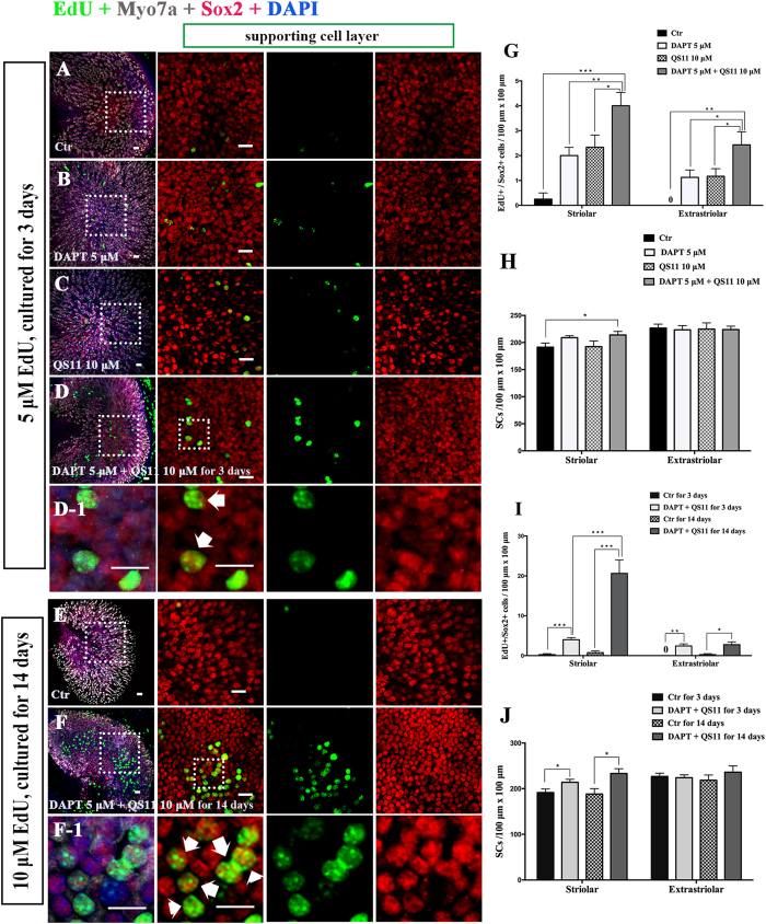
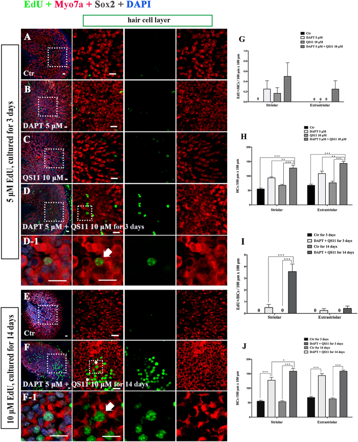
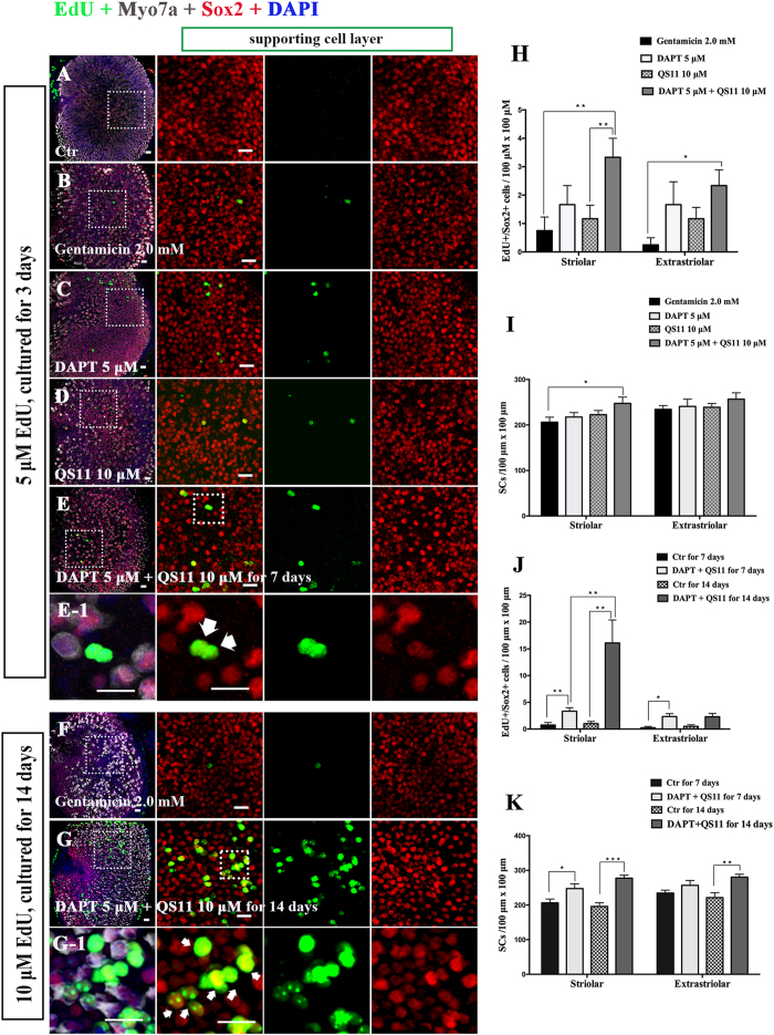
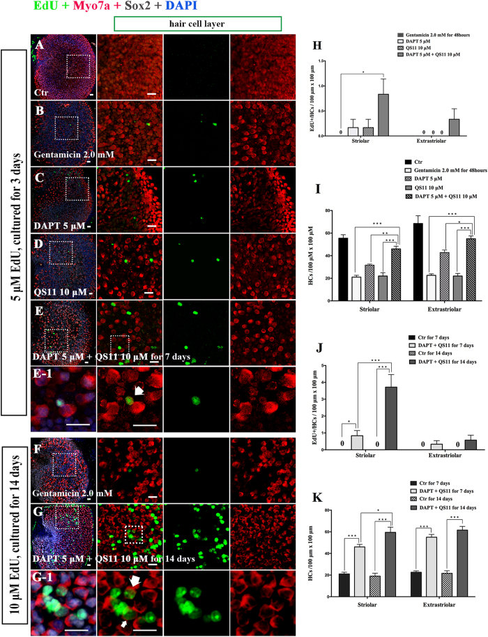
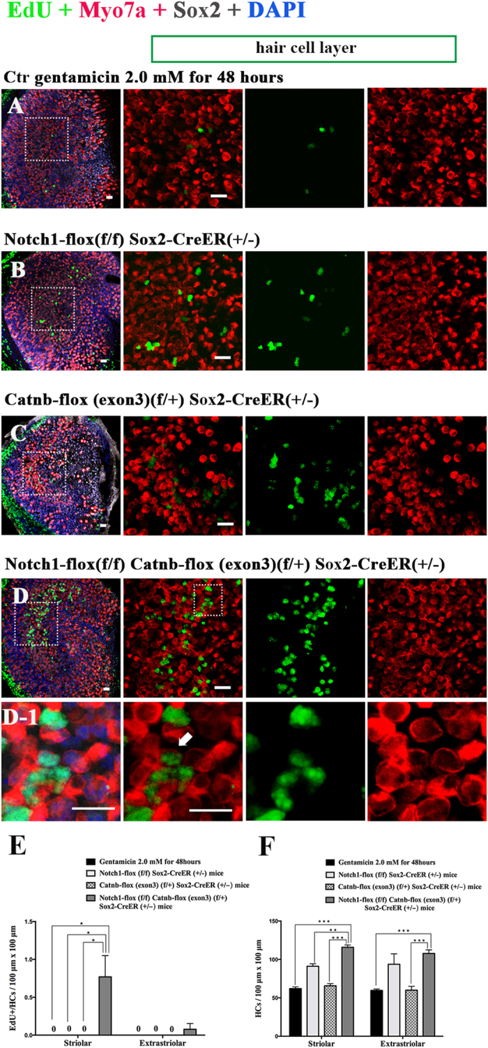
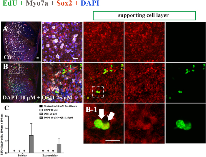
This work sought to determine the crosstalk between the Notch and Wnt signaling pathways in regulating supporting cell (SC) proliferation and hair cell (HC) regeneration in mouse utricles. We cultured postnatal day (P)3 and P60 mouse utricles, damaged the HCs with gentamicin, and treated the utricles with the γ-secretase inhibitor DAPT to inhibit the Notch pathway and with the Wnt agonist QS11 to active the Wnt pathway. We also used Sox2-CreER, Notch1-flox (exon 1), and Catnb-flox (exon 3) transgenic mice to knock out the Notch pathway and activate the Wnt pathway in Sox2+ SCs. Notch inhibition alone increased SC proliferation and HC number in both undamaged and damaged utricles. Wnt activation alone promoted SC proliferation, but the HC number was not significantly increased. Here we demonstrated the cumulative effects of Notch inhibition and Wnt activation in regulating SC proliferation and HC regeneration. Simultaneously inhibiting Notch and overexpressing Wnt led to significantly greater SC proliferation and greater numbers of HCs than manipulating either pathway alone. Similar results were observed in the transgenic mice. This study suggests that the combination of Notch inhibition and Wnt activation can significantly promote SC proliferation and increase the number of regenerated HCs in mouse utricle.


Figures

Figure 1
Figure 1. The P3 mouse utricles were cultured with DAPT, QS11, or a combination of DAPT and QS11 for 3 days or 14 days.
(Scale bar = 10 μm) (A) The untreated P3 mouse utricles cultured for 3 days showed very few EdU+/Sox2+ cells in the sensory epithelium. (B) When utricles were treated with DAPT for 3 days, there were some EdU+/Sox2+ cells in the sensory epithelium. (C) When utricles were treated with QS11 for 3 days, there were few EdU+/Sox2+ cells in the sensory epithelium and the number of SCs did not change significantly (p > 0.05). (D) In the utricles treated with a combination of DAPT and QS11 for 3 days, there were more EdU+/Sox2+ cells compared to the DAPT-only and the QS11-only group (p < 0.05). The number of SCs in the striolar region was greater than the control group (p < 0.05). (D-1) The high magnifications of image D show two of EdU+/Sox2+ cells in the utricles. (G) The histograms show differences in the number of EdU+/Sox2+ cells between the groups cultured for 3 days. (H) The histograms show differences in the number of SCs between the groups cultured for 3 days. (E) The untreated P3 mouse utricles cultured for 14 days showed very few EdU+/Sox2+ cells in the sensory epithelium. (F) In the cultured utricles co-treated with DAPT and QS11 and supplied with 10 μM EdU, there were many EdU+/Sox2+ cells in the utricles and a large number of Sox2+ SCs. (F-1) The EdU+/Sox2+ cells in the utricles are shown at high magnification. (I) The number of EdU+/Sox2+ SCs in the 14-day culture was significantly greater than in the 3-day cultured. (J) The histograms show differences in the number of SCs between the utricles co-treated with DAPT+QS11 for 3 days and 14 days. The cells were counted per 100 μm × 100 μm in the striolar or extrastriolar region of the utricles. (*p < 0.05, **p < 0.01, ***p < 0.001).
Figure 2
Figure 2. The P3 mouse utricles were cultured with DAPT, QS11, or a combination of DAPT and QS11 for 3 days or for 14 days.
(Scale bar = 10 μm) (A) The untreated P3 mouse utricles cultured for 3 days. (B) When utricles were treated with DAPT for 3 days, there were few EdU+/Myo7a+ cells in the sensory epithelium and the number of HCs increased (p < 0.05 vs. the control). (C) When the utricles were treated with QS11 for 3 days, there were few EdU+/Myo7a+ cells in the sensory epithelium and the number of HCs did not change significantly (p > 0.05). (D) In the DAPT+QS11 combination-treated utricles, there were some EdU+/Myo7a+ cells in the utricles, which were mostly in the striolar region. There were more HCs than the other groups (p < 0.05). (D-1) The high magnifications of the image in (D) show the EdU+/Myo7a+ cell in the utricle. (G) The histograms show differences in the number of EdU+ HCs between these groups cultured for 3 days. (H) The histograms show the differences in the number of HCs between these groups cultured for 3 days. (E) The untreated P3 mouse utricles cultured for 14 days. (F) In the utricles treated with 10 μM EdU and a combination of DAPT and QS11 for 14 days, there were some EdU+/Myo7a+ cells, which were found mostly in the striolar region. The number of HCs in the utricles had clearly increased. (F-1) The high magnifications of image F show EdU+/Myo7a+ cells in the utricles. (I) In the cultured utricles co-treated with DAPT and QS11 and supplied with 10 μM EdU for 14 days, the number of EdU+/Myo7a+ cells was significantly increased compared with the utricles co-treated for 3 days. (J) The histograms show the differences in the number of HCs between the utricles co-treated with DAPT + QS11 for 3 days or 14 days. The cells were counted per 100 μm × 100 μm in the striolar or extrastriolar region of the utricles (*p < 0.05, **p < 0.01, ***p < 0.001).
Figure 3
Figure 3. The P3 mouse utricles were treated with gentamicin for 48 hours then treated with DAPT and QS11 separately or in combination for 7 days or for 14 days.
(Scale bar = 10 μm) (A) The untreated P3 mouse utricles cultured for 7 days. (B) The P3 mouse utricles were treated with gentamicin for 48 hours and then cultured for another 7 days. (C) When utricles were treated with DAPT for 7 days after being treated with gentamicin, there were some EdU+/Sox2+ cells in the sensory epithelium. (D) When utricles were treated with QS11 for 7 days after being treated with gentamicin, there were few EdU+/Sox2+ cells in the sensory epithelium. (E) When utricles were treated with a combination of DAPT+QS11 for 7 days after being treated with gentamicin, there were more EdU+/Sox2+ cells in the sensory epithelium. (E-1) The high magnifications of picture E show EdU+/Sox2+ cells in the utricles. (H) The histograms show differences in the number of EdU+/Sox2+ cells between these groups cultured for 7 days. (I) The histograms show differences in the number of SCs between these groups cultured for 7 days. (F) The P3 mouse utricles were treated with gentamicin for 48 hours and cultured for 14 days. The HCs were clearly damaged. (G) In the cultured utricles co-treated with DAPT and QS11 and supplied with 10 μM EdU, there were many more EdU+/Sox2+ cells in the utricles compared with the utricles co-treated for 7 days. (G-1) The high magnifications of picture G show EdU+/Sox2+ cells in the utricles. (J) The histograms show that the number of EdU+/SCs in the 14-day culture group was significantly greater than in the 7-day culture group. (K) The histograms show the differences in the number of SCs between the utricles co-treated with DAPT+QS11 for 7 days and 14 days. The cells were counted per 100 μm × 100 μm in the striolar or extrastriolar region of the utricles. (*p < 0.05, **p < 0.01, ***p < 0.001)
Figure 4
Figure 4. Sox2-CreER and Notch1-flox (exon 1) mice were mated with Catnb-flox (exon 3) mice to generate pups.
The P3 mouse utricles were dissected and cultured with 2 mM gentamicin for 48 hours to damage the HCs of the utricles. The cultured utricles were treated with tamoxifen and cultured for an additional 7 days. (Scale bar = 10 μm) (A) In the control group, there were no obvious EdU+/Sox2+ cells in the sensory epithelium of the utricles. (B) In the Notch1-flox (f/f) Sox2-CreER (+/−) mouse utricles, there were some EdU+/ Sox2+ cells in the sensory epithelium. (C) In the Catnb-flox (exon3) (f/+) Sox2-CreER (+/−) mouse utricles, there were few EdU+/Sox2+ cells in the sensory epithelium of the utricles. (D) In the Notch1-flox (f/f) Catnb-flox (exon3) (f/+) Sox2-CreER (+/−) group, there were significantly more EdU+/Sox2+ cells in the sensory epithelium of the utricles. (D1) The high magnification of picture D shows the EdU+/Sox2+ cells in the utricular sensory epithelium of the Notch1-flox (f/f) Catnb-flox (exon3) (f/+) Sox2-CreER (+/−) group. (E) The histograms show the differences in the number of EdU+/Sox2+ cells between these groups. The cells were counted per 100 μm × 100 μm in the striolar or extrastriolar region of the utricles. In the Notch1-flox (f/f); Catnb-flox (exon3) (f/+); Sox2-CreER (+/−) mice, there were significantly more EdU+/Sox2+ SCs in the utricle sensory epithelium than in the other groups (*p < 0.05). (F) The histograms show the differences in the number of SCs between these groups. The cells were counted per 100 μm × 100 μm in the striolar or extrastriolar region of the utricles. The total number of SCs was also significantly increased compared to other groups (*p < 0.05, **p < 0.01, ***p < 0.001).
Figure 5
Figure 5. The P3 mouse utricles were treated with gentamicin for 48 hours then treated with DAPT and QS11 separately or in combination for 7 days or 14 days.
(Scale bar = 10 μm) (A) The untreated P3 mouse utricles were cultured for 7 days. (B) The P3 mouse utricles were treated with gentamicin for 48 hours and cultured for 7 days. (C) When utricles were treated with DAPT for 7 days after HC loss, there were few EdU+/Myo7a+ cells in the utricles. (D) When utricles were treated with QS11 for 7 days after being treated with gentamicin, there were few EdU+/Myo7a+ cells in the utricles. (E) When utricles were treated with a combination of DAPT + QS11 for 7 days after being treated with gentamicin, there were some EdU+/Myo7a+ cells, and the number of HCs was greater than the other groups (p < 0.05). (E1) The high magnifications of picture E show the EdU+/Myo7a+ cell in the utricle. (H) The histograms show differences in the number of EdU+/HCs between these groups cultured for 7 days. (I) The histograms show differences in the number of HCs between these groups cultured for 7 days. (F) The P3 mouse utricles were treated with gentamicin for 48 hours and cultured for 14 days. (G) In the cultured utricles co-treated with DAPT and QS11 and supplied with 10 μM EdU for 14 days, there were some EdU+/Myo7a+ cells in the utricles. (G-1) The high magnifications of picture image G show the EdU+/Myo7a+ cells in the utricles. (J) The histograms show that the number of EdU+/Myo7a+ cells in the 14-day culture group was significantly greater than in the 7-day culture group. (K) The histograms show the differences in the number of HCs between the utricles co-treated with DAPT and QS11 for 7 days and those treated for 14 days. The cells were counted per 100 μm × 100 μm in the striolar or extrastriolar region of the utricles (*p < 0.05, **p < 0.01, ***p < 0.001).
Figure 6
Figure 6. Sox2-CreER and Notch1-flox (exon 1) mice were mated with Catnb-flox (exon 3) mice to generate pups.
The P3 mouse utricles were dissected and cultured with 2 mM gentamicin for 48 hours to damage the HCs of the utricles. The cultured utricles were treated with tamoxifen and cultured for an additional 7 days. (Scale bar = 10 μm) (A) In the control group, there were no obvious EdU+/ Myo7a+ cells in the sensory epithelium of the utricles. The number of HCs did not change significantly. (B) In the Notch1-flox (f/f) Sox2-CreER (+/−) mouse utricles, the number of HCs was increased compared to the control group (p < 0.05). (C) In the Catnb-flox (exon3) (f/+) Sox2-CreER (+/−) mouse utricles, the number of HCs did not change significantly. (D) In the Notch1-flox (f/f) Catnb-flox (exon3) (f/+) Sox2-CreER (+/−) group, there were some EdU+/Myo7a+ cells in the sensory epithelium of the utricles. The number of HCs also increased significantly. (**p < 0.01, ***p < 0.001). (D1) The high magnification of image D shows one of the EdU+/Myo7a+ cells in the utricular sensory epithelium of the Notch1-flox (f/f) Catnb-flox (exon3) (f/+) Sox2-CreER (+/−) mice. (E) The histograms show the differences in the number of EdU+/HCs between these groups. The cells were counted per 100 μm × 100 μm in the striolar or extrastriolar region of the utricles. No EdU+/Myo7a+ cells were observed in control and single-regulated groups. In the Notch1-flox (f/f) Catnb-flox (exon3) (f/+) Sox2-CreER (+/−) mice, we observed some EdU+/Myo7a+ cells in the utricular sensory epithelium. (*p < 0.05) (F) The histograms show the differences in the number of HCs between these groups. The cells were counted per 100 μm × 100 μm in the striolar or extrastriolar region of the utricles. The number of HCs in the Notch1-flox (f/f) Catnb-flox (exon3) (f/+) Sox2-CreER (+/−) mice utricles was greater than the other group (**p < 0.01, ***p < 0.001).
Figure 7
Figure 7. Cultured adult (2 months old) mouse cultured utricles were treated with 1 mM gentamicin for 24 hours then treated with 10 μM DAPT and 25 μM QS11 for 7 days.
There were some EdU+/Sox2+ cells in the DAPT and QS11 co-treated adult mouse utricles. (Scale bar = 10 μm).

Similar articles

Cited by

References

    1. Jung J. Y. et al.. siRNA targeting Hes5 augments hair cell regeneration in aminoglycoside-damaged mouse utricle. Molecular therapy : the journal of the American Society of Gene Therapy 21, 834–841 (2013). - PMC - PubMed
    1. Dye B. J., Frank T. C., Newlands S. D. & Dickman J. D. Distribution and time course of hair cell regeneration in the pigeon utricle. Hear Res 133, 17–26 (1999). - PubMed
    1. Weisleder P. & Rubel E. W. Hair cell regeneration after streptomycin toxicity in the avian vestibular epithelium. The Journal of comparative neurology 331, 97–110 (1993). - PubMed
    1. Cafaro J., Lee G. S. & Stone J. S. Atoh1 expression defines activated progenitors and differentiating hair cells during avian hair cell regeneration. Dev Dyn 236, 156–170 (2007). - PubMed
    1. Stone J. S. & Cotanche D. A. Hair cell regeneration in the avian auditory epithelium. Int J Dev Biol 51, 633–647 (2007). - PubMed

Publication types

LinkOut - more resources