

The chemomodulatory effects of glufosfamide on docetaxel cytotoxicity in prostate cancer cells
- PMID: 27413637
- PMCID: PMC4933087
- DOI: 10.7717/peerj.2168
The chemomodulatory effects of glufosfamide on docetaxel cytotoxicity in prostate cancer cells
Abstract
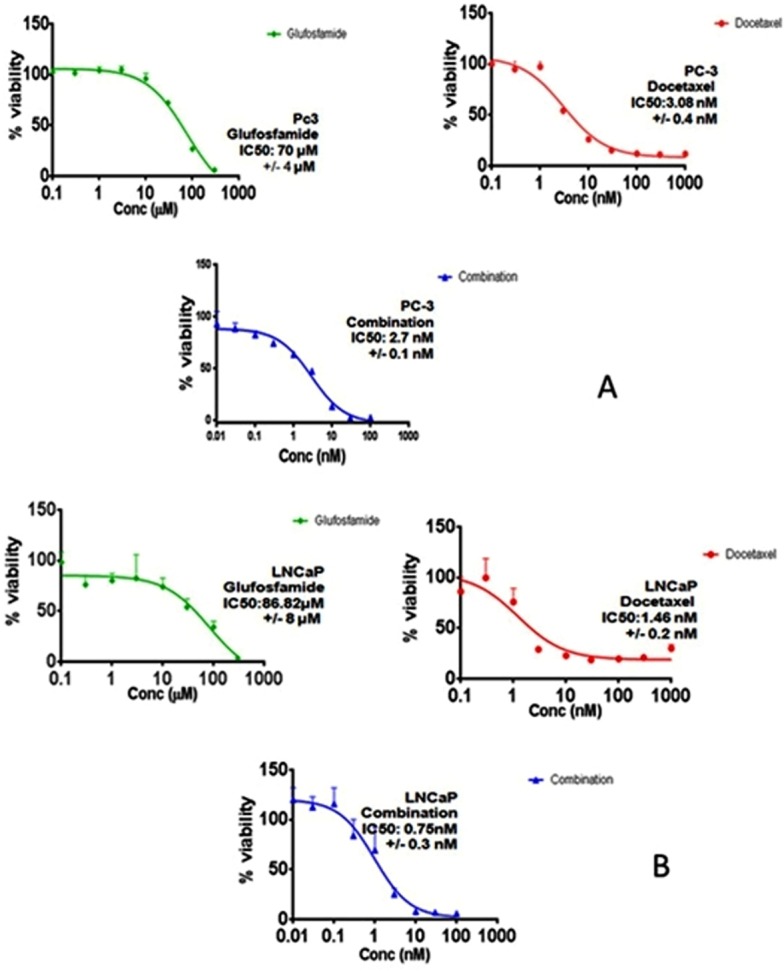
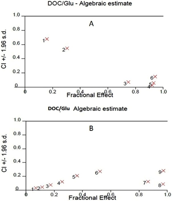
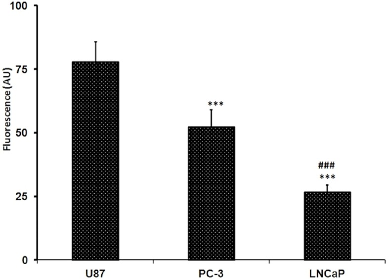
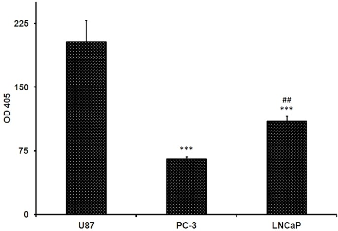
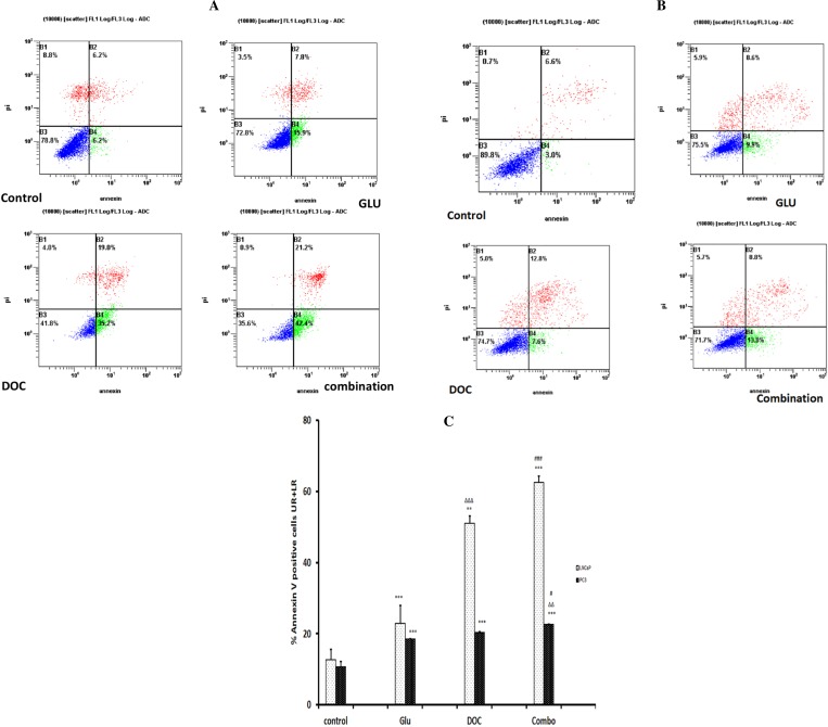
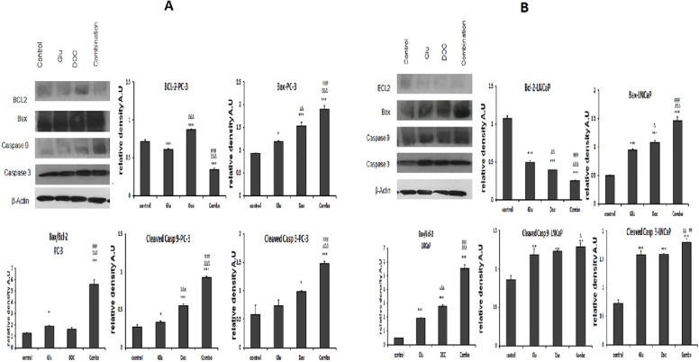
Background. Glufosfamide (GLU) is a glucose conjugate of ifosfamide in which isophosphoramide mustard is glycosidically linked to the β-D-glucose molecule. Based on GLU structure, it is considered a targeted chemotherapy with fewer side effects. The main objective of the current study is to assess the cytotoxic potential of GLU for the first time in prostate cancer (PC) cells representing different stages of the tumor. Furthermore, this study examined the potential synergistic activity of GLU in combination with docetaxel (DOC). Methods. Two different cell lines were used, LNCaP and PC-3. Concentration-response curves were assessed. The tested groups per cell line were, control, GLU, DOC and combination. Treatment duration was 72 h. Cytotoxicity was assessed using sulforhodamine B (SRB) assay and half maximal inhibitory concentration (IC50) was calculated. Synergy analyses were performed using Calcusyn(®)software. Subsequent mechanistic studies included β-glucosidase activity assay, glucose uptake and apoptosis studies, namely annexin V-FITC assay and the protein expression of mitochondrial pathway signals including Bcl-2, Bax, Caspase 9 and 3 were assessed. Data are presented as mean ± SD; comparisons were carried out using one way analysis of variance (ANOVA) followed by Tukey-Kramer's test for post hoc analysis. Results. GLU induced cytotoxicity in both cell lines in a concentration-dependent manner. The IC50 in PC-3 cells was significantly lower by 19% when compared to that of LNCaP cells. The IC50 of combining both drugs showed comparable effect to DOC in PC-3 but was tremendously lowered by 49% compared to the same group in LNCaP cell line. β-glucosidase activity was higher in LNCaP by about 67% compared to that determined in PC-3 cells while the glucose uptake in PC-3 cells was almost 2 folds that found in LNCaP cells. These results were directly correlated to the efficacy of GLU in each cell line. Treatment of PC cells with GLU as single agent or in combination with DOC induced significantly higher apoptosis as evidenced by Annexin V-staining. Apoptosis was significantly increased in combination group by 4.9 folds and by 2.1 Folds when compared to control in LNCaP cells and PC-3 cells; respectively. Similarly, the expression of Bcl-2 was significantly decreased while Bax, caspase 9 and 3 were significantly increased in the combined treatment groups compared to the control. Conclusion. GLU has a synergistic effect in combination with DOC as it increases the cell kill which can be attributed at least partially to apoptosis in both the tested cell lines and it is suggested as a new combination regimen to be considered in the treatment of the prostate cancer. Further experiments and clinical investigations are needed for assessment of that regimen.
Keywords: Apoptosis; Docetaxel; Glufosfamide; LNCaP; PC-3; Prostate cancer.
Conflict of interest statement
The authors declare there are no competing interests.
Figures
Similar articles
-
An in vitro cytotoxicity of glufosfamide in HepG2 cells relative to its nonconjugated counterpart.J Egypt Natl Canc Inst. 2021 Aug 23;33(1):22. doi: 10.1186/s43046-021-00080-6. J Egypt Natl Canc Inst. 2021. PMID: 34423383
-
Caffeic acid phenethyl ester synergistically enhances docetaxel and paclitaxel cytotoxicity in prostate cancer cells.IUBMB Life. 2013 Aug;65(8):716-29. doi: 10.1002/iub.1188. Epub 2013 Jul 11. IUBMB Life. 2013. PMID: 23847086
-
Rapamycin enhances the susceptibility of both androgen-dependent and -independent prostate carcinoma cells to docetaxel.Chin Med J (Engl). 2010 Feb 5;123(3):356-60. Chin Med J (Engl). 2010. PMID: 20193259
-
Glufosfamide as a new oxazaphosphorine anticancer agent.Anticancer Drugs. 2011 Jul;22(6):488-93. doi: 10.1097/CAD.0b013e328345e1e0. Anticancer Drugs. 2011. PMID: 21427562 Review.
-
Glufosfamide: beta-D-Glc-IPM, D 19575.Drugs R D. 2005;6(1):49-52. doi: 10.2165/00126839-200506010-00006. Drugs R D. 2005. PMID: 15801867 Review.
Cited by
-
Cross-Talk between Wnt Signaling and Src Tyrosine Kinase.Biomedicines. 2022 May 11;10(5):1112. doi: 10.3390/biomedicines10051112. Biomedicines. 2022. PMID: 35625853 Free PMC article. Review.
-
Docetaxel/cabazitaxel and fatty acid binding protein 5 inhibitors produce synergistic inhibition of prostate cancer growth.Prostate. 2020 Jan;80(1):88-98. doi: 10.1002/pros.23921. Epub 2019 Oct 29. Prostate. 2020. PMID: 31661167 Free PMC article.
-
Docetaxel and Lidocaine Co-Loaded (NLC-in-Hydrogel) Hybrid System Designed for the Treatment of Melanoma.Pharmaceutics. 2021 Sep 24;13(10):1552. doi: 10.3390/pharmaceutics13101552. Pharmaceutics. 2021. PMID: 34683846 Free PMC article.
-
The Effects and Mechanism of YK-4-279 in Combination with Docetaxel on Prostate Cancer.Int J Med Sci. 2017 Apr 7;14(4):356-366. doi: 10.7150/ijms.18382. eCollection 2017. Int J Med Sci. 2017. PMID: 28553168 Free PMC article.
References
-
- Becker R, Ritter A, Eichhorn U, Lips J, Bertram B, Wiessler M, Zdzienicka MZ, Kaina B. Induction of DNA breaks and apoptosis in crosslink-hypersensitive v79 cells by the cytostatic drug beta-D-glucosyl-ifosfamide mustard. British Journal of Cancer. 2002;86(1):130–135. doi: 10.1038/sj.bjc.6600027. - DOI - PMC - PubMed
-
- Beckner ME, Gobbel GT, Abounader R, Burovic F, Agostino NR, Laterra J, Pollack IF. Glycolytic glioma cells with active glycogen synthase are sensitive to PTEN and inhibitors of PI3K and gluconeogenesis. Laboratory Investigation. 2005;85(12):1457–1470. - PubMed
Grants and funding
LinkOut - more resources
Full Text Sources
Other Literature Sources
Research Materials
