

Development of Droplet Microfluidics Enabling High-Throughput Single-Cell Analysis
- PMID: 27399651
- PMCID: PMC6272933
- DOI: 10.3390/molecules21070881
Development of Droplet Microfluidics Enabling High-Throughput Single-Cell Analysis
Abstract
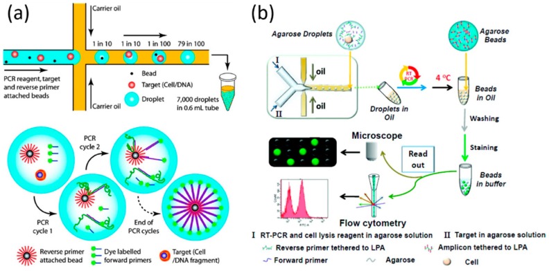
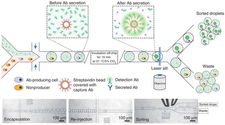
This article reviews recent developments in droplet microfluidics enabling high-throughput single-cell analysis. Five key aspects in this field are included in this review: (1) prototype demonstration of single-cell encapsulation in microfluidic droplets; (2) technical improvements of single-cell encapsulation in microfluidic droplets; (3) microfluidic droplets enabling single-cell proteomic analysis; (4) microfluidic droplets enabling single-cell genomic analysis; and (5) integrated microfluidic droplet systems enabling single-cell screening. We examine the advantages and limitations of each technique and discuss future research opportunities by focusing on key performances of throughput, multifunctionality, and absolute quantification.
Keywords: droplet microfluidics; high-throughput; single-cell encapsulation; single-cell genetic analysis; single-cell proteomic analysis; single-cell screening.
Conflict of interest statement
The authors declare no conflict of interest.
Figures
Similar articles
-
NOVAsort for error-free droplet microfluidics.Nat Commun. 2024 Nov 1;15(1):9444. doi: 10.1038/s41467-024-52932-z. Nat Commun. 2024. PMID: 39487108 Free PMC article.
-
Microfluidic impedance flow cytometry enabling high-throughput single-cell electrical property characterization.Int J Mol Sci. 2015 Apr 29;16(5):9804-30. doi: 10.3390/ijms16059804. Int J Mol Sci. 2015. PMID: 25938973 Free PMC article. Review.
-
High throughput single cell counting in droplet-based microfluidics.Sci Rep. 2017 May 2;7(1):1366. doi: 10.1038/s41598-017-01454-4. Sci Rep. 2017. PMID: 28465615 Free PMC article.
-
Recent developments in microfluidics for cell studies.Adv Mater. 2014 Aug 20;26(31):5525-32. doi: 10.1002/adma.201305348. Epub 2014 Feb 17. Adv Mater. 2014. PMID: 24536032 Review.
-
Droplet microfluidics--a tool for single-cell analysis.Angew Chem Int Ed Engl. 2012 Dec 3;51(49):12176-92. doi: 10.1002/anie.201200460. Epub 2012 Nov 23. Angew Chem Int Ed Engl. 2012. PMID: 23180509 Review.
Cited by
-
Deterministic trapping, encapsulation and retrieval of single-cells.Lab Chip. 2017 Jun 27;17(13):2186-2192. doi: 10.1039/c7lc00283a. Lab Chip. 2017. PMID: 28585962 Free PMC article.
-
The Physics and Manipulation of Dean Vortices in Single- and Two-Phase Flow in Curved Microchannels: A Review.Micromachines (Basel). 2023 Dec 1;14(12):2202. doi: 10.3390/mi14122202. Micromachines (Basel). 2023. PMID: 38138371 Free PMC article. Review.
-
Generation and manipulation of hydrogel microcapsules by droplet-based microfluidics for mammalian cell culture.Lab Chip. 2017 May 31;17(11):1913-1932. doi: 10.1039/c7lc00262a. Lab Chip. 2017. PMID: 28509918 Free PMC article. Review.
-
Recent advances in understanding noroviruses.F1000Res. 2017 Jan 26;6:79. doi: 10.12688/f1000research.10081.1. eCollection 2017. F1000Res. 2017. PMID: 28163914 Free PMC article. Review.
-
Synthesis and active manipulation of magnetic liquid beads.Biomed Microdevices. 2024 May 6;26(2):24. doi: 10.1007/s10544-024-00708-z. Biomed Microdevices. 2024. PMID: 38709370 Free PMC article.
References
Publication types
MeSH terms
LinkOut - more resources
Full Text Sources
Other Literature Sources
