. 2016 Jul 7;12(7):e1005748.
doi: 10.1371/journal.ppat.1005748. eCollection 2016 Jul.

Human Cytomegalovirus Immediate-Early 1 Protein Rewires Upstream STAT3 to Downstream STAT1 Signaling Switching an IL6-Type to an IFNγ-Like Response

Affiliations

Human Cytomegalovirus Immediate-Early 1 Protein Rewires Upstream STAT3 to Downstream STAT1 Signaling Switching an IL6-Type to an IFNγ-Like Response

Thomas Harwardt et al. PLoS Pathog. .

Abstract

The human cytomegalovirus (hCMV) major immediate-early 1 protein (IE1) is best known for activating transcription to facilitate viral replication. Here we present transcriptome data indicating that IE1 is as significant a repressor as it is an activator of host gene expression. Human cells induced to express IE1 exhibit global repression of IL6- and oncostatin M-responsive STAT3 target genes. This repression is followed by STAT1 phosphorylation and activation of STAT1 target genes normally induced by IFNγ. The observed repression and subsequent activation are both mediated through the same region (amino acids 410 to 445) in the C-terminal domain of IE1, and this region serves as a binding site for STAT3. Depletion of STAT3 phenocopies the STAT1-dependent IFNγ-like response to IE1. In contrast, depletion of the IL6 receptor (IL6ST) or the STAT kinase JAK1 prevents this response. Accordingly, treatment with IL6 leads to prolonged STAT1 instead of STAT3 activation in wild-type IE1 expressing cells, but not in cells expressing a mutant protein (IE1dl410-420) deficient for STAT3 binding. A very similar STAT1-directed response to IL6 is also present in cells infected with a wild-type or revertant hCMV, but not an IE1dl410-420 mutant virus, and this response results in restricted viral replication. We conclude that IE1 is sufficient and necessary to rewire upstream IL6-type to downstream IFNγ-like signaling, two pathways linked to opposing actions, resulting in repressed STAT3- and activated STAT1-responsive genes. These findings relate transcriptional repressor and activator functions of IE1 and suggest unexpected outcomes relevant to viral pathogenesis in response to cytokines or growth factors that signal through the IL6ST-JAK1-STAT3 axis in hCMV-infected cells. Our results also reveal that IE1, a protein considered to be a key activator of the hCMV productive cycle, has an unanticipated role in tempering viral replication.


Conflict of interest statement

The authors have declared that no competing interests exist.

Figures
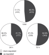
Fig 1
Fig 1. IE1 is both an activator and a repressor of human gene expression.
Relative proportions and absolute counts (in brackets) of unique probe sets significantly up- or down-regulated in GeneChip analyses following induction of IE1 for 24 h, 72 h or 24 h and 72 h combined (total). The numbers are based on S1 Data.
Fig 2
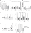
Fig 2. Human genes repressed by IE1 are IL6- and OSM-responsive pSTAT3 target genes.
(A) TetR (w/o) and TetR-IE1 (IE1) cells were treated with dox for 72 h. Relative mRNA levels were determined by RT-qPCR with primers specific for the C4A, CHL1, CXCL12, IFI16, RASL11A or SOCS3 genes. Results were normalized to TUBB, and means and standard deviations of biological triplicates are shown in comparison to TetR cells (set to 1). (B) TetR cells were treated with solvent or IL6 plus IL6R (IL6/Rα) for 6 h or 24 h. Relative mRNA levels in comparison to solvent-treated cells (set to 1) were determined by RT-qPCR with primers specific for the indicated genes. Results were normalized to TUBB, and means and standard deviations of biological triplicates are shown. (C) TetR cells were treated with solvent or OSM for 24 h. Relative mRNA levels in comparison to solvent-treated cells (set to 1) were determined by RT-qPCR with primers specific for the indicated genes. Results were normalized to TUBB, and means and standard deviations of biological triplicates are shown. (D) TetR cells were transfected with the indicated siRNA duplexes. Two and five days post transfection (p.t.), whole cell protein extracts were prepared and subjected to immunoblotting for STAT3α and GAPDH (left panel). Five days post transfection, relative mRNA levels were determined by RT-qPCR with primers specific for the indicated genes. Results were normalized to TUBB, and means and standard deviations of two biological and two technical replicates are shown in comparison to control siRNA-transfected cells (set to 1) (right panel). (E) Whole cell protein extracts from TetR cells without (w/o) or with stable expression of the indicated STAT3α-Myc proteins were subjected to immunoblotting for STAT3 (Myc tag), TUBA, pSTAT3 (Y705) and pSTAT3 (S727) (left panel). Total RNA samples from TetR cells overexpressing either wild-type STAT3α-Myc or STAT3α_Y705F-Myc were subjected to RT-qPCR with primers specific for the indicated genes. Results were normalized to TUBB, and means and standard deviations of biological triplicates are shown in comparison to cells expressing wild-type STAT3α-Myc (set to 1) (right panel).
Fig 3
Fig 3. Systematic deletion analysis of C-terminal IE1 residues 373–491.
(A) Schematic overview of amino acids 373–491 in the tested wild-type and mutant IE1 proteins. Positions of the low-complexity motifs (acidic domains AD1-3 and serine/proline-rich region S/P), the SUMOylation site (K450) and the chromatin tethering domain (CTD) are shown. (B) TetR cells without (w/o) or with inducible expression of the indicated HA-tagged wild-type or mutant IE1 proteins were treated with dox for 72 h. Whole cell protein extracts were prepared and analyzed by immunoblotting for IE1 (HA tag) and GAPDH. (C) TetR cells without (w/o) or with inducible expression of the indicated HA-tagged wild-type or mutant IE1 proteins were treated with dox for 72 h. Whole cell extracts prepared in the presence of N-ethylmaleimide were used for immunoprecipitation with anti-HA-agarose, and samples were analyzed by immunoblotting for IE1 (HA tag) and SUMO1.
Fig 4
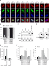
Fig 4. Residues within IE1 region 410–445 are required for targeting of STAT3 and down-regulation of STAT3-responsive genes.
(A) TetR cells without (w/o) or with inducible expression of the indicated HA-IE1 proteins were treated with dox for 48 h. During the final 24 h of dox treatment, cells were kept in medium with 0.5% FBS. Subcellular localization of endogenous STAT3α in IE1 expressing cells was analyzed by indirect immunofluorescence microscopy. Samples were simultaneously reacted with a rabbit monoclonal antibody to STAT3α and a mouse monoclonal antibody to HA-tagged IE1, followed by incubation with a rabbit-specific Alexa Fluor 594 conjugate and a mouse-specific Alexa Fluor 488 conjugate. Host cell nuclei were visualized by 4',6-diamidino-2-phenylindole (DAPI) staining. Additionally, merge images of STAT3α, IE1 and DAPI signals are presented. (B) The percentage of cells with i) predominantly nuclear STAT3α staining (N>C), ii) equally strong nuclear and cytoplasmic STAT3α staining (N = C) and iii) predominantly cytoplasmic STAT3α staining (C>N) was determined for 100 randomly selected cells per sample described in (A). (C) TetR cells without or with inducible expression of HA-tagged wild-type IE1 or IE1dl410-420 were treated with dox for 72 h and with solvent (w/o) or IL6 plus IL6R (IL6/Rα) for 30 min. Cytoplasmic and nuclear extracts were prepared and analyzed by immunoblotting for histone H2B, STAT2, STAT3α and IE1. (D) TetR cells without (w/o) or with inducible expression of HA-tagged wild-type IE1 or IE1dl410-420 were treated with dox for 72 h. Whole cell extracts were prepared and used for immunoprecipitations (IPs) with anti-HA-agarose. Samples of lysates and immunoprecipitates were analyzed by immunoblotting for IE1 and STAT3α. (E) TetR cells without (w/o) or with inducible expression of HA-tagged wild-type IE1 or IE1dl410-420 were treated with dox for 72 h and with IL6 plus IL6R for 30 min. Samples were subjected to ChIP with rabbit polyclonal antibodies to STAT3 or normal rabbit IgG and primers specific for sequences in the SOCS3 promoter or coding region. The percentage of output versus input DNA is presented as the difference between STAT3 and normal IgG ChIPs. Means and standard deviations of two biological and two technical replicates are shown. (F) TetR cells without (w/o) or with inducible expression of the indicated HA-tagged wild-type or mutant IE1 proteins were treated with dox for 72 h. Relative mRNA expression levels were determined by RT-qPCR with primers specific for the STAT3 target genes CXCL12 and SOCS3. Results were normalized to TUBB, and means and standard deviations of two biological and two technical replicates are shown in comparison to IE1-negative TetR cells (set to 1).
Fig 5
Fig 5. Residues within IE1 region 410–445 are required for phosphorylation of STAT1 and up-regulation of IFNγ-responsive genes.
(A) TetR cells without (w/o) or with inducible expression of the indicated HA-tagged wild-type or mutant IE1 proteins were treated with dox for 72 h. Whole cell protein extracts were prepared and analyzed by immunoblotting for IE1 (HA tag), pSTAT1 (Y701), pSTAT1 (S727), total STAT1 and GAPDH. (B) TetR cells without (w/o) or with inducible expression of the indicated HA-tagged wild-type or mutant IE1 proteins were treated with dox for 72 h. Relative mRNA expression levels were determined by RT-qPCR with primers specific for the STAT1 target genes CXCL10 and CXCL11. Results were normalized to TUBB, and means and standard deviations of two biological and two technical replicates are shown in comparison to IE1-negative TetR cells (set to 1).
Fig 6
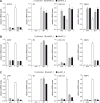
Fig 6. Knock-down of STAT3 recapitulates, while knock-down of IL6ST or JAK1 disrupts IE1-mediated induction of IFNγ-stimulated genes.
(A) TetR (w/o) or TetR-IE1 (IE1) cells were transfected with a control siRNA or two different siRNAs specific for STAT3. From 48 h post siRNA transfection, cells were treated with dox for 72 h. Relative mRNA levels were determined by RT-qPCR for STAT3, IE1 and the STAT1 target genes CXCL10 and GBP4. Results were normalized to TUBB, and means and standard deviations of two biological and two technical replicates are shown in comparison to control siRNA-transfected TetR-IE1 cells (set to 1). (B) TetR (w/o) or TetR-IE1 (IE1) cells were transfected with a control siRNA or two different siRNAs specific for IL6ST. From 48 h post siRNA transfection, cells were treated with dox for 72 h. Relative mRNA levels were determined by RT-qPCR for IL6ST, IE1, CXCL10 and GBP4. Results were normalized to TUBB, and means and standard deviations of two biological and two technical replicates are shown in comparison to control siRNA-transfected TetR-IE1 cells (set to 1). (C). TetR (w/o) or TetR-IE1 (IE1) cells were transfected with a control siRNA or two different siRNAs specific for JAK1. From 48 h post siRNA transfection, cells were treated with dox for 72 h. Relative mRNA levels were determined by RT-qPCR for JAK1, IE1, CXCL10 and GBP4. Results were normalized to TUBB, and means and standard deviations of two biological and two technical replicates are shown in comparison to control siRNA-transfected TetR-IE1 cells (set to 1).
Fig 7
Fig 7. IE1 switches an IL6-type to an IFNγ-like response.
(A) TetR (w/o) or TetR-IE1 (IE1) cells were treated with dox for 72 h and with IL6 plus IL6R (IL6/Rα) for the indicated times. Whole cell protein extracts were prepared and analyzed by immunoblotting for IE1, total STAT1, pSTAT1 (Y701), total STAT3α, pSTAT3 (Y705) and GAPDH. (B) TetR (w/o) or TetR-IE1 cells expressing HA-tagged wild-type IE1 or IE1dl410-420 were treated with dox for 72 h and with solvent, IFNα, IFNγ or IL6 plus IL6R (IL6/Rα) for 24 h. Whole cell protein extracts were analyzed by immunoblotting for IE1, total STAT1, pSTAT1 (Y701) and GAPDH. (C) TetR (w/o) or TetR-IE1 cells expressing HA-tagged wild-type IE1 or IE1dl410-420 were treated with dox for 72 h and with solvent, IFNα, IFNγ or IL6 plus IL6R (IL6/Rα) for 24 h. Relative mRNA levels were determined by RT-qPCR for the type I IFN/STAT2 target genes OAS1 and EIF2AK2 (protein kinase R) (left panels), the type II IFN/STAT1 target genes CXCL10 and CXCL11 (middle panels) and the IL6/STAT3 target genes CXCL12 and SOCS3 (right panels). Results were normalized to TUBB, and means and standard deviations of biological triplicates are shown in comparison to solvent-treated TetR cells (set to 1).
Fig 8
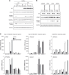
Fig 8. IE1 rewires IL6 signaling to STAT1 activation during hCMV infection.
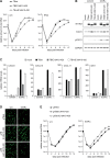
(A) MRC-5 cells were infected with TBwt, TBIE1dl410-420 or TBrvIE1dl410-420 at low input multiplicity (0.1 PFU/cell) in the absence (w/o) or presence of exogenous IFNβ. Culture media were replaced every 24 h, and viral replication was assessed by qPCR-based relative quantification of hCMV DNA from culture supernatants at the indicated times post infection with primers specific for the viral UL86 sequence. Data are presented as means and standard deviations from three independent infections. (B) MRC-5 cells were mock-infected or infected with TBwt, TBIE1dl410-420 or TBrvIE1dl410-420 at a high input multiplicity (5 PFU/cell). At 6 h post infection, cultures were treated with solvent or IL6 plus IL6R (IL6/Rα). At 24 h post infection, whole cell protein extracts were prepared and analyzed by immunoblotting for IE1/IE2, total STAT1, pSTAT1 (Y701) and GAPDH. (C) MRC-5 cells were mock-infected or infected with TBwt, TBIE1dl410-420 or TBrvIE1dl410-420 at a high input multiplicity (5 PFU/cell). At 6 h post infection, cultures were treated with solvent or IL6 plus IL6R (IL6/Rα). At 24 h post infection, relative mRNA levels were determined by RT-qPCR for the STAT1 target genes CXCL9, CXCL10, CXCL11 and IDO. Results were normalized to TUBB, and means and standard deviations of biological triplicates are shown in comparison to solvent-treated mock-infected cells (set to 1). (D) STAT2-deficient human skin fibroblasts were infected with gTBwt, gTBIE1dl410-420 or gTBrvIE1dl410-420 at low input multiplicity (0.1 PFU/cell) in the absence (w/o) or presence of IL6 plus IL6Rα (IL6/Rα). Every 48 h, half of the culture media was replaced and viral replication was assessed at day 6 post infection by fluorescence microscopy. (E) STAT2-deficient human skin fibroblasts were infected with gTBwt, gTBIE1dl410-420 or gTBrvIE1dl410-420 at low input multiplicity (0.1 PFU/cell) in the absence (w/o) or presence of IL6 plus IL6Rα (IL6/Rα). Every 48 h, half of the culture media was replaced and viral replication was assessed by qPCR-based relative quantification of hCMV DNA from culture supernatants with primers specific for the viral UL86 sequence. Data are presented as means and standard deviations from two biological and two technical replicates.
Fig 9
Fig 9. Model linking repression of STAT3-responsive to activation of STAT1-responsive genes by IE1.
(Left panel) Binding of IL6 family cytokines (or growth factors) to the extracellular IL6R subunits leads to tyrosine phosphorylation of JAK1 (and other JAK family kinases) associated with the IL6ST receptor subunits. In turn, JAK1 phosphorylates tyrosine residues in IL6ST creating binding sites for STAT3. Following tyrosine phosphorylation by JAK1, pSTAT3 forms active dimers capable of binding DNA. These STAT3 dimers accumulate in the nucleus and activate target gene transcription. STAT1 remains mostly unphosphorylated, cytoplasmic and excluded from binding to the receptor in the absence of IE1. (Right panel) In the presence of IE1, cytoplasmic STAT3 pools become depleted due to formation of a nuclear IE1-STAT3 complex. In the absence of cytoplasmic STAT3, STAT1 binds to the activated IL6ST and undergoes JAK1-mediated tyrosine phosphorylation resulting in active dimers. These STAT1 dimers accumulate in the nucleus, bind to DNA and activate target gene expression resulting in an IFNγ-like response triggered by IL6-type cytokines.

Similar articles

Cited by

References

    1. Abroun S, Saki N, Ahmadvand M, Asghari F, Salari F, Rahim F. STATs: An old story, yet mesmerizing. Cell J. 2015;17(3):395–411. - PMC - PubMed
    1. O'Shea JJ, Schwartz DM, Villarino AV, Gadina M, McInnes IB, Laurence A. The JAK-STAT pathway: impact on human disease and therapeutic intervention. Annu Rev Med. 2015;66:311–28. 10.1146/annurev-med-051113-024537 - DOI - PMC - PubMed
    1. Guschin D, Rogers N, Briscoe J, Witthuhn B, Watling D, Horn F, et al. A major role for the protein tyrosine kinase JAK1 in the JAK/STAT signal transduction pathway in response to interleukin-6. EMBO J. 1995;14(7):1421–9. - PMC - PubMed
    1. Rodig SJ, Meraz MA, White JM, Lampe PA, Riley JK, Arthur CD, et al. Disruption of the Jak1 gene demonstrates obligatory and nonredundant roles of the Jaks in cytokine-induced biologic responses. Cell. 1998;93(3):373–83. - PubMed
    1. Reich NC. STATs get their move on. JAKSTAT. 2013;2(4):e27080 10.4161/jkst.27080 - DOI - PMC - PubMed

Publication types

MeSH terms