. 2016 Jul 6;36(27):7079-94.
doi: 10.1523/JNEUROSCI.1069-16.2016.

Selective Deletion of Astroglial FMRP Dysregulates Glutamate Transporter GLT1 and Contributes to Fragile X Syndrome Phenotypes In Vivo

Affiliations

Selective Deletion of Astroglial FMRP Dysregulates Glutamate Transporter GLT1 and Contributes to Fragile X Syndrome Phenotypes In Vivo

Haruki Higashimori et al. J Neurosci. .

Abstract

How the loss of fragile X mental retardation protein (FMRP) in different brain cell types, especially in non-neuron glial cells, induces fragile X syndrome (FXS) phenotypes has just begun to be understood. In the current study, we generated inducible astrocyte-specific Fmr1 conditional knock-out mice (i-astro-Fmr1-cKO) and restoration mice (i-astro-Fmr1-cON) to study the in vivo modulation of FXS synaptic phenotypes by astroglial FMRP. We found that functional expression of glutamate transporter GLT1 is 40% decreased in i-astro-Fmr1-cKO somatosensory cortical astrocytes in vivo, which can be fully rescued by the selective re-expression of FMRP in astrocytes in i-astro-Fmr1-cON mice. Although the selective loss of astroglial FMRP only modestly increases spine density and length in cortical pyramidal neurons, selective re-expression of FMRP in astrocytes significantly attenuates abnormal spine morphology in these neurons of i-astro-Fmr1-cON mice. Moreover, we found that basal protein synthesis levels and immunoreactivity of phosphorylated S6 ribosomal protein (p-s6P) is significantly increased in i-astro-Fmr1-cKO mice, while the enhanced cortical protein synthesis observed in Fmr1 KO mice is mitigated in i-astro-Fmr1-cON mice. Furthermore, ceftriaxone-mediated upregulation of surface GLT1 expression restores functional glutamate uptake and attenuates enhanced neuronal excitability in Fmr1 KO mice. In particular, ceftriaxone significantly decreases the growth rate of abnormally accelerated body weight and completely corrects spine abnormality in Fmr1 KO mice. Together, these results show that the selective loss of astroglial FMRP contributes to cortical synaptic deficits in FXS, presumably through dysregulated astroglial glutamate transporter GLT1 and impaired glutamate uptake. These results suggest the involvement of astrocyte-mediated mechanisms in the pathogenesis of FXS.

Significance statement: Previous studies to understand how the loss of function of fragile X mental retardation protein (FMRP) causes fragile X syndrome (FXS) have largely focused on neurons; whether the selective loss of astroglial FMRP in vivo alters astrocyte functions and contributes to the pathogenesis of FXS remain essentially unknown. This has become a long-standing unanswered question in the fragile X field, which is also relevant to autism pathogenesis. Our current study generated astrocyte-specific Fmr1 conditional knock-out and restoration mice, and provided compelling evidence that the selective loss of astroglial FMRP contributes to cortical synaptic deficits in FXS, likely through the dysregulated astroglial glutamate transporter GLT1 expression and impaired glutamate uptake. These results demonstrate previously undescribed astrocyte-mediated mechanisms in the pathogenesis of FXS.

Keywords: FMRP; astrocyte; autism; fragile X; glutamate transporter; protein synthesis.


Figures

Figure 1.
Figure 1.
Generation of inducible astrocyte-specific Fmr1 conditional KO (i-astro-Fmr1-cKO) and restoration (i-astro-Fmr1-cON) mice. A, A representative immunoblot of FMRP from acutely isolated (through FACS) cortical astrocytes (P30). B, C, Breeding diagrams for generating i-astro-Fmr1-cKO (B) and i-astro-Fmr1-cON (C) mice. D, Representative immunostaining images of CNS cell type-specific makers on CreERT+ Ai14 mice. Scale bar, 25 μm. E, Representative tdT reporter expression in cortical astrocytes of GLAST CreERT+ Ai14 mice. Scale bar, 2 mm. F, A representative immunoblot of FMRP expression from cultured astrocytes derived from control and astrocyte-specific cKO mice following 4-OHT treatment; n = 4 mice/group. G, A representative immunoblot of FMRP from acutely isolated (through FACS) cortical astrocytes of eGFP+ CreERT− Fmr1f/y and eGFP+ CreERT+ Fmr1f/y mice following 4-OHT administration; n = 3 mice/group. H, I, A representative immunoblot (H) and quantification (I) of FMRP from cortical tissues of astrocyte-specific cON and control mice following 4-OHT administration. The gray arrow indicates the correct FMRP immunoreactivity band; n = 7 mice/group. J, K, Immunostaining of FMRP in cortical sections from astrocyte-specific cKO (J) or cON (K) mice following 4-OHT administration. White arrows indicate the FMRP immunoreactivity in eGFP+ or eYFP+ astrocytes. Scale bar, 100 μm.
Figure 2.
Figure 2.
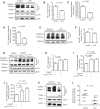
Astroglial FMRP regulates functional expression of GLT1 in vivo. A, B, A representative immunoblot of GLT1 (A) and its quantification (B) from cortex of astrocyte-specific cKO and control mice (P40). n = 6–7 mice/group. C, D, Total (C) and GLT1-mediated (D) functional glutamate uptake in the cortex of astrocyte-specific cKO and control mice; n = 6–8 mice/group. E, F, A representative immunoblot of GLAST (E) and its quantification (F) from the cortex of astrocyte-specific cKO and control mice (P40); n = 5 mice/group. G, H, A representative immunoblot of GLT1 (G) and its quantification (H) from the cortex of astrocyte-specific cON (CreERT+ Fmr1loxP-neo/y), control (CreERT− Fmr1loxP-neo/y), and WT mice (P40); n = 5 mice/group. I, J, Total (I) and GLT1-mediated (J) functional glutamate uptake in the cortex of astrocyte-specific cON (CreERT+ Fmr1loxP-neo/y), control (CreERT− Fmr1loxP-neo/y), and WT mice; n = 5 mice/group. DHK, 500 μm; TBOA, 500 μm. K, L, A representative immunoblot of human glutamate transporter EAAT2 (K) and its quantification (L) from human FXS and age-matched control postmortem cortical tissues; n = 5 patients/group. The age of each tissue sample is labeled next to the data point. The p values were determined using the Student's t test or one-way ANOVA with post hoc Bonferroni's test.
Figure 3.
Figure 3.
Dysregulated functional GLT1 expression enhances pyramidal neuronal excitability in cortex of astrocyte-specific cKO mice. A, A representative trace of neuronal firing recordings in layer 5 somatosensory neocortical neurons in astrocyte-specific cKO (CreERT+ Fmr1f/y) and control (CreERT− Fmr1f/y) cortical slices before and during bath application of GLT1 inhibitor DHK (10 μm). B, Quantitative summary of the neuronal firing rate in CreERT− Fmr1f/y and CreERT+ Fmr1f/y slices with 10 or 50 μm DHK; n = 14–28 neurons from 7–10 mice per group. C, A representative trace of neuronal firing recordings in layer 5 somatosensory neocortical neurons in GLT1+/+ and GLT1+/− cortical slices before and during the bath application of GLT1 inhibitor DHK (10 μm). D, Quantitative summary of neuronal firing rate in GLT1+/+ and GLT1+/− slices with 10 or 50 μm DHK; n = 16–21 neurons from 10 mice per group. E, A representative trace of neuronal firing recordings in layer 5 somatosensory neocortical neurons in CreERT− Fmr1loxP-neo/y and CreERT+ Fmr1loxP-neo/y cortical slices before and during the bath application of GLT1 inhibitor DHK (10 μm). F, Quantitative summary of the neuronal firing rate in CreERT− Fmr1loxP-neo/y and CreERT+ Fmr1loxP-neo/y slices with 10 or 50 μm DHK; n = 15–29 neurons from 8 to 10 mice per group. Segment of trace from CreERT+ Fmr1f/y, GLT1+/−, and CreERT− Fmr1loxP-neo/y slices shown in more expanded time scale. The p values were calculated using one-way ANOVA with post hoc Tukey's test.
Figure 4.
Figure 4.
Dendritic spine morphology and density in cortical pyramidal neurons of astrocyte-specific cKO mice. A, Representative apical and basal dendrites from cortical layers 2/3 of CreERT− Fmr1f/y and CreERT+ Fmr1f/y mice illustrated by Golgi staining. Scale bar, 2.5 μm. B, C, Spine density of apical (B) and basal (C) dendrites from CreERT− Fmr1f/y and CreERT+ Fmr1f/y cortical neurons; n = 23–24 dendrites/group from four mice. D, E, Cumulative probability curves of apical (D) and basal (E) spine length from CreERT− Fmr1f/y and CreERT+ Fmr1f/y cortical neurons; n = 46–50 spines from four mice per group. F, G, Length distribution of apical (F) and basal (G) spines of cortical pyramidal neurons from CreERT− Fmr1f/y and CreERT+ Fmr1f/y mice; n = 46–50 spines from four mice per group. H, Representative confocal and filament tracing Imaris images of basal dendritic spines of cortical pyramidal neuron dye-filled with neurobiotin-488 tracer. Scale bar, 10 μm. I, Quantification of basal dendritic spines of dye-filled cortical neurons from CreERT− Fmr1f/y and CreERT+ Fmr1f/y mice; n = 6 dye-filled dendrites/group. J, Cumulative probability curves of basal spine length from dye-filled pyramidal neurons of CreERT− Fmr1f/y and CreERT+ Fmr1f/y cortical slices; n = 52 spines from four mice per group. The p values were calculated using the Student's t test and two-way ANOVA with post hoc Bonferroni's test.
Figure 5.
Figure 5.
Dendritic spine morphology and density in cortical pyramidal neurons of astrocyte-specific cON mice. A, Representative apical and basal dendrites from cortical layers 2/3 of CreERT− Fmr1loxP-neo/y and CreERT+ Fmr1loxP-neo/y mice illustrated by Golgi staining. Scale bar, 2.5 μm. B, C, Spine density of apical (B) and basal (C) dendrites from CreERT− Fmr1loxP-neo/y and CreERT+ Fmr1loxP-neo/y cortical neurons; n = 14 dendrites/group from three mice. D, E, Cumulative probability curve of apical (D) and basal (E) spine length from CreERT− Fmr1loxP-neo/y and CreERT+ Fmr1loxP-neo/y cortical neurons; n = 56–69 spines from three mice per group. Both the trend line and individual sample points were shown on a cumulative probability curve. F, G, Length distribution of apical (F) and basal (G) spines of cortical pyramidal neurons from CreERT− Fmr1loxP-neo/y and CreERT+ Fmr1loxP-neo/y mice; n = 56–69 spines/group. The p values were determined using the Student's t test and two-way ANOVA with post hoc Bonferroni's test.
Figure 6.
Figure 6.
Altered protein synthesis in cortex of astrocyte-specific cKO and cON mice. A, Protein synthesis levels measured by metabolic labeling in Fmr1 KO, astrocyte-specific cKO, and control cortical slices; n = 10–11 slices from six mice per group. B, Representative confocal images of the immunostaining of p-s6P in CreERT− Fmr1f/y and CreERT+ Fmr1f/y cortical sections. White arrows, Prominent p-s6P-positive neurons. Scale bar, 50 μm. C, Quantification of p-s6P immunoreactivity in CreERT− Fmr1f/y and CreERT+ Fmr1f/y cortical neurons; n = 65–124 neurons from three to four mice per group. D, Immunoreactivity of p-s6P in cortical neurons from individual CreERT− Fmr1f/y and CreERT+ Fmr1f/y mice. E, Protein synthesis levels measured by metabolic labeling in astrocyte-specific cON and control mice; n = 10 slices from more than five mice per group. F, Representative confocal images of the immunostaining of p-s6P in CreERT− Fmr1loxP-neo/y and CreERT+ Fmr1loxP-neo/y cortical sections. White arrows, Prominent p-s6P-positive neurons. Scale bar, 50 μm. G, Quantification of p-s6P immunoreactivity in CreERT− Fmr1loxP-neo/y and CreERT+ Fmr1loxP-neo/y cortical neurons; n = 55–58 neurons from two to three mice per group. H, Immunoreactivity of p-s6P in cortical neurons from individual CreERT− Fmr1loxP-neo/y and CreERT+ Fmr1loxP-neo/y mice. A level of 10 A.U. is defined as the background p-s6P immunoreactivity. The p values were calculated using the Student's t test and one-way ANOVA with post hoc Tukey's test: *p < 0.05; **p < 0.01.
Figure 7.
Figure 7.
Pharmacological upregulation of functional GLT1 expression attenuates enhanced neuronal excitability in cortex of Fmr1 KO mice. A, Body weight changes of saline- and Cef-injected Fmr1 KO mice during early postnatal development; n = 14 mice/group. B, A representative immunoblot of GLT1 protein levels in total (T), intracellular (I), and membrane (M) fractions prepared from saline- or Cef-injected Fmr1 KO cortical tissues. C, Quantification of total and surface GLT1 protein levels in cortex of saline- or Cef-injected Fmr1 KO mice; n = 3 mice/group. D, E, Total (D) and GLT1-mediated (E) glutamate uptake from saline- or Cef-injected Fmr1 KO cortical tissues; n = 7–10 mice/group. F, GLT1-mediated functional glutamate uptake from saline- and Cef-injected WT (Fmr1+/y) cortical tissues; n = 8 mice/group. G, A representative trace of neuronal firing recordings in layer 5 somatosensory neocortical neurons in saline- or Cef-injected Fmr1 KO cortical slices before and during the bath application of GLT1 inhibitor DHK (10 μm). H, Quantitative summary of neuronal firing rates in saline- or Cef-injected Fmr1 KO cortical slices with 10 or 50 μm DHK; n = 12–28 neurons from 7 to 10 mice per group. The p values were determined using the Student's t test, one-way ANOVA with post hoc Tukey's test, and two-way ANOVA with post hoc Bonferroni's test.
Figure 8.
Figure 8.
Pharmacological upregulation of functional GLT1 expression corrects spine abnormalities in cortex of Fmr1 KO mice. A, Representative apical and basal dendrites from cortical layer 2/3 of saline- or Cef-injected Fmr1 KO cortical slices illustrated by Golgi staining. Scale bar, 2.5 μm. B, C, Spine density of apical (B) and basal (C) dendrites from saline- or Cef-injected Fmr1 KO cortical neurons; n = 13 dendrites from four mice per group. D, E, Cumulative probability curve of apical (D) and basal (E) spine length from saline- or Cef-injected Fmr1 KO cortical neurons; n = 79–140 spines from four mice per group. Both the trend line and individual sample points are shown on cumulative probability curve. F, G, Length distribution of apical (F) and basal (G) spines of cortical pyramidal neurons in saline- and Cef-injected Fmr1 KO mice; n = 79–140 spines from four mice per group. The p values were determined using the Student's t test, two-way ANOVA, and post hoc Bonferroni's test.

Similar articles

Cited by

References

    1. Amiri A, Sanchez-Ortiz E, Cho W, Birnbaum SG, Xu J, McKay RM, Parada LF. Analysis of FMR1 deletion in a subpopulation of post-mitotic neurons in mouse cortex and hippocampus. Autism Res. 2014;7:60–71. doi: 10.1002/aur.1342. - DOI - PubMed
    1. Bakker CE, Verheij C, Willemsen R, van der Helm R, Oerlemans F, Vermey M, Bygrave A, Hoogeveen AT, Oostra BA, Reyniers E, D'Hooge R, Cras P, Van Velzen D, Nagels G, Martin JJ, De Deyn PP, Darby JK, Willems PJ. Fmr1 knockout mice: a model to study fragile X mental retardation. Cell. 1994;78:23–33. - PubMed
    1. Bassell GJ, Warren ST. Fragile X syndrome: loss of local mRNA regulation alters synaptic development and function. Neuron. 2008;60:201–214. doi: 10.1016/j.neuron.2008.10.004. - DOI - PMC - PubMed
    1. Broekman ML, Comer LA, Hyman BT, Sena-Esteves M. Adeno-associated virus vectors serotyped with AAV8 capsid are more efficient than AAV-1 or -2 serotypes for widespread gene delivery to the neonatal mouse brain. Neuroscience. 2006;138:501–510. doi: 10.1016/j.neuroscience.2005.11.057. - DOI - PubMed
    1. Cahoy JD, Emery B, Kaushal A, Foo LC, Zamanian JL, Christopherson KS, Xing Y, Lubischer JL, Krieg PA, Krupenko SA, Thompson WJ, Barres BA. A transcriptome database for astrocytes, neurons, and oligodendrocytes: a new resource for understanding brain development and function. J Neurosci. 2008;28:264–278. doi: 10.1523/JNEUROSCI.4178-07.2008. - DOI - PMC - PubMed

Publication types

MeSH terms