. 2016 Jul 5;7(4):e00518-16.
doi: 10.1128/mBio.00518-16.

Shutdown of HIV-1 Transcription in T Cells by Nullbasic, a Mutant Tat Protein

Affiliations

Shutdown of HIV-1 Transcription in T Cells by Nullbasic, a Mutant Tat Protein

Hongping Jin et al. mBio. .

Abstract

Nullbasic is a derivative of the HIV-1 transactivator of transcription (Tat) protein that strongly inhibits HIV-1 replication in lymphocytes. Here we show that lentiviral vectors that constitutively express a Nullbasic-ZsGreen1 (NB-ZSG1) fusion protein by the eEF1α promoter led to robust long-term inhibition of HIV-1 replication in Jurkat cells. Although Jurkat-NB-ZSG1 cells were infected by HIV-1, no virus production could be detected and addition of phorbol ester 12-myristate 13-acetate (PMA) and JQ1 had no effect, while suberanilohydroxamic acid (SAHA) modestly stimulated virus production but at levels 300-fold lower than those seen in HIV-1-infected Jurkat-ZSG1 cells. Virus replication was not recovered by coculture of HIV-1-infected Jurkat-NB-ZSG1 cells with uninfected Jurkat cells. Latently infected Jurkat latent 6.3 and ACH2 cells treated with latency-reversing agents produced measurable viral capsid (CA), but little or none was made when they expressed NB-ZSG1. When Jurkat cells chronically infected with HIV-1 were transduced with lentiviral virus-like particles conveying NB-ZSG1, a >3-log reduction in CA production was observed. Addition of PMA increased virus CA production but at levels 500-fold lower than those seen in nontransduced Jurkat cells. Transcriptome sequencing analysis confirmed that HIV-1 mRNA was strongly inhibited by NB-ZSG1 but indicated that full-length viral mRNA was made. Analysis of HIV-1-infected Jurkat cells expressing NB-ZSG1 by chromatin immunoprecipitation assays indicated that recruitment of RNA polymerase II (RNAPII) and histone 3 lysine 9 acetylation were inhibited. The reduction of HIV-1 promoter-associated RNAPII and epigenetic changes in viral nucleosomes indicate that Nullbasic can inhibit HIV-1 replication by enforcing viral silencing in cells.

Importance: HIV-1 infection is effectively controlled by antiviral therapy that inhibits virus replication and reduces measurable viral loads in patients below detectable levels. However, therapy interruption leads to viral rebound due to latently infected cells that serve as a source of continued viral infection. Interest in strategies leading to a functional cure of HIV infection by permanent viral suppression, which may be achievable, is growing. Here we show that a mutant form of the HIV-1 Tat protein, referred to as Nullbasic, can inhibit HIV-1 transcription in infected Jurkat T cell to undetectable levels. Analysis shows that Nullbasic alters the epigenetic state of the HIV-1 long terminal repeat promoter, inhibiting its association with RNA polymerase II. This study indicates that key cellular proteins and pathways targeted here can silence HIV-1 transcription. Further elucidation could lead to functional-cure strategies by suppression of HIV transcription, which may be achievable by a pharmacological method.


Figures

FIG 1
FIG 1
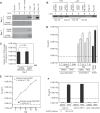
HIV-1 replication is not detected in Jurkat-NB-ZSG1 cells. (A) The Jurkat-NB-ZSG1 and Jurkat-ZSG1 stable cell lines or parental Jurkat cells were infected with HIV-1, and the infected cells were cultured for up to 64 days. Supernatant was sampled as indicated, and the concentration of CA was measured by ELISA with a threshold of detection of 7.8 pg/ml (•, solid line). Jurkat cells stably expressed ZSG1 (Δ, dotted line, right y axis) or NB-ZSG1 (•, dotted line, right y axis). Representative data from six independent experiments with similar results are shown. The symbol † indicates experiment was ended. (B) Cell lysates made from Jurkat-NB-ZSG1 or Jurkat-ZSG1 cells on the days shown were assayed by Western blotting with an anti-Tat (top panel), anti-tubulin (bottom panel), or anti-HIV-1 Gag antibody. (C) Total cellular RNA was isolated from Jurkat-ZSG1, Jurkat-NB-ZSG1, and Jurkat cells. HIV-1 mRNA was measured by qRT-PCR, and the values were normalized to the levels of endogenous GAPDH mRNA in the sample. A plus sign indicates that HIV-1 mRNA was not detected in the sample. Representative data from six independent experiments with similar results are shown. The RT-PCR assays were performed three times in triplicate. The data presented are mean values ± the standard deviations.
FIG 2
FIG 2
Jurkat-NB-ZSG1 cells harbor proviral HIV-1 DNA but no detectable viral mRNA. (A) PCR amplification of an HIV-1 env gene segment with total cellular DNA obtained from uninfected Jurkat, Jurkat-ZSG1, and Jurkat-NB-ZSG1 cells (lane 1) or from the same cell lines incubated with heat-inactivated (H.I.) virus (lane 2) or treated with or without nevirapine (NVP) for 2 h and infected with HIV-1NL4.3 (lanes 3 and 4). A PCR master mix alone was used as a negative (Neg.) control (lane 5) or with proviral DNA added as a positive (Pos.) control (Ctrl.) (lane 6). (B) Total cellular DNA obtained on days 28 and 64 from HIV-1-infected cell lines and assayed by endpoint PCR for env DNA. No viral DNA was detected in uninfected cells processed simultaneously (lanes 4 to 6). Negative and positive controls as in panel A are shown (lanes 7 and 8). (C) Total cellular DNA from day 3 HIV-1-infected or uninfected Jurkat-NB-ZSG1 and Jurkat-ZSG1 cells was assayed by Alu-PCR for integrated proviral DNA. The relative copy number was normalized to the CCR5 gene level in each sample. (D) On the days indicated, 1 nM PMA or DMSO (carrier) was added to Jurkat-NB-ZSG1 and Jurkat-ZSG1 cell cultures and incubated for 24 h. The soluble CA in the culture supernatant was then quantified by ELISA (the dotted line shows the limit of detection). ACH2 cell were used as a control for activation of HIV-1 gene expression by PMA. (E) Coculture of HIV-1-infected Jurkat-NB-ZSG1 or Jurkat-ZSG1 cells with uninfected Jurkat cells at a ratio of 1:1 or 1:100, respectively, for 28 days. The CA level in the supernatant was assayed by ELISA. (F) JLat 6.3, JLat 6.3-ZSG1, and JLat 6.3-NB-ZSG1 cells were incubated with 10 nM PMA for 24 h, and the CA levels in the supernatants were assayed by ELISA (the dotted line shows the limit of detection). The GFP-positive cell population was measured by flow cytometry. The assays in panels E and F were performed three times in triplicate, and the mean values and standard deviations are shown. Experiments representative of at least three independent experiments with similar results are shown. The P value in panel C was calculated with a Student t test.
FIG 3
FIG 3
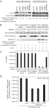
Postinfection treatment of HIV-1-infected Jurkat cells with NB-ZSG1 VLPs inhibits HIV-1 gene expression. (A) Jurkat cells were infected with HIV-1 and cultured for 30 days. The infected cells were transduced with VLPs conveying NB-ZSG1 or ZSG1 so that >95% of the cells were positive for ZSG1 or NB-SZG1 (three successive transductions [TD1, TD2, and TD3], as shown). HIV-1 CA in supernatant from HIV-1-infected Jurkat cells or NB-ZSG1-treated or ZSG1-treated, HIV-1-infected Jurkat cells was quantified by ELISA (left y axis, solid lines) or NB-ZSG1 and ZSG1 expression level in cells by flow cytometry (right y axis, dotted lines). An experiment representative of at least three independent experiments with similar results is shown. (B) Western blot analysis of the cell lysates from panel A with anti-Tat and anti-β-tubulin antibodies. The molecular masses of the NB-ZSG1 fusion protein and β-tubulin are indicated. (C) Total cellular RNA was isolated from HIV-1-infected Jurkat; NB-ZSG1-treated or ZSG1-treated, HIV-1-infected Jurkat; or uninfected Jurkat cells. qRT-PCR assays were performed with primers specific for the 5′ UTR or for sequences in env. The relative level of viral 5′ UTR RNA in each sample was normalized to the amount of GAPDH mRNA in the sample. (D) Total cellular DNA was isolated and subjected to Alu-PCR analysis for the cell lines. In panels C and D, mean values and standard deviations from experiments representative of three independent experiments performed in triplicate are shown. A two-tailed Student t test with equal variance was used to calculate the P value. (E) PMA stimulation of HIV-1 gene expression in an NB-ZSG1-treated, HIV-1-infected Jurkat cell population. Uninfected Jurkat cells, HIV-1-infected Jurkat cells treated with ZSG1 or NB-ZSG1, and U1 cells were incubated with 1 nM PMA for 24 h, and the concentration of CA in the supernatant was measured by ELISA. The experiment was performed in triplicate and repeated three times with similar results. The mean values and standard deviations of a representative experiment are shown.
FIG 4
FIG 4
Virus production is restored in NB-ZSG1-treated, HIV-1-infected Jurkat cells by knockdown of NB-ZSG1. (A) Stable HeLa cell lines expressing NB-mCherry or Tat-mCherry were transfected alone or together with two siRNAs (siNB1i and siNB2i) or with a scrambled negative-control (shCTL) siRNA sequence. Cell lysates were analyzed by Western blotting with anti-Tat and anti-β-tubulin antibodies as indicated. A digital image of each Western blot assay was processed with ImageJ software, and the relative Tat signal level was normalized to the β-tubulin signal level in the same sample. (B) Lentiviral vectors that expressed shRNA based on siNB1i or siCTL sequences or the siCTL sequences were used to transduce Jurkat-NB-ZSG1 cells as indicated. Cell lysates were prepared from transduced cells and probed by Western blotting with anti-Tat, anti-mCherry, and anti-β-tubulin antibodies. The relative Tat signal in each sample was measured as described for panel A. (C) The HIV-1 CA present in culture supernatant was quantified by ELISA. Mean values and standard deviations from three independent experiments are shown. (D) Total cellular RNA was isolated from HIV-1-infected Jurkat; NB-ZSG1- or ZSG1-treated, HIV-1-infected Jurkat; or uninfected Jurkat cells. qRT-PCR assays were performed with primers specific for the 5′ UTR. The relative level of viral 5′ UTR RNA in each sample was normalized to the amount of GAPDH mRNA in the sample.
FIG 5
FIG 5
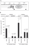
ChIP assays for a DNA-protein complex in HIV-1-infected Jurkat cells treated with ZSG1 or NB-ZSG1. (A) Schematic of the HIV-1 LTR promoter from −200 to +200. The binding sites for NF-κB, SP1, and the TATA binding protein (TBP) and the location of nuc-1 are indicated. Viral DNA in the U5 region was detected with primers specific for HIVNL43 (indicated by arrowheads). (B) ChIP assays were performed with an anti-RNAPII or anti-H3K9ac antibody and primers for nuc-1. The gene for GAPDH was used as a reference. The relative enrichment of viral DNA in the immunoprecipitation reaction was calculated as a signal over the background with the same DNA-protein complex and an isotype-matched anti-GFP antibody. The experiments were performed three times, and the mean values and stand deviations are shown. The P values were calculated with a Student t test.

Similar articles

Cited by

References

    1. Van Lint C, Bouchat S, Marcello A. 2013. HIV-1 transcription and latency: an update. Retrovirology 10:67. doi:10.1186/1742-4690-10-67. - DOI - PMC - PubMed
    1. Coffin JM, Huges SH, Varmus HE. 1997. Retroviruses. Cold Spring Harbor Laboratory Press, Cold Spring Harbor, NY. - PubMed
    1. Garber ME, Wei P, Jones KA. 1998. HIV-1 Tat interacts with cyclin T1 to direct the P-TEFb CTD kinase complex to TAR RNA. Cold Spring Harb Symp Quant Biol 63:371–380. doi:10.1101/sqb.1998.63.371. - DOI - PubMed
    1. Sobhian B, Laguette N, Yatim A, Nakamura M, Levy Y, Kiernan R, Benkirane M. 2010. HIV-1 Tat assembles a multifunctional transcription elongation complex and stably associates with the 7SK snRNP. Mol Cell 38:439–451. doi:10.1016/j.molcel.2010.04.012. - DOI - PMC - PubMed
    1. Ott M, Geyer M, Zhou Q. 2011. The control of HIV transcription: keeping RNA polymerase II on track. Cell Host Microbe 10:426–435. doi:10.1016/j.chom.2011.11.002. - DOI - PMC - PubMed

Publication types

MeSH terms

Substances

LinkOut - more resources