

Phenylboronic Acid-Mediated Tumor Targeting of Chitosan Nanoparticles
- PMID: 27375786
- PMCID: PMC4924506
- DOI: 10.7150/thno.15156
Phenylboronic Acid-Mediated Tumor Targeting of Chitosan Nanoparticles
Abstract
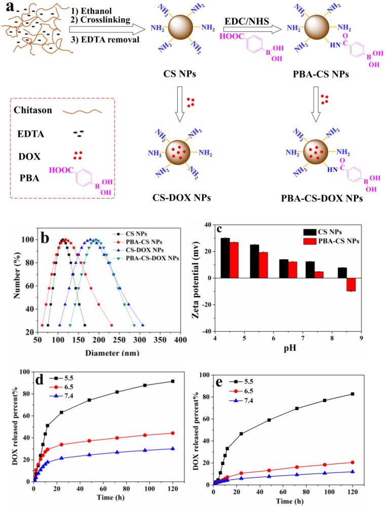
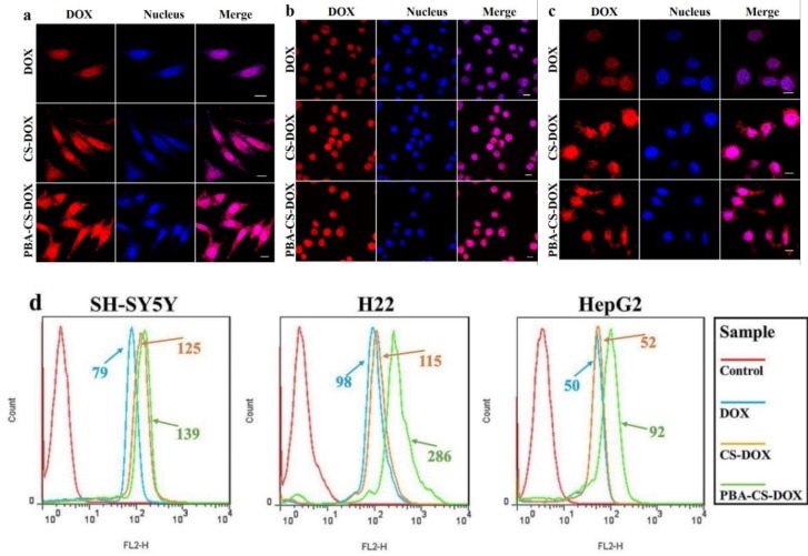
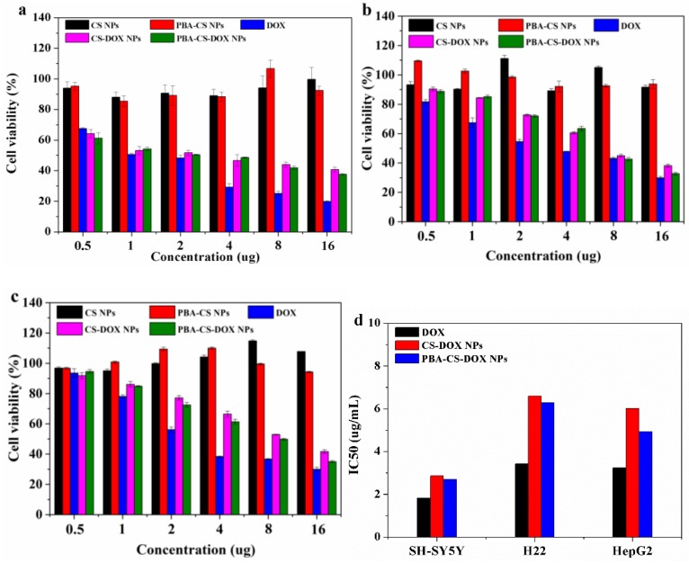
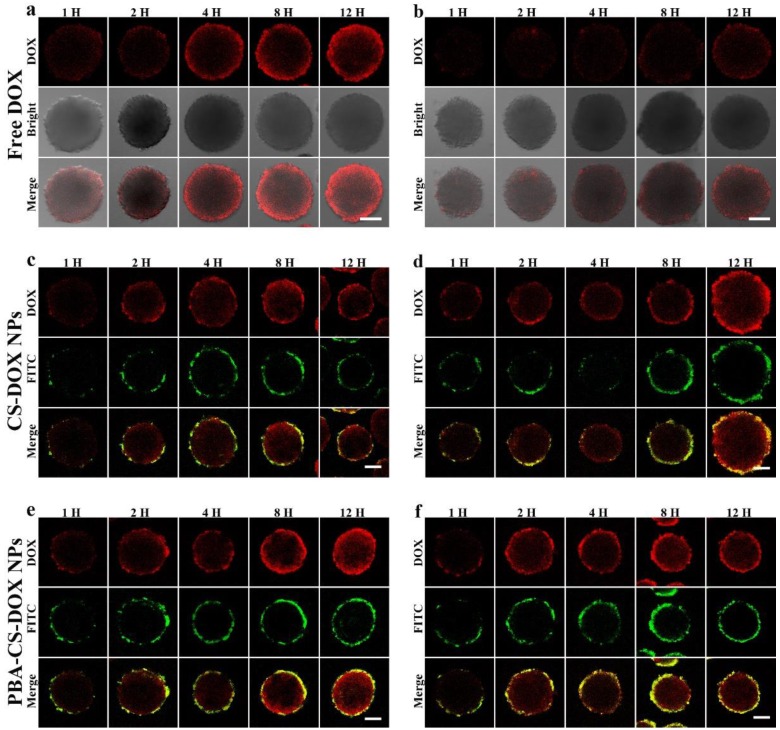
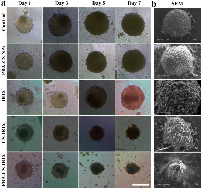
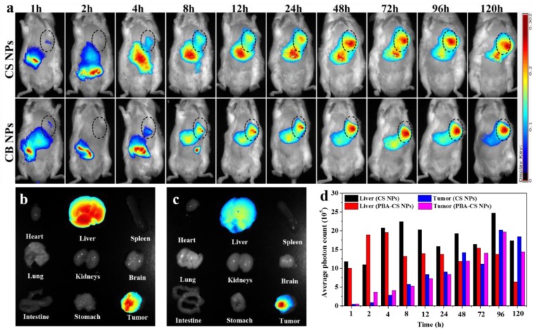
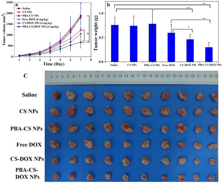
The phenylboronic acid-conjugated chitosan nanoparticles were prepared by particle surface modification. The size, zeta potential and morphology of the nanoparticles were characterized by dynamic light scattering, zeta potential measurement and transmission electron microscopy. The cellular uptake, tumor penetration, biodistribution and antitumor activity of the nanoparticles were evaluated by using monolayer cell model, 3-D multicellular spheroid model and H22 tumor-bearing mice. The incorporation of phenylboronic acid group into chitosan nanoparticles impart a surface charge-reversible characteristic to the nanoparticles. In vitro evaluation using 2-D and 3-D cell models showed that phenylboronic acid-decorated nanoparticles were more easily internalized by tumor cells compared to non-decorated chitosan nanoparticles, and could deliver more drug into tumor cells due to the active targeting effect of boronic acid group. Furthermore, the phenylboronic acid-decorated nanoparticles displayed a deeper penetration and persistent accumulation in the multicellular spheroids, resulting in better inhibition growth to multicellular spheroids than non-decorated nanoparticles. Tumor penetration, drug distribution and near infrared fluorescence imaging revealed that phenylboronic acid-decorated nanoparticles could penetrate deeper and accumulate more in tumor area than non-decorated ones. In vivo antitumor examination demonstrated that the phenylboronic acid-decorated nanoparticles have superior efficacy in restricting tumor growth and prolonging the survival time of tumor-bearing mice than free drug and drug-loaded chitosan nanoparticles.
Keywords: 3-D multicellular spheroids; Chitosan nanoparticles; antitumor.; phenylboronic acid.
Conflict of interest statement
Competing Interests: The authors have no competing interests.
Figures


Similar articles
-
Drug delivery to solid tumors: the predictive value of the multicellular tumor spheroid model for nanomedicine screening.Int J Nanomedicine. 2017 Oct 31;12:7993-8007. doi: 10.2147/IJN.S146927. eCollection 2017. Int J Nanomedicine. 2017. PMID: 29184400 Free PMC article. Review.
-
Doxorubicin delivery to 3D multicellular spheroids and tumors based on boronic acid-rich chitosan nanoparticles.Biomaterials. 2013 Jun;34(19):4667-79. doi: 10.1016/j.biomaterials.2013.03.008. Epub 2013 Mar 26. Biomaterials. 2013. PMID: 23537667
-
3-Carboxyphenylboronic acid-modified carboxymethyl chitosan nanoparticles for improved tumor targeting and inhibitory.Eur J Pharm Biopharm. 2017 Apr;113:168-177. doi: 10.1016/j.ejpb.2016.12.034. Epub 2017 Jan 13. Eur J Pharm Biopharm. 2017. PMID: 28089786
-
Phenylboronic acid-decorated gelatin nanoparticles for enhanced tumor targeting and penetration.Nanotechnology. 2016 Sep 23;27(38):385101. doi: 10.1088/0957-4484/27/38/385101. Epub 2016 Aug 11. Nanotechnology. 2016. PMID: 27514078
-
Dextran-doxorubicin/chitosan nanoparticles for solid tumor therapy.Wiley Interdiscip Rev Nanomed Nanobiotechnol. 2009 Jul-Aug;1(4):415-25. doi: 10.1002/wnan.43. Wiley Interdiscip Rev Nanomed Nanobiotechnol. 2009. PMID: 20049807 Review.
Cited by
-
mTHPC-Loaded Extracellular Vesicles Significantly Improve mTHPC Diffusion and Photodynamic Activity in Preclinical Models.Pharmaceutics. 2020 Jul 17;12(7):676. doi: 10.3390/pharmaceutics12070676. Pharmaceutics. 2020. PMID: 32709026 Free PMC article.
-
ATP-activated decrosslinking and charge-reversal vectors for siRNA delivery and cancer therapy.Theranostics. 2018 Sep 9;8(17):4604-4619. doi: 10.7150/thno.26889. eCollection 2018. Theranostics. 2018. PMID: 30279726 Free PMC article.
-
Systematic Review of Cancer Targeting by Nanoparticles Revealed a Global Association between Accumulation in Tumors and Spleen.Int J Mol Sci. 2021 Dec 1;22(23):13011. doi: 10.3390/ijms222313011. Int J Mol Sci. 2021. PMID: 34884816 Free PMC article.
-
Drug delivery to solid tumors: the predictive value of the multicellular tumor spheroid model for nanomedicine screening.Int J Nanomedicine. 2017 Oct 31;12:7993-8007. doi: 10.2147/IJN.S146927. eCollection 2017. Int J Nanomedicine. 2017. PMID: 29184400 Free PMC article. Review.
-
In vitro and in vivo antitumour effects of phenylboronic acid against mouse mammary adenocarcinoma 4T1 and squamous carcinoma SCCVII cells.J Enzyme Inhib Med Chem. 2017 Dec;32(1):1299-1304. doi: 10.1080/14756366.2017.1384823. J Enzyme Inhib Med Chem. 2017. PMID: 29072095 Free PMC article.
References
-
- Alexis F, Pridgen EM, Langer R, Farokhzad OC. Nanoparticle technologies for cancer therapy. Drug Delivery: Springer. 2010;197:55–86. - PubMed
-
- Gref R, Minamitake Y, Peracchia MT, Trubetskoy V, Torchilin V, Langer R. Biodegradable long-circulating polymeric nanospheres. Science. 1994;263:1600–3. - PubMed
-
- Farokhzad OC, Langer R. Impact of Nanotechnology on Drug Delivery. ACS Nano. 2009;3:16–20. - PubMed
-
- Youan B-BC. Impact of nanoscience and nanotechnology on controlled drug delivery. Nanomedicine (Lond) 2008;3:401–6. - PubMed
Publication types
MeSH terms
Substances
LinkOut - more resources
Full Text Sources
Other Literature Sources
Medical
