

Selective depletion of CD11c+ CD11b+ dendritic cells partially abrogates tolerogenic effects of intravenous MOG in murine EAE
- PMID: 27338697
- PMCID: PMC5514845
- DOI: 10.1002/eji.201546274
Selective depletion of CD11c+ CD11b+ dendritic cells partially abrogates tolerogenic effects of intravenous MOG in murine EAE
Abstract
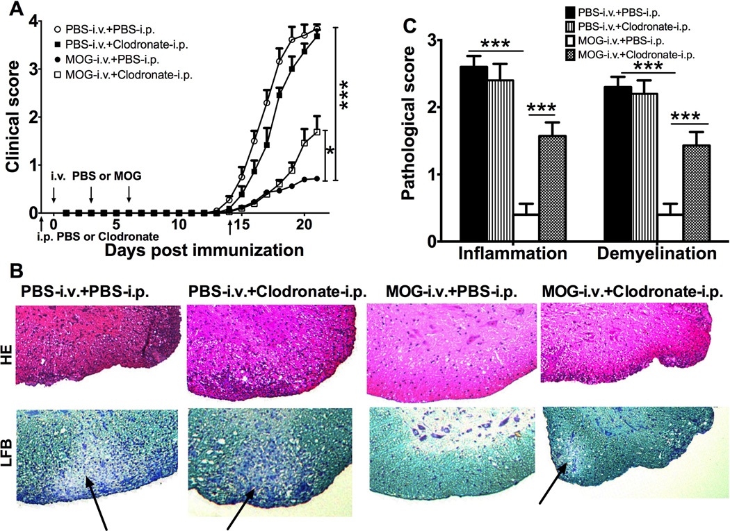
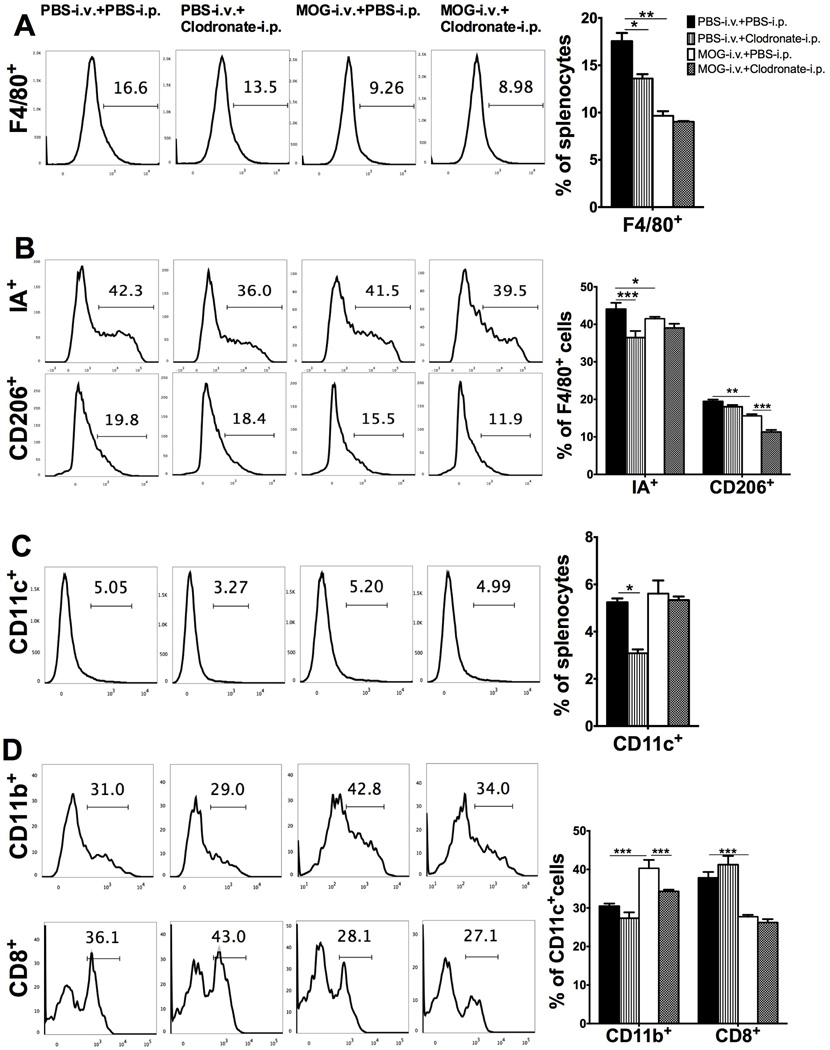
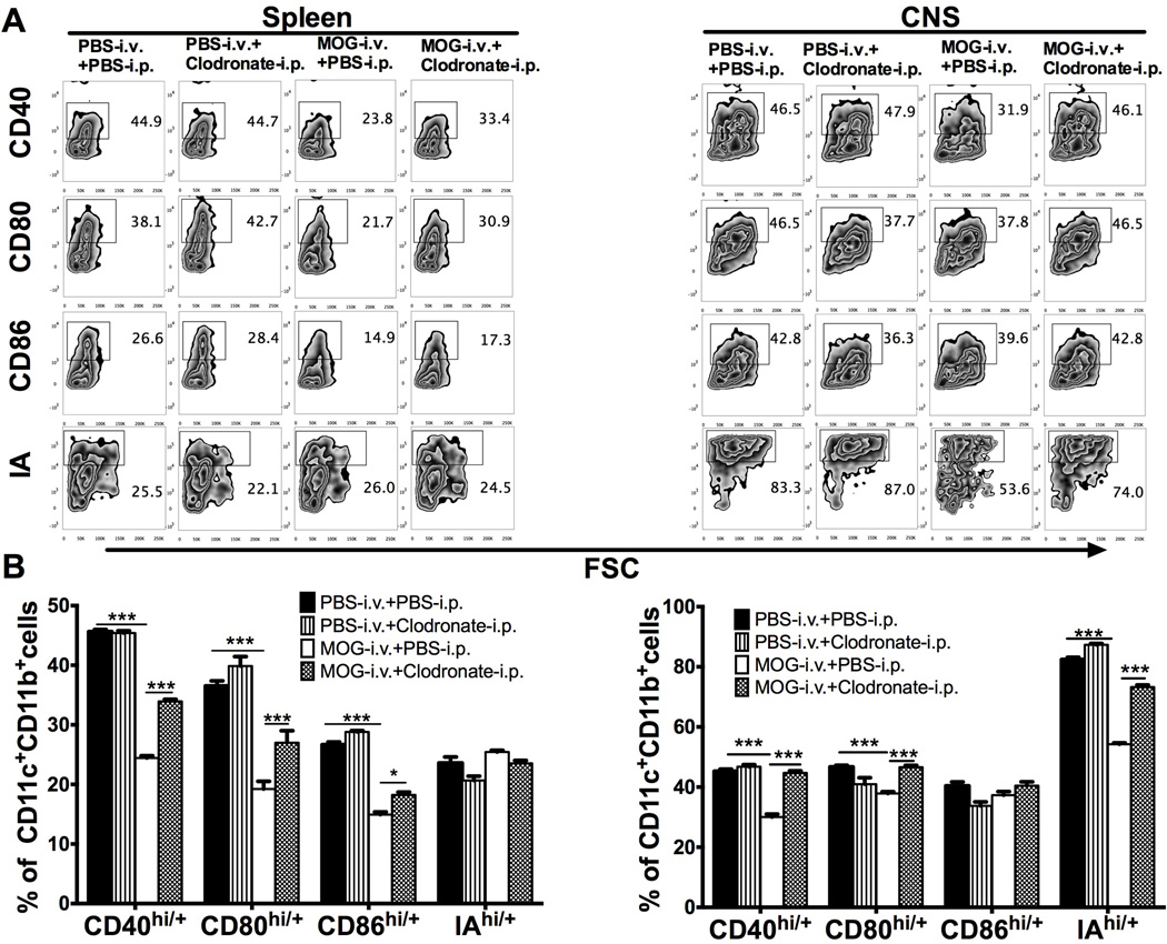
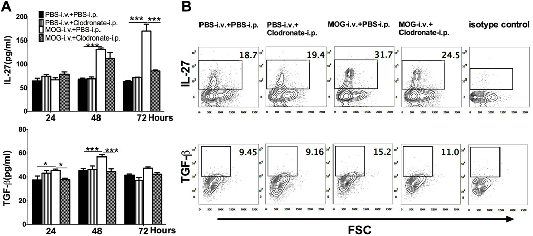
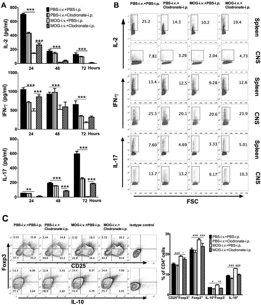
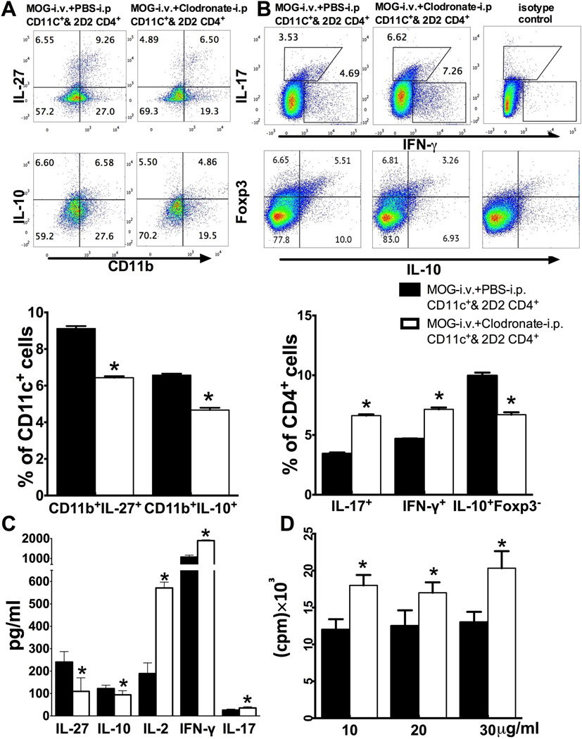
Intravenous (i.v.) injection of a soluble myelin antigen can induce tolerance, which effectively ameliorates experimental autoimmune encephalomyelitis (EAE). We have previously shown that i.v. myelin oligodendrocyte glycoprotein (MOG) induces tolerance in EAE and expands a subpopulation of tolerogenic CD11c+ CD11b+ dendritic cells (DCs) with an immature phenotype having low expression of IA and co-stimulatory molecules CD40, CD86, and CD80. Here, we further investigate the role of tolerogenic DCs in i.v. tolerance by injecting clodronate-loaded liposomes, which selectively deplete CD11c+ CD11b+ and immature DCs, but not CD11c+ CD8+ DCs and mature DCs. I.v. MOG-induced suppression of EAE was partially, yet significantly, blocked by CD11c+ CD11b+ DC depletion. While i.v. MOG inhibited IA, CD40, CD80, CD86 expression and induced TGF-β, IL-27, IL-10 production in CD11c+ CD11b+ DCs, these effects were abrogated after injection of clodronate-loaded liposomes. Depletion of CD11c+ CD11b+ DCs also precluded i.v. autoantigen-induced T-cell tolerance, such as decreased production of IL-2, IFN-γ, IL-17 and numbers of IL-2+ , IFN-γ+ , and IL-17+ CD4+ T cells, as well as an increased proportion of CD4+ CD25+ Foxp3+ regulatory T cells and CD4+ IL-10+ Foxp3- Tr1 cells. CD11c+ CD11b+ DCs, through low expression of IA and costimulatory molecules as well as high expression of TGF-β, IL-27, and IL-10, play an important role in i.v. tolerance-induced EAE suppression.
Keywords: Dendritic cell; Experimental autoimmune encephalomyelitis; Immune tolerance; Multiple sclerosis.
Published 2016. This article is a U.S. Government work and is in the public domain in the USA.
Conflict of interest statement
The authors declare no financial or commercial conflict of interest.
Figures

Similar articles
-
CD11c+CD11b+ dendritic cells play an important role in intravenous tolerance and the suppression of experimental autoimmune encephalomyelitis.J Immunol. 2008 Aug 15;181(4):2483-93. doi: 10.4049/jimmunol.181.4.2483. J Immunol. 2008. PMID: 18684939 Free PMC article.
-
Immune tolerance induced by intravenous transfer of immature dendritic cells via up-regulating numbers of suppressive IL-10(+) IFN-γ(+)-producing CD4(+) T cells.Immunol Res. 2013 May;56(1):1-8. doi: 10.1007/s12026-012-8382-7. Immunol Res. 2013. PMID: 23292714 Free PMC article.
-
Galectin-1 is essential for the induction of MOG35-55 -based intravenous tolerance in experimental autoimmune encephalomyelitis.Eur J Immunol. 2016 Jul;46(7):1783-96. doi: 10.1002/eji.201546212. Epub 2016 May 25. Eur J Immunol. 2016. PMID: 27151444 Free PMC article.
-
Aspirin and the induction of tolerance by dendritic cells.Handb Exp Pharmacol. 2009;(188):197-213. doi: 10.1007/978-3-540-71029-5_9. Handb Exp Pharmacol. 2009. PMID: 19031027 Review.
-
Tolerogenic dendritic cells induced by vitamin D receptor ligands enhance regulatory T cells inhibiting autoimmune diabetes.Ann N Y Acad Sci. 2003 Apr;987:258-61. doi: 10.1111/j.1749-6632.2003.tb06057.x. Ann N Y Acad Sci. 2003. PMID: 12727648 Review.
Cited by
-
Impact of disease-modifying therapy on dendritic cells and exploring their immunotherapeutic potential in multiple sclerosis.J Neuroinflammation. 2022 Dec 12;19(1):298. doi: 10.1186/s12974-022-02663-z. J Neuroinflammation. 2022. PMID: 36510261 Free PMC article. Review.
-
Tolerogenic Dendritic Cell-Based Approaches in Autoimmunity.Int J Mol Sci. 2021 Aug 5;22(16):8415. doi: 10.3390/ijms22168415. Int J Mol Sci. 2021. PMID: 34445143 Free PMC article. Review.
-
Dendritic Cells As Inducers of Peripheral Tolerance.Trends Immunol. 2017 Nov;38(11):793-804. doi: 10.1016/j.it.2017.07.007. Epub 2017 Aug 18. Trends Immunol. 2017. PMID: 28826942 Free PMC article. Review.
-
Antigen-specific airway IL-33 production depends on FcγR-mediated incorporation of the antigen by alveolar macrophages in sensitized mice.Immunology. 2018 Sep;155(1):99-111. doi: 10.1111/imm.12931. Epub 2018 Apr 19. Immunology. 2018. PMID: 29569388 Free PMC article.
-
Activation of Transcription Factor 4 in Dendritic Cells Controls Th1/Th17 Responses and Autoimmune Neuroinflammation.J Immunol. 2021 Sep 1;207(5):1428-1436. doi: 10.4049/jimmunol.2100010. Epub 2021 Aug 4. J Immunol. 2021. PMID: 34348977 Free PMC article.
References
-
- Grigoriadis N, van Pesch V. A basic overview of multiple sclerosis immunopathology. Eur J Neurol. 2015;22(Suppl 2):3–13. - PubMed
-
- Hilliard BA, Kamoun M, Ventura E, Rostami A. Mechanisms of suppression of experimental autoimmune encephalomyelitis by intravenous administration of myelin basic protein: role of regulatory spleen cells. Exp Mol Pathol. 2000;68:29–37. - PubMed
-
- Luchtman DW, Ellwardt E, Larochelle C, Zipp F. IL-17 and related cytokines involved in the pathology and immunotherapy of multiple sclerosis: Current and future developments. Cytokine Growth Factor Rev. 2014;25:403–413. - PubMed
Publication types
MeSH terms
Substances
Grants and funding
LinkOut - more resources
Full Text Sources
Other Literature Sources
Research Materials
