. 2016 Jul 5;113(27):7557-62.
doi: 10.1073/pnas.1603310113. Epub 2016 Jun 22.

E3 ubiquitin ligase RFWD2 controls lung branching through protein-level regulation of ETV transcription factors

Affiliations

E3 ubiquitin ligase RFWD2 controls lung branching through protein-level regulation of ETV transcription factors

Yan Zhang et al. Proc Natl Acad Sci U S A. .

Abstract

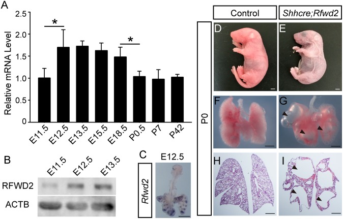
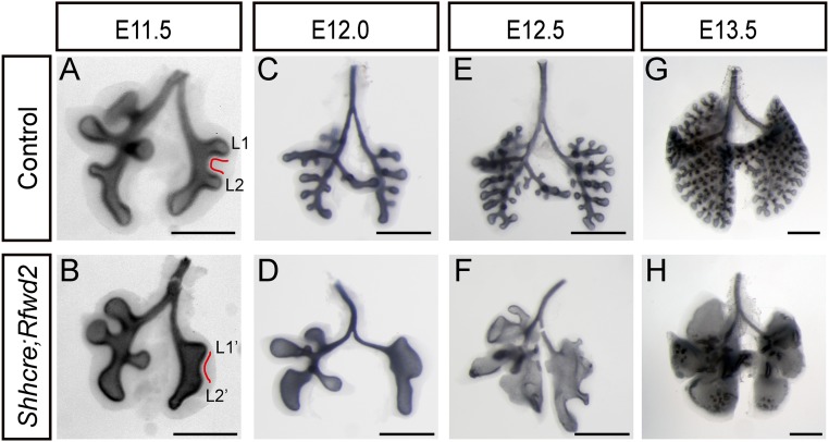
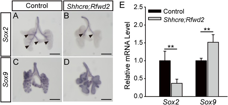
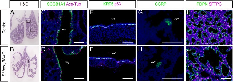
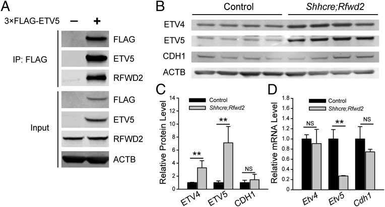
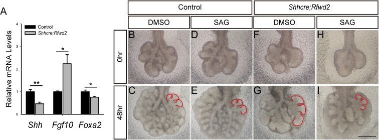
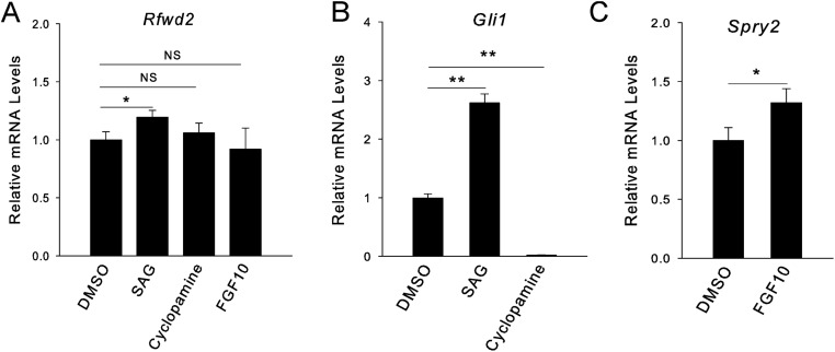
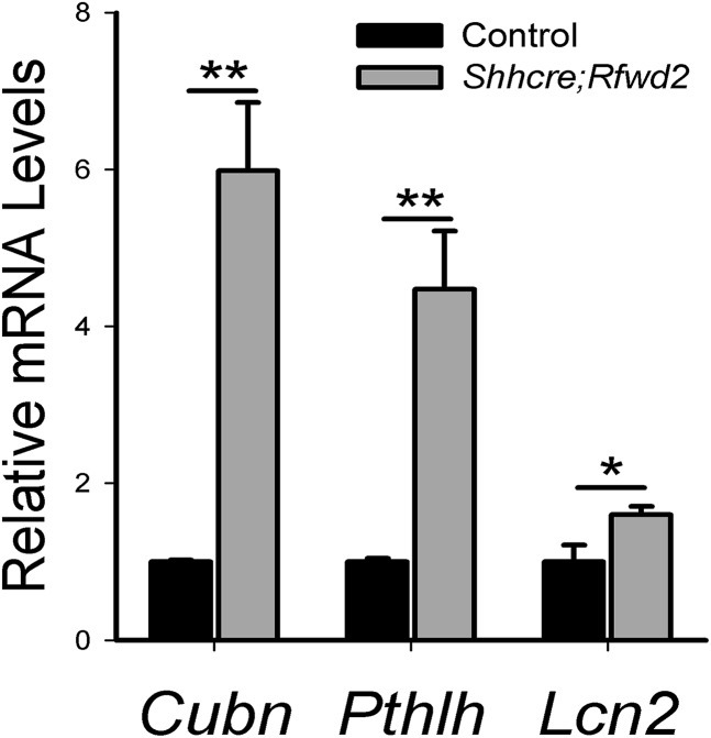
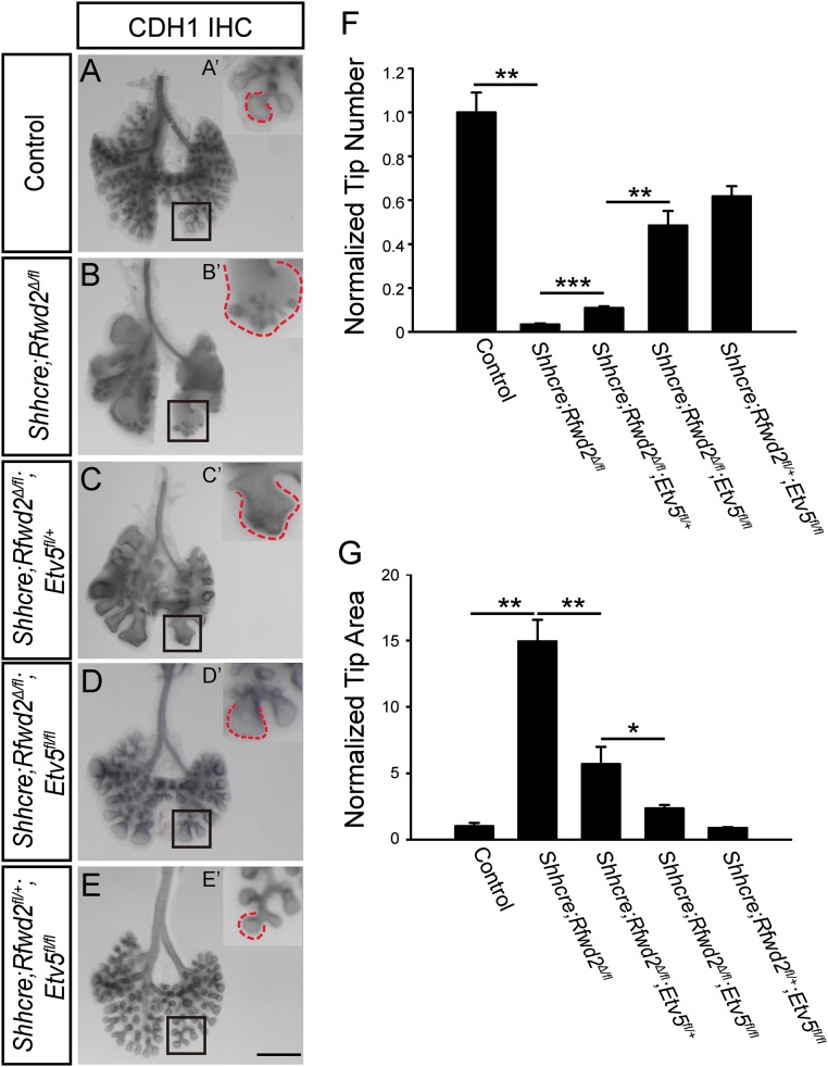
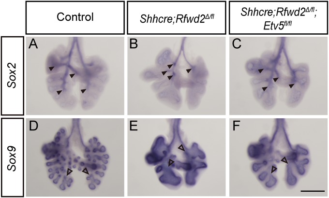
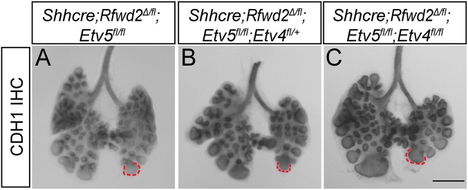
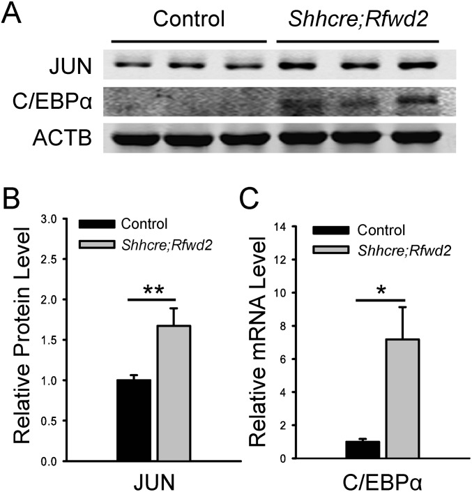
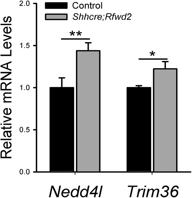
The mammalian lung is an elaborate branching organ, and it forms following a highly stereotypical morphogenesis program. It is well established that precise control at the transcript level is a key genetic underpinning of lung branching. In comparison, little is known about how regulation at the protein level may play a role. Ring finger and WD domain 2 (RFWD2, also termed COP1) is an E3 ubiquitin ligase that modifies specific target proteins, priming their degradation via the ubiquitin proteasome system. RFWD2 is known to function in the adult in pathogenic processes such as tumorigenesis. Here, we show that prenatal inactivation of Rfwd2 gene in the lung epithelium led to a striking halt in branching morphogenesis shortly after secondary branch formation. This defect is accompanied by distalization of the lung epithelium while growth and cellular differentiation still occurred. In the mutant lung, two E26 transformation-specific (ETS) transcription factors essential for normal lung branching, ETS translocation variant 4 (ETV4) and ETV5, were up-regulated at the protein level, but not at the transcript level. Introduction of Etv loss-of-function alleles into the Rfwd2 mutant background attenuated the branching phenotype, suggesting that RFWD2 functions, at least in part, through degrading ETV proteins. Because a number of E3 ligases are known to target factors important for lung development, our findings provide a preview of protein-level regulatory network essential for lung branching morphogenesis.

Keywords: COP1; E3 ubiquitin ligases; ETV transcription factors; RFWD2; lung branching.


Conflict of interest statement

The authors declare no conflict of interest.

Figures

Fig. 1.
Fig. 1.
Rfwd2 is expressed in the developing lung epithelium, and inactivation leads to lung defects and lethality at birth. (A) qRT-PCR of Rfwd2 mRNA levels in wild-type lungs from E11.5 to postnatal day (P) 42 (adult). Data were normalized to Actb (also termed β-actin) and then to the expression level at E11.5. The quantification was carried out in n ≥ 3 samples for each stage. *P < 0.05. (B) Western blot of RFWD2 protein levels in wild-type lungs at E11.5, E12.5, and E13.5. ACTB was used as a loading control. (C) Representative whole-mount RNA in situ hybridization of Rfwd2 in E12.5 wild-type lung indicating expression primarily in the lung epithelium. Intact pups (D and E), whole lung (F and G), or H&E-stained sections (H and I) of controls and Shhcre;Rfwd2 mutants at birth are shown. Arrowheads indicate large air sacs. (Scale bars: 500 μm.)
Fig. 2.
Fig. 2.
Inactivation of Rfwd2 in the lung epithelium results in defects in lung branching morphogenesis. Representative control (A, C, E, and G) and Shhcre;Rfwd2 mutant (B, D, F, and H) whole lungs from E11.5 to E13.5 with the epithelium outlined by anti-CDH1 (also termed E-cadherin) immunohistochemical staining. (A and B) Red lines outline the epithelium between lateral secondary branch (L) 1 and L2. (Scale bars: 500 μm.)
Fig. 3.
Fig. 3.
Inactivation of Rfwd2 altered proximal–distal airway patterning. (AD) Representative RNA in situ hybridization for Sox2 or Sox9 at E12.5 in Shhcre;Rfwd2 mutants and littermate controls. Arrowheads indicate the distal ends of the Sox2 domain. Sox9 expression in the trachea and extrapulmonary bronchi is in the mesenchymal precursors of the cartilage but not in the epithelial cells, as is its distal expression domain. (Scale bars: 500 μm.) (E) mRNA quantification by qRT-PCR of E12.5 lungs (Sox2: 1.00 ± 0.26 for controls, 0.37 ± 0.12 for Shhcre;Rfwd2 mutants, P = 0.004; Sox9: 1.00 ± 0.068 for controls, 1.51 ± 0.21 for Shhcre;Rfwd2 mutants, P = 0.005). Quantification was carried out in n ≥ 3 samples. **P < 0.01.
Fig. S1.
Fig. S1.
Cell differentiation occurs in Shhcre;Rfwd2 mutants. (A and B) H&E-stained lung sections from E18.5 lungs. Boxes delineate approximate areas stained for C and D (box with solid lines) or I and J (box with dashed lines). (CJ) Immunostaining of markers for club cells (SCGB1A1), ciliated cells (Ace-Tub), basal cells (KRT5, p63), and pulmonary neuroendocrine cells (CGRP) in the airways, and for type I (PDPN) and type II (SFTPC) cells in the alveolar regions at E18.5. AW, airway. (Scale bars: 100 μm.)
Fig. 4.
Fig. 4.
Inactivation of Rfwd2 led to increased expression of ETV4 and ETV5 proteins. (A) ETV5 is immunoprecipitated in the same complex as RFWD2. MLE-12 cells were transfected with 3× FLAG-ETV5 (+) or 3× FLAG control (−) expression vector, immunoprecipitated with an anti-FLAG antibody. Either the immunoprecipitated products or inputs before immunoprecipitation (IP) were immunoblotted with the indicated antibodies. (B) Western blot of ETV4, ETV5, and CDH1 proteins in E12.5 lungs with the indicated genotypes. ACTB was used as a loading control. (C) Quantification of Western blot normalized to ACTB. The density of bands was measured using the Image Studio Lite program. **P < 0.01. NS, not significant. (D) Quantification of mRNA expression of Etv4, Etv5, and Cdh1 by qRT-PCR in E12.5 lungs. Quantification was carried out in n ≥ 3 samples. **P < 0.01.
Fig. S2.
Fig. S2.
SAG treatment of Shhcre;Rfwd2 mutant lungs led to attenuation of the tip dilation phenotype. (A) qRT-PCR analysis of genes Shh, Fgf10, and Foxa2 in Shhcre;Rfwd2 mutants compared with control at E13.5. n = 3 in each group. *P < 0.05; **P < 0.01. Representative images of E11.5 lungs cultured for 48 h in either DMSO (B, C, F, and G) or SAG (50 nM) (D, E, H, and I) are shown. Red lines outline the branch tips. (Scale bars: 500 μm.)
Fig. S3.
Fig. S3.
SHH-induced Rfwd2 expression in wild-type cultured lungs. A qRT-PCR analysis of the transcript level of Rfwd2 (A), Gli1 (B), and Spry2 (C) of E11.5 wild-type lungs cultured for 48 h in SAG (1 μM), SHH signaling inhibitor cyclopamine (500 nM), or FGF10 recombinant protein (500 ng/mL) was performed. Gli1 and Spry2 transcripts served as readouts of SHH or FGF activity, respectively. *P < 0.05; **P < 0.01. NS, not significant.
Fig. S4.
Fig. S4.
Expression of some ETV-regulated genes shows opposite change in Shhcre;Rfwd2 mutant lungs. A qRT-PCR analysis of genes Cubn, Pthlh, and Lcn2 in Shhcre;Rfwd2 mutants compared with control at E13.5 is shown. n = 3 in each group. *P < 0.05; **P < 0.01.
Fig. 5.
Fig. 5.
Introduction of Etv5 mutation allele partially reversed the Shhcre;Rfwd2 branching phenotype. (AE) Representative E13.5 lungs of indicated genotypes with the epithelium outlined by anti-CDH1 immunohistochemical staining. The boxed areas in AE are shown at high magnification in A′–E′. Dashed lines indicated the baselines of branch tips. (Scale bar: 500 μm.) (F) Introducing the Etv5 mutant allele attenuated the branch tip number phenotype. For quantifying tip number, the epithelial tips of the left lobe were manually counted. Data were normalized to control samples. Quantification was carried out in n = 3 samples for each genotype (tip number: 1.00 ± 0.09 for controls, 0.03 ± 0.004 for Shhcre/+;Rfwd2Δ/fl, 0.11 ± 0.01 for Shhcre/+;Rfwd2Δ/fl;Etv5fl/+, 0.48 ± 0.07 for Shhcre/+;Rfwd2Δ/fl;Etv5fl/fl, 0.62 ± 0.05 for Shhcre/+;Rfwd2fl/+;Etv5fl/fl; P = 0.0014 for control versus Shhcre/+;Rfwd2Δ/fl, P = 0.0002 for Shhcre/+;Rfwd2Δ/fl versus Shhcre/+;Rfwd2Δ/fl;Etv5fl/+, P = 0.005 for Shhcre/+;Rfwd2Δ/fl;Etv5fl/+ versus Shhcre/+;Rfwd2Δ/fl;Etv5fl/fl). **P < 0.01; ***P < 0.001. (G) Introducing the Etv5 mutant allele also attenuated the branch tip area phenotype. For quantifying the lung tip areas, lungs were imaged following whole-mount CDH1 staining. ImageJ (NIH) was used to draw a free-form trace around each tip of left lobe such as the ones outlined, and the average area of the tips within the trace was measured. Data were normalized to control samples. Quantification was carried out in n = 3 samples for each genotype (tip area: 1.0 ± 0.26 for controls, 14.94 ± 1.63 for Shhcre/+;Rfwd2Δ/fl, 5.69 ± 1.31 for Shhcre/+;Rfwd2Δ/fl;Etv5fl/+, 2.36 ± 0.24 for Shhcre/+;Rfwd2Δ/fl;Etv5fl/fl, 0.88 ± 0.05 for Shhcre/+;Rfwd2fl/+;Etv5fl/fl; P = 0.002 for control versus Shhcre/+;Rfwd2Δ/fl, P = 0.0009 for Shhcre/+;Rfwd2Δ/fl versus Shhcre/+;Rfwd2Δ/fl;Etv5fl/+, P = 0.022 for Shhcre/+;Rfwd2Δ/fl;Etv5fl/+ versus Shhcre/+;Rfwd2Δ/fl;Etv5fl/fl). *P < 0.05; **P < 0.01.
Fig. 6.
Fig. 6.
Introduction of Etv5 mutant allele into the Shhcre;Rfwd2 mutant attenuated the proximal–distal patterning defect. RNA in situ hybridization for Sox2 or Sox9 at E12.5 in representative control (A and D), Shhcre;Rfwd2 mutant (B and E), and Shhcre;Rfwd2;Etv5 (C and F) mice. Filled arrowheads indicate the distal points of Sox2 expression domain. Open arrowheads indicate the proximal points of Sox9 expression domain. (Scale bar: 500 μm.)
Fig. S5.
Fig. S5.
Branching phenotype was not further attenuated by introduction of Etv4 mutant allele in the Shhcre;Rfwd2;Etv5 mutant background. (AC) Representative E13.5 lungs of the indicated genotypes are shown, with the epithelium outlined by anti-CDH1 immunohistochemical staining. Red dashed lines outline equivalent branch tips. (Scale bar: 500 μm.)
Fig. S6.
Fig. S6.
Inactivation of Rfwd2 led to increased expression of JUN and C/EBPα proteins. (A) Western blot of JUN and C/EBPα proteins in E12.5 lungs with indicated genotypes. ACTB was used as a loading control. (B and C) Quantification of Western blot normalized to ACTB. The density of bands was measured using the Image Studio Lite program. *P < 0.05; **P < 0.01.
Fig. S7.
Fig. S7.
Expression of other E3 ligase genes are altered in Shhcre;Rfwd2 mutant lungs. qRT-PCR analysis of genes Nedd4l and Trim36 in Shhcre;Rfwd2 mutants compared with control at E13.5. n = 3 in each group. *P < 0.05; **P < 0.01.

Similar articles

Cited by

References

    1. Cardoso WV, Lü J. Regulation of early lung morphogenesis: Questions, facts and controversies. Development. 2006;133(9):1611–1624. - PubMed
    1. Morrisey EE, Hogan BL. Preparing for the first breath: Genetic and cellular mechanisms in lung development. Dev Cell. 2010;18(1):8–23. - PMC - PubMed
    1. Metzger RJ, Klein OD, Martin GR, Krasnow MA. The branching programme of mouse lung development. Nature. 2008;453(7196):745–750. - PMC - PubMed
    1. Short K, Hodson M, Smyth I. Spatial mapping and quantification of developmental branching morphogenesis. Development. 2013;140(2):471–478. - PubMed
    1. Shi W, et al. Overexpression of Smurf1 negatively regulates mouse embryonic lung branching morphogenesis by specifically reducing Smad1 and Smad5 proteins. Am J Physiol Lung Cell Mol Physiol. 2004;286(2):L293–L300. - PubMed

Publication types