Review
. 2016 Jul;96(3):1127-68.
doi: 10.1152/physrev.00019.2015.

Rebuilding the Damaged Heart: Mesenchymal Stem Cells, Cell-Based Therapy, and Engineered Heart Tissue

Affiliations
Review

Rebuilding the Damaged Heart: Mesenchymal Stem Cells, Cell-Based Therapy, and Engineered Heart Tissue

Samuel Golpanian et al. Physiol Rev. 2016 Jul.

Abstract

Mesenchymal stem cells (MSCs) are broadly distributed cells that retain postnatal capacity for self-renewal and multilineage differentiation. MSCs evade immune detection, secrete an array of anti-inflammatory and anti-fibrotic mediators, and very importantly activate resident precursors. These properties form the basis for the strategy of clinical application of cell-based therapeutics for inflammatory and fibrotic conditions. In cardiovascular medicine, administration of autologous or allogeneic MSCs in patients with ischemic and nonischemic cardiomyopathy holds significant promise. Numerous preclinical studies of ischemic and nonischemic cardiomyopathy employing MSC-based therapy have demonstrated that the properties of reducing fibrosis, stimulating angiogenesis, and cardiomyogenesis have led to improvements in the structure and function of remodeled ventricles. Further attempts have been made to augment MSCs' effects through genetic modification and cell preconditioning. Progression of MSC therapy to early clinical trials has supported their role in improving cardiac structure and function, functional capacity, and patient quality of life. Emerging data have supported larger clinical trials that have been either completed or are currently underway. Mechanistically, MSC therapy is thought to benefit the heart by stimulating innate anti-fibrotic and regenerative responses. The mechanisms of action involve paracrine signaling, cell-cell interactions, and fusion with resident cells. Trans-differentiation of MSCs to bona fide cardiomyocytes and coronary vessels is also thought to occur, although at a nonphysiological level. Recently, MSC-based tissue engineering for cardiovascular disease has been examined with quite encouraging results. This review discusses MSCs from their basic biological characteristics to their role as a promising therapeutic strategy for clinical cardiovascular disease.


Figures

FIGURE 1.
FIGURE 1.
Mesenchymal stem cell (MSC) expansion from P-0 to P-1. First step of MSC manufacturing involves bone marrow (BM) harvesting from healthy donors. MSC manufacturing begins with mononuclear enrichment (MNC) using Ficoll gradient. Cells are cultured for 3–4 wk in tissue culture flasks and then cryopreserved until they are ready to be infused. Representative fluorescence-activated cell sorting (FACS) analysis of CD105+ expression of MSCs isolated from BM. CD105 expression >90%.
FIGURE 2.
FIGURE 2.
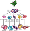
Multipotential fate of mesenchymal stem cells. MSCs have the ability to self-renew within the bone marrow (curved arrow). They can also differentiate into cell types of the mesodermal lineage (solid arrows) as well as the ectodermal and endodermal lineages (dashed arrows), although their in vivo transdifferentiation capacity is debatable. [Adapted from Uccelli et al. (363), with permission from Nature Publishing Group.]
FIGURE 3.
FIGURE 3.
Regulation of MSC osteogenesis and adipogenesis. Deficiency of S-nitrosoglutathione reductase (GSNOR) leads to increased levels of S-nitrosylated PPAR-γ with a decreased affinity for fatty acid-binding protein 4 (FABP4), its target promoter. Alterations in the PPAR-γ transcriptome modify expression of various genes responsible for regulating the balance between osteogenesis and adipogenesis. [Adapted from Cao et al. (44), with permission from American Society of Clinical Investigation.]
FIGURE 4.
FIGURE 4.
Cardiogenic potential of transplanted MSCs. A: cluster of Ypos/BrdUpos cells (white arrowheads) located in infarct and border zones of treated hearts 12 wk after MSC implantation. Some of the transplanted MSCs do not exhibit BrdUpos signal (green), but maintain Ypos signal (red, yellow arrowheads). Conversely, another group shows BrdUpos signal (white arrows) and negative Y chromosome signal due to technique sensitivity. B: cluster of Ypos cells (green, white arrows) in the border zone of MSC-treated animals colocalizing with tropomyosin (red). C: evidence of cardiac differentiation in a panoramic view of an infarct border zone of MSC-treated hearts. Inset depicts one Ypos (green) myocyte costained with tropomyosin. High magnification of the square is shown in the inset. D: confocal microscopy analysis of the same cell by orthogonal section of a z-stack (arrows point the cell analyzed in xy-plane). E: two transplanted Ypos cells (green, arrows) coupled with the resident cardiomyocytes by expressing connexin-43 (orange). F: evidence of cardiac commitment in the transplanted cell by the colocalization of Ypos signal with the cardiac transcription factor Nkx2.5 (green, arrow). Nuclei were counterstained with DAPI in all of the immunofluorescence assays. G: cluster of BrdUpos cells (green) in the border zone of MSC-treated animals exhibiting colocalization with transcription factor GATA-4 (red, arrows). H: quantitation of transplanted cells according to Y chromosome cell tracking. Ypos cells show no preference in distribution according to LV areas (top). Importantly, at 12 wk posttransplantation, implanted MSCs showed commitment to repopulate the three major cardiac cell lineages and maintain a reservoir of nondifferentiated cells (bottom). Cell quantification per unit area for the Y chromosome (n = 6 for MSC-treated hearts, n = 4 for placebo-treated hearts). At least four tissue sections for infarct, border, and remote zone per heart were evaluated. Total area evaluated is 2,673.34 mm2. CM, cardiomyocyte; End, endothelial cells; VSM, vascular smooth muscle. [From Quevedo et al. (290).]
FIGURE 5.
FIGURE 5.
Vascular differentiation of transplanted MSCs. A: representative image of a vessel containing numerous Ypos cells colocalized with smooth muscle actinin (a-sma in green, arrowheads) and endothelial cells (factor VIII-related antigen in white, arrows). High magnification of the inset to visualize the Ypos cells that colocalize with sma (arrowheads) and factor VIII-related antigen (white, arrows) demonstrating vascular smooth muscle and endothelial commitment, respectively. B and C: confirmation of Ypos cells commitment into vascular structures as depicted by colocalization with SM22-α (B) and calponin (C, arrowheads in both pictures). Ypos cells also commit to endothelial cell lineages (arrows). D: capillary formation with the incorporation of Ypos cell (arrow) costained with factor VIII-related antigen depicting the luminal surface of the vessel. E: assessment of vessel number per unit area according to their respectively size. F: Ypos cells also reside in the interstitial compartment (arrows) of border myocardium in a nondifferentiated stage (n = 6 for MSC-treated hearts, n = 4 for placebo). At least 4 tissue sections from infarct, border, and remote zone were evaluated per animal. [From Quevedo et al. (290).]
FIGURE 6.
FIGURE 6.
Transcription factors and signaling molecules participating in regulation of MSC differentiation. Various molecules and factors induce MSC differentiation into several cell lines. BMP-2, bone morphogenetic protein-2; EGF, epidermal growth factor; FGF-2, fibroblast growth factor-2; LRP5/6, low-density lipoprotein receptor-related protein-5/6; MEF2, myocyte enhancer factor-2; MRF, myogenic regulatory factors; Osx, Osterix; PDGF, platelet-derived growth factor; RUNX2, runt-related transcription factor-2; TGF-β, transforming growth factor-β; VEGF, vascular endothelial growth factor. [Adapted from Karantalis et al. (173).]
FIGURE 7.
FIGURE 7.
Major molecular pathways of MSC differentiation. The Wnt and TGF-β signaling pathways are responsible for regulating the differentiation of MSCs into osteoblasts and chondrocytes while promoting their proliferation and survival through activation of intracellular cascades with subsequent modification of gene expression. [Adapted from Williams et al. (393), with permission from American Heart Association.]
FIGURE 8.
FIGURE 8.
Immune profile of mesenchymal stem cells (MSCs). Graphic summary of the interactions between MSC and the immune system. MSCs can suppress proliferation of both T helper (TH) and cytotoxic T cells (Tc) through multiple pathways. Differentiation of MSCs to TH2 and regulatory T-cells (Treg) is triggered, resulting in an anti-inflammatory environment. Interleukin (IL)-6 blocks the maturation of dendritic cells (DC) by inhibiting upregulation of CD40, CD80, and CD86, which subsequently reduces T-cell activation. Monocytes are stimulated by MSCs to preferentially differentiate towards the M2 phenotype. IL-10, produced by M2 macrophages, can boost the formation of Treg, and simultaneously reduces neutrophil tissue migration. Neutrophils (polymorphonuclear granulocytes; PMN) have a longer life span; however, production of reactive oxygen species (ROS) is decreased. Natural killer (NK) cell proliferation and cytotoxic activity are both suppressed. B-cell proliferation is inhibited, and production of antibodies is reduced. HGF, hepatocyte growth factor; IDO, indoleamine-pyrrole-2-3-dioxygenase; PGE2, prostaglandin E2; and TGF-β, transforming growth factor-β. (Adapted from van den Akker F, de Jager SC, Sluijter JP. Mesenchymal stem cell therapy for cardiac inflammation: iummunomodulatory properties and the influence of toll-like receptors. Mediators Inflamm 2013: 181020, 2013.)
FIGURE 9.
FIGURE 9.
MSC cardiomyogenesis. A: graph depicting the contribution of cardiomyocyte precursors following exogenous administration of MSCs (green line) and endogenous CSCs (orange line), during cardiac repair after MI. MSC differentiation occurs rapidly after delivery. At 2 wk, MSCs activate endogenous expansion of c-kit+ CSCs (orange line). B: 2 wk following TEI, the number of c-kit+ cells coexpressing GATA-4 is greater in MSCs versus non-MSCs treated hearts. The cardiac precursors are preferentially located in the IZ and BZ of the MI, indicating an active process of endogenous regeneration (‡P = 0.019; †P < 0.0001) C and D: the 2-wk-old chimeric myocardium contains mature cardiomyocytes (open arrow), immature MSCs (arrowheads, inset), and cardiac precursors of MSCs origin (arrow), coupled to host myocardium by connexin-43 gap junctions. Interestingly, endogenous c-kit+ CSCs are found in close proximity to MSCs (D). E: cluster of c-kit+ CSCs in an MSC-treated heart; numerous CSCs are committed to cardiac lineage documented by GATA-4 and MDR-1 coexpression (arrows). F: few, isolated c-kit+ cells were found in non-MSC-treated animals. [From Hatzistergos et al. (144), with permission from American Heart Association.]
FIGURE 10.
FIGURE 10.
Delivery methods of MSC therapy. A: intravenous (IV) infusion of mesenchymal stem cells (MSCs; peripheral IV not shown). B: administration of MSCs through transendocardial injection (TESI). C: direct epicardial injection of MSCs. D: delivery of MSCs via intracoronary infusion.
FIGURE 11.
FIGURE 11.
Infarct size assessment and regional myocardial function. A and B: sequential short axis heart sections from base (top) to apex (bottom) of delayed gadolinium enhancement MRI images depicting the infarct extension (white) before treatment and 12 wk following MSC therapy (A) compared with the placebo (B). Comparable gross pathology sections are shown adjacent to the MRI images. Arrows delineate the infarct extension, and the asterisk illustrates the presence of a thrombus near the apex in a placebo animal. C: reduction in infarct size following 8 and 12 wk post MSC transplantation versus placebo (*P < 0.001 within MSC group ANOVA, †P < 0.05 between groups ANOVA, ‡P < 0.001 vs. preinjection status by Student-Newman-Keuls test). D: myocardial strain analysis represented by peak Ecc decreased in response to MSC treatment in infarct (left) and border (middle) areas but remained constant in the remote uninfarcted zone (right) (*P < 0.05 for ANOVA within MSC group, †P < 0.05 ANOVA between groups, ‡P < 0.05 vs. preinjection status by Student-Newman-Keuls test). At least five MRI time points were analyzed for MSC-treated hearts (n = 6) and placebo-treated hearts (n = 4). For week 4 tagged MRI images and week 8 delayed contrast enhancement MRI analysis, MSCs (n = 4) and placebo (n = 4). [From Quevedo et al. (290).]
FIGURE 12.
FIGURE 12.
Myocardial blood flow and segmental contractility showed improvement related to the cell engraftment. A: quantitative analysis of first-pass perfusion MRI in the infarct (left), border (middle), and remote (right) myocardium demonstrating the improvement in the MSC-treated group compared with placebo group (*P < 0.05 for ANOVA within MSC, †P < 0.001 ANOVA between groups, ‡P < 0.05 vs. preinjection status by Student-Newman-Keuls test). LVBP, left ventricular blood pool. B: LV ejection fraction improves in MSC group at 12 wk posttransplantation (*P < 0.05 for ANOVA within MSC, †P < 0.05 ANOVA between groups, ‡P < 0.05 vs. preinjection status by Student-Newman-Keuls). C–H: the functional outcomes in heart function (i.e., infarct size reduction, increase in contractility, and increase in tissue perfusion) showed related interaction between them (C–E) and with the magnitude of cells detected (F–H) at 12 wk after injection (P < 0.05 for all Pearson correlations). At least five time points for first-pass perfusion MRI image analysis. MSCs-treated hearts (n = 6) and placebo (n = 4) hearts for most of the time points except at 4 wk for ejection fraction (n = 8, 4 MSC and 4 placebo). [From Quevedo et al. (290).]
FIGURE 13.
FIGURE 13.
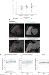
Impact of autologous MSC therapy versus whole bone marrow or placebo in patients with ischemic cardiomyopathy. Panels depict results from the TAC-HFT trial: percent change in scar size as a percentage of left ventricular mass and impact on functional capacity. A: the 14 patients treated with mesenchymal stem cells (MSCs) exhibited a significant reduction in scar size (P = .004) as a percentage of left ventricular mass with no differences for the 15 patients treated with bone marrow cells or the 16 patients in the placebo group. B: short-axis and long-axis views of the basal area of a patient's heart, with delayed tissue enhancement delineated at the septal wall (short-axis) and anterior and inferior walls (long-axis) as well as the entire apex (long-axis). Delayed tissue enhancement corresponds to scarred tissue and is depicted brighter than the nonscarred tissue. The red, green, and white lines demarcate the endocardial, epicardial contours, and borders of the segments, respectively. Twelve months after injection of mesenchymal stem cells, scar mass was reduced from 30.85 g at baseline to 21.17 g (short axis) and delayed tissue enhancement receded in the midinferior and basal anterior walls (long axis). C: patients in the mesenchymal stem cell group exhibited a significant increase in 6-min walk distance when 6-mo and 12-mo time points were compared with baseline in a repeated measures model (P = 0.03). No significant difference was observed for patients in the bone marrow cell group (P = 0.73) or in the placebo group (P = 0.25). [From Heldman et al. (147), with permission from American Medical Association.]
FIGURE 14.
FIGURE 14.
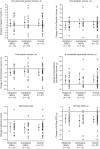
Impact of allogeneic versus autologous MSC therapy in patients with ischemic cardiomyopathy. Panels depict results from the POSEIDON trial: computer tomography (CT) parameters change from baseline. MSCs, mesenchymal stem cells; EED, early enhancement effect. Mean changes from baseline to 13 mo are noted by triangles and depict change in cardiac phenotype assessed by cardiac CT scan. Error bars indicate 95% confidence intervals (CIs). Individual patient changes from baseline are shown as circles. Shown are changes in cardiac structural and functional parameters from baseline to 13-mo follow-up in allogeneic, autologous, and combined patient groups. Within-group P values are noted as aP < 0.05, bP < 0.01, and cP < 0.001. [From Hare et al. (137).]
FIGURE 15.
FIGURE 15.
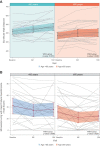
Patient functional capacity and quality of life following MSC therapy in old and young patients. A: graphic representation shows the estimated mean 6-min walk distance values and individual patient values in each age group at each time point, using a repeated-measures model. Both age groups increased similarly until 6 mo post-TESI. Although the older age group plateaus by 1 yr post-TESI, average distances between age groups do not differ significantly (P = 0.954 and P = 0.288 at 6 mo and 1 yr post-TESI, respectively; between-group comparison). B: in this graphic representation of the estimated mean Minnesota Living With Heart Failure Questionnaire, the total score values and individual patient values in each age group at each time point are depicted, using a repeated-measures model. The total scores of both age groups improved in a parallel fashion at 6 mo post-TESI. Both groups plateaued by 1 yr post-TESI with similar mean total scores on between-group comparison (P = 0.524 and P = 0.871 at 6 mo and 1 yr post-TESI, respectively; between-group comparison). TESI, transendocardial stem cell injection. [From Golpanian et al. (119), with permission from Elsevier.]

Similar articles

Cited by

References

    1. Stem the tide. Nature : 163–164, 2015. - PubMed
    1. Abarbanell AM, Wang Y, Herrmann JL, Weil BR, Poynter JA, Manukyan MC, Meldrum DR. Toll-like receptor 2 mediates mesenchymal stem cell-associated myocardial recovery and VEGF production following acute ischemia-reperfusion injury. Am J Physiol Heart Circ Physiol : H1529–H1536, 2010. - PMC - PubMed
    1. Abdel-Latif A, Bolli R, Tleyjeh IM, Montori VM, Perin EC, Hornung CA, Zuba-Surma EK, Al-Mallah M, Dawn B. Adult bone marrow-derived cells for cardiac repair: a systematic review and meta-analysis. Arch Int Med : 989–997, 2007. - PubMed
    1. Aggarwal S, Pittenger MF. Human mesenchymal stem cells modulate allogeneic immune cell responses. Blood : 1815–1822, 2005. - PubMed
    1. Ahmad T, Mukherjee S, Pattnaik B, Kumar M, Singh S, Kumar M, Rehman R, Tiwari BK, Jha KA, Barhanpurkar AP, Wani MR, Roy SS, Mabalirajan U, Ghosh B, Agrawal A. Miro1 regulates intercellular mitochondrial transport and enhances mesenchymal stem cell rescue efficacy. EMBO J : 994–1010, 2014. - PMC - PubMed