. 2016 Aug 12;90(17):7934-42.
doi: 10.1128/JVI.00712-16. Print 2016 Sep 1.

ICAM-1 Binding Rhinoviruses A89 and B14 Uncoat in Different Endosomal Compartments

Affiliations

ICAM-1 Binding Rhinoviruses A89 and B14 Uncoat in Different Endosomal Compartments

Rick Conzemius et al. J Virol. .

Erratum in

Abstract

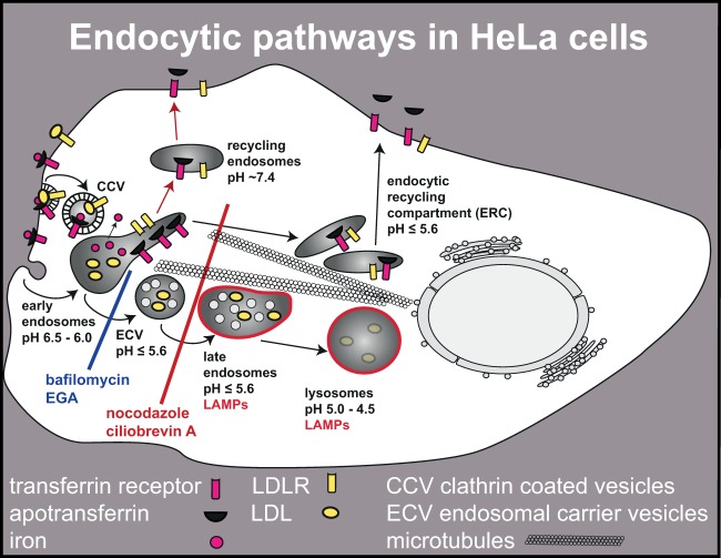
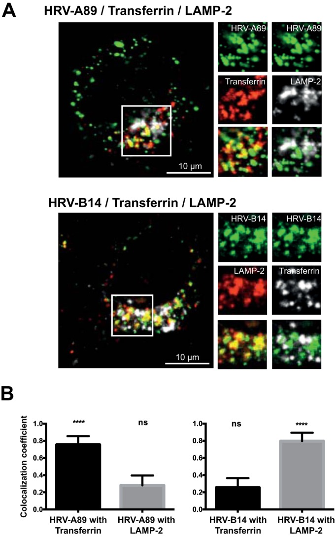
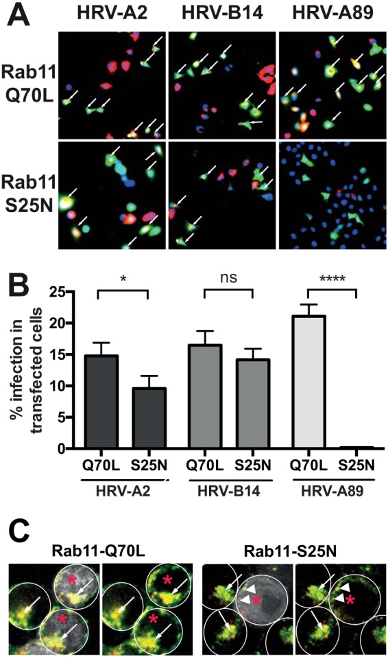
Human rhinovirus A89 (HRV-A89) and HRV-B14 bind to and are internalized by intercellular adhesion molecule 1 (ICAM-1); as demonstrated earlier, the RNA genome of HRV-B14 penetrates into the cytoplasm from endosomal compartments of the lysosomal pathway. Here, we show by immunofluorescence microscopy that HRV-A89 but not HRV-B14 colocalizes with transferrin in the endocytic recycling compartment (ERC). Applying drugs differentially interfering with endosomal recycling and with the pathway to lysosomes, we demonstrate that these two major-group HRVs productively uncoat in distinct endosomal compartments. Overexpression of constitutively active (Rab11-GTP) and dominant negative (Rab11-GDP) mutants revealed that uncoating of HRV-A89 depends on functional Rab11. Thus, two ICAM-1 binding HRVs are routed into distinct endosomal compartments for productive uncoating.

Importance: Based on similarity of their RNA genomic sequences, the more than 150 currently known common cold virus serotypes were classified as species A, B, and C. The majority of HRV-A viruses and all HRV-B viruses use ICAM-1 for cell attachment and entry. Our results highlight important differences of two ICAM-1 binding HRVs with respect to their intracellular trafficking and productive uncoating; they demonstrate that serotypes belonging to species A and B, but entering the cell via the same receptors, direct the endocytosis machinery to ferry them along distinct pathways toward different endocytic compartments for uncoating.


Figures

FIG 1
FIG 1
Endocytic pathways in HeLa cells and effect of inhibitors. After clathrin-mediated internalization of LDLR-bound LDL, the complex dissociates in the mildly acidic environment of early endosomes. Whereas LDL are transported via ECV and late endosomes to lysosomes for degradation, the LDLR recycles to the plasma membrane following the pathway also taken by transferrin receptor-bound apotransferrin. Recycling from early endosomes occurs by fast (short, red arrows) and slow (long arrows) routes. The slow route directs apotransferrin and various receptors to the ERC that has, in HeLa cells, a similarly low pH as ECV/late endosomes. Transport of ligands to lysosomes can be arrested in early endosomes by bafilomycin or EGA. In contrast, depolymerization of microtubules by nocodazole or inhibition of cytoplasmic dynein by ciliobrevin blocks transport to lysosomes as well as recycling via the slow route.
FIG 2
FIG 2
HRV-A89 uncoats much more slowly than HRV-B14. (A) Time course of RNA release. Cells were incubated with HRV-A89 at 100 TCID50/cell and with HRV-B14 at 300 TCID50/cell at 4°C for 60 min; unbound virus was removed, and cells were transferred into infection medium. At the specified times, uncoating was halted via addition of NH4Cl, and incubation was continued for 16 h. Infected cells were then determined in a TissueFaxs. The number of infected cells at 120 min was arbitrarily set to 100% for each virus. Data shown are the means ± standard deviations from three independent experiments, each carried out in quadruplicate. (B) Time course of loss of infectivity. Viruses as specified were allowed to bind to the cells for 1 h at 4°C. Unbound virus was removed by washing three times with cold PBS. At time zero the cells were returned to 37°C in infection medium; they were frozen at the times indicated. Virus was released by freeze-thawing, debris was removed by centrifugation, and viral titer was determined in the supernatant as the TCID50 per milliliter. Values are expressed in percentages of the initial viral titer. The means ± standard deviations from two independent experiments each carried out in six replicates is shown. Note that no further decrease of infectivity was observed between 120 min and 180 min (data not shown).
FIG 3
FIG 3
HRV-A89 is routed to the endocytic recycling compartment, and HRV-B14 is routed to late endosomes. (A) HRV-A89 was bound (at 100 TCID50/cell) to HeLa cells at 4°C; cells were transferred into medium containing Alexa Fluor 647-transferrin and incubated for 60 min at 37°C. HRV-B14 (at 100 TCID50/cell) and Alexa Fluor 647-transferrin were internalized for 60 min at 37°C to obtain a stronger signal. Cells were cooled, transferrin remaining at the plasma membrane was removed, and cells were fixed with PFA, quenched, and permeabilized. HRV-A89 was detected with rabbit antiserum P5, and HRV-B14 was detected with rat anti-HRV-B14 antiserum followed by the respective Alexa Fluor 488-labeled secondary antibodies. LAMP-2 was detected with mouse anti-LAMP-2 antibody H4B4 and Alexa Fluor 568 goat anti mouse IgG. Images are confocal. To facilitate detection of colocalization between HRV-A89 and transferrin, transferrin is shown in red. (B) Colocalization was determined by analyzing single confocal layers of 20 cells with ImageJ. Means ± standard deviations as determined with GraphPad Prism, version 6, are shown. ****, P ≤ 0.0001; ns, not significant.
FIG 4
FIG 4
Uncoating and infection of HRV-A89 are inhibited by ciliobrevin A and nocodazole but not by EGA. (A) Experimental setup. Cells were preincubated with and without (control) the respective drug for 30 min and challenged with the respective HRV for 1 h. Unabsorbed virus was washed away at 4°C with buffer containing NH4Cl to halt uncoating, and cells were further incubated for 16 h without drug but in the presence of NH4Cl. As a negative control, NH4Cl was kept present throughout the experiment. Percent infected cells was determined in a TissueFaxs and related to the number of cells infected when NH4Cl was added at t = 120 min (control arbitrarily set to 100%). (B) Nocodazole and ciliobrevin A inhibit HRV-A89 but not HRV-B14 or HRV-A2. EGA inhibits HRV-A2 and HRV-B14 but not HRV-A89. Note the following: (i) that the lack of inhibition of HRV-A2 by nocodazole was deduced from previous experiments in Bayer et al. (37), and the bar was just added for completeness; ii) that both major-group viruses are not completely inhibited by NH4Cl. Averages and standard deviations were calculated with GraphPad Prism, version 6, from 3 to 14 independent experiments carried out in quadruplicate, and the significance was determined by ordinary one-way ANOVA with Dunnett's test as a post hoc multiple comparison procedure by comparing every inhibitor to the positive control. *, P ≤ 0.05; **, P ≤ 0.01; ***, P ≤ 0.001; ****, P ≤ 0.0001; ns, not significant. (C and D) Virus (at 100 TCID50/cell) was internalized at 37°C in the absence and presence of nocodazole for 20 min (HRV-A2) or 60 min (HRV-B14). Cells were then cooled, washed, and processed for detection of HRV-A2 (shown in green) and LAMP-1 (shown in red) or HRV-B14 (shown in green) and LAMP-2 (shown in red), as described in Materials and Methods. (E) HRV-A89 at 100 TCID50/cell was bound to HeLa cells for 60 min at 4°C. Unbound virus was removed, and cells were warmed to 37°C for 60 min in the presence of 5 μg/ml Alexa Fluor 647-transferrin. Subsequently, cells were cooled, transferrin at the plasma membrane was removed, and HRV-A89 (shown in green) was detected by indirect immunofluorescence microscopy (see Materials and Methods). To facilitate detection of colocalization between HRV-A89 and transferrin, transferrin is shown in red. (F) Schematic of HRV localization in the absence and presence of nocodazole. Confocal images are shown in panels C to E. Arrows point to colocalization of virus with LAMP (C, D, and F) or with transferrin (E and F) in large perinuclear endosomes in the absence of nocodazole. In the presence of nocodazole, arrowheads indicate colocalization of the respective virus with LAMPs (C and D) or transferrin (E) in peripheral vesicles. Previous publications (21, 37) have shown that in the presence of nocodazole HRV-A2 and most likely HRV-B14 are arrested in ECV where uncoating can occur, whereas transferrin and thus HRV-A89 are not transported to the ERC, preventing virus uncoating (F).
FIG 5
FIG 5
Rab11 GTPase is required for infection with HRV-A89 but not for infection with HRV-A2 and HRV-B14. Cells were seeded in 24-well plates on coverslips, grown until 70% confluent, and transfected with EGFP-tagged plasmids for the expression of constitutively active (Q70L) or dominant negative (S25N) Rab11 mutants as indicated. (A and B) Twenty-four hours later, the respective virus (at 100 TCID50/cell) was allowed to internalize for 60 min; unbound virus was washed away with cold PBS+ containing 25 mM NH4Cl for 15 min, and replication was allowed to proceed for 6.5 h at 37°C in infection medium containing NH4Cl. Cells were fixed, quenched, permeabilized, and blocked, and de novo-synthesized viral protein was detected with the respective antibodies, followed by suitable fluorescent secondary antibodies. Images were viewed and recorded on a TissueFaxs, and cells expressing EGFP and/or viral proteins were determined by using the TissueQuest software. The numbers of transfected and infected cells (A, white arrows) are shown in panel B as the means ± standard deviations from one typical experiment carried out in triplicate. The significance was calculated as described in the legend of Fig. 3. *, P ≤ 0.05; **, P ≤ 0.01; ***, P ≤ 0.001; ****, P ≤ 0.0001; ns, not significant. (C) HRV-A89 at 100 TCID50/cell and Alexa Fluor 647-transferrin (5 μg/ml MEM+) were internalized for 60 min at 37°C. After removal of transferrin from the plasma membrane, HRV-A89 was detected by indirect immunofluorescence (see Materials and Methods). Colocalization of HRV-A89 (red) and transferrin (green) in cells expressing the respective Rab11-EGFP by confocal fluorescence microscopy. Cells expressing Rab-EGF are marked with red asterisks; on the respective left panels they are also shown in white. Individual cells are encircled with white lines. In control and Rab11-Q70L-expressing cells, virus and transferrin accumulate in the ERC (white arrows), whereas in Rab11-S25N-expressing cells, virus and transferrin accumulate in peripheral vesicles (white arrowheads).

Similar articles

Cited by

References

    1. Uncapher CR, Dewitt CM, Colonno RJ. 1991. The major and minor group receptor families contain all but one human rhinovirus serotype. Virology 180:814–817. doi:10.1016/0042-6822(91)90098-V. - DOI - PubMed
    1. Vlasak M, Roivainen M, Reithmayer M, Goesler I, Laine P, Snyers L, Hovi T, Blaas D. 2005. The minor receptor group of human rhinovirus (HRV) includes HRV23 and HRV25, but the presence of a lysine in the VP1 HI loop is not sufficient for receptor binding. J Virol 79:7389–7395. doi:10.1128/JVI.79.12.7389-7395.2005. - DOI - PMC - PubMed
    1. Bochkov YA, Watters K, Ashraf S, Griggs TF, Devries MK, Jackson DJ, Palmenberg AC, Gern JE. 2015. Cadherin-related family member 3, a childhood asthma susceptibility gene product, mediates rhinovirus C binding and replication. Proc Natl Acad Sci U S A 112:5485–5490. doi:10.1073/pnas.1421178112. - DOI - PMC - PubMed
    1. Fuchs R, Blaas D. 2012. Productive entry pathways of human rhinoviruses. Adv Virol 2012:826301. doi:10.1155/2012/826301. - DOI - PMC - PubMed
    1. Goldstein JL, Brown MS. 1974. Binding and degradation of low density lipoproteins by cultured human fibroblasts. Comparison of cells from a normal subject and from a patient with homozygous familial hypercholesterolemia. J Biol Chem 249:5153–5162. - PubMed

Publication types

Substances

Grants and funding

Funding was provided by Austrian Science Fund (FWF) projects P27444-B13 and P23308-B13) to Dieter Blaas.

LinkOut - more resources