. 2016 Jun 22;11(6):e0157305.
doi: 10.1371/journal.pone.0157305. eCollection 2016.

Aurora-C Interactions with Survivin and INCENP Reveal Shared and Distinct Features Compared with Aurora-B Chromosome Passenger Protein Complex

Affiliations

Aurora-C Interactions with Survivin and INCENP Reveal Shared and Distinct Features Compared with Aurora-B Chromosome Passenger Protein Complex

Kaori Sasai et al. PLoS One. .

Abstract

Aurora-C, a member of the Aurora kinase family that can complement Aurora-B function in mitosis is either moderately expressed or repressed in most adult somatic tissues but is active in early embryonic development and expressed at elevated levels in multiple human cancers. Aurora-C overexpression reportedly plays a role in tumorigenic transformation. We performed detailed characterization of Aurora-C interactions with members of the Chromosome Passenger Complex (CPC), Survivin and Inner Centromere Protein (INCENP) in reference to known Aurora-B interactions to understand the functional significance of Aurora-C overexpression in human cancer cells. The results revealed that silencing of Aurora-C or -B individually does not affect localization of the other kinase and the two kinases exist predominantly in independent complexes in vivo. Presence of Aurora-C and -B in molecular complexes of varying as well as overlapping sizes and co-existence in INCENP overexpressing cells indicated oligomerization of ternary complexes under different physiological conditions in vivo. Furthermore, Aurora-C and -B stabilized INCENP through interaction with and phosphorylation of the IN box domain while Aurora-C was activated following Survivin phosphorylation on Serine 20. Phosphorylation of Survivin residue Serine 20 by Aurora-C and -B appears important for proper chromosome segregation. Taken together, our study suggests that Aurora-C, expressed at low levels in somatic cells, functions as a catalytic component of the CPC together with Aurora-B through mitosis. Elevated expression of Aurora-C in cancer cells alters the structural and functional characteristics of the Aurora-B-CPC leading to chromosomal instability.


Conflict of interest statement

Competing Interests: The authors have declared that no competing interests exist.

Figures

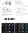
Fig 1
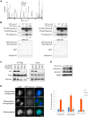
Fig 1. C-terminus end of Aurora-C binds with Survivin and is responsible for its localization.
(A) PC3 cells were treated with 500 ng/ml nocodazole for 24 h and lysed with L buffer containing 250 mM NaCl. 1 mg of lysate was immunoprecipitated with 1 μg of anti-Aurora-C, anti-Survivin, or normal IgG antibodies respectively. Immunoprecipitates were immunoblotted with indicated antibodies. The asterisk indicates non-specific bands. (B) 35S-labeled Survivin was incubated with beads bound to any of the following: GST, GST-Aurora-C full-length (Full), N-terminus amino acids 1–40 (N), or C-terminus amino acids 41–309 (C). Beads were resolved by SDS-PAGE, and visualized by autoradiography (for binding, top) or CBB stained (bottom). (C) 35S-labeled Aurora-C full length or deletion mutants were incubated with beads bound to GST or GST-Survivin, and analyzed as in B. The panel shows autoradiography (for binding, top; for input, bottom) or CBB stained gel (middle). (D) Alignment of the C-terminus sequences of Aurora-C and Aurora-B. (E) Localization of GFP-Aurora-C full length or GFP-Aurora-C deletion mutant (amino acids 1–291) in HeLa cells. Transfected cells were immunostained with indicated antibodies (red). DNA was stained with DAPI (blue). Scale bars indicate 10 μm. Each experiment was repeated at least twice with two to three biological replicates and the figures show representative images.
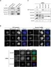
Fig 2
Fig 2. Localization following gene silencing and immunoprecipitation indicate independent Aurora-C and Aurora-B complexes.
(A) Nocodazole (500 ng/ml) treated 1.7 mg of PC-3 cell lysate were immunoprecipitated with 2 μg of anti-Aurora-C and anti-Aurora-B antibodies. Immunoprecipitates were immunoblotted with the indicated antibodies. (B) GFP-Aurora-C expressing Tet-on HeLa cells was transfected with control siRNA (GL2) or with siRNA against Aurora-C, Aurora-B, INCENP, or Survivin for 48 h. The cells were immunostained with anti-INCENP (top), anti-Survivin (middle), or anti-Aurora-B (bottom) antibodies, respectively (red). DNA was stained with DAPI (blue). Scale bars indicate 10 μm. (C) Representative immunoblots treated as in B. Cells were lysed and immunoblotted with anti-GFP (Aurora-C), anti-Aurora-B, anti-INCENP, anti-Survivin, and anti-β-actin antibodies. The experiments were repeated three times with three biological replicates and the figures show representative images.
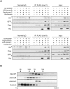
Fig 3
Fig 3. Aurora-C and Aurora-B Complexes in immune precipitates and in Sucrose Density Gradients.
(A) 293T cells were transfected with HA-INCENP, Flag-Aurora-C, GFP-Aurora-B, and His-Survivin (top panel). Flag-Aurora-B and GFP-Aurora-C were co-transfected with HA-INCENP and His-Survivin in 293T cells (bottom panel). 24 h after transfection, cells were lysed and immunoprecipitated by anti-Flag antibody or normal mouse IgG. Immunoprecipitates were immunoblotted with indicated antibodies. Absence or presence of each protein are indicated with (-) and (+) signs, respectively. Asterisks indicate non-specific bands. (B) Nocodazole-treated SK-OV-3 cell lysate was sedimented on 5–30% sucrose density gradients. Equal volumes of each fraction were run on SDS-PAGE gels and immunoblotted with anti-INCENP, anti-Aurora-B, anti-Aurora-C, and anti-Survivin antibodies. Thyroglobulin (19S), γ-globulin (7S), ovalbumin (3.5S) and myoglobin (2S) were sedimented in a parallel gradient as markers. Each experiment was repeated twice with three biological replicates and the figures show representative images.
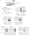
Fig 4
Fig 4. Direct binding and phosphorylation dependent Aurora-C activation through conserved IN box domain of INCENP.
(A) Schematic represents INCENP full-length and carboxyl terminal deletion mutants (C3-C8) [5]. The deduced regions of INCENP involved in Aurora-B and Aurora-C binding (aa 822–897) and activation (aa 822–892), as well as the domain serving as a substrate for phosphorylation (aa 893–919) are indicated. 3A indicates three alanine mutants of TSS motif (T893, S894, S895) for Aurora-B and Aurora-C phosphorylation sites. (B) 35S-labeled in vitro translated Aurora-C was incubated with beads bound to either GST or GST-ΔINCENP deletions. Bound Aurora-C and GST-ΔINCENP deletions were visualized by autoradiography and CBB staining, respectively. Relative Aurora-C binding to GST-INCENP deletions was measured and quantified by ImageJ software. Arrowheads show GST fusion proteins. (C) GST-Aurora-C was incubated with GST-ΔINCENP deletions and histone H3 in vitro. Protein amount was determined by CBB stain and kinase activity was measured using histone H3 as substrate. [γ32P]-ATP incorporation in proteins was visualized by autoradiography. (D) GST-Aurora-C WT or KD mutants were incubated with histone H3, either WT or 3A mutant of GST-ΔINCENP C3 (left panel) or full length His-INCENP (right panel) in vitro, and analyzed as in C. (E) GST-Aurora-C or GST-Aurora-B was co-infected with His-INCENP and GST-Survivin in Sf-9 cells and proteins were isolated on glutathione sepharose. Proteins were detected by immunoblot with anti-INCENP antibody or CBB stain, and kinase activity was measured using histone H3 as substrate as in C. Absence or presence of each protein are indicated with (-) and (+) signs, respectively. (F) GST-Aurora-C WT, T198A or T202A mutants were incubated with full-length His-INCENP and histone H3 in vitro, and analyzed as in C. Experiments were repeated twice and representative images are shown in each figure.
Fig 5
Fig 5. Stabilization of INCENP by Aurora-C phosphorylation.
(A) Flag-Aurora-C WT, KD, T198A, or T202A mutants were co-transfected with HA-INCENP WT or 3A mutant and GFP empty vector in 293T cells. After 24 h transfection, cells were analyzed. GFP was used as internal control for transfection. Quantification of HA-INCENP protein levels were determined by using the ImageJ software. Relative fold change with normalization by GFP expression is shown. (B) 24 h after transfection, 293T cells were split and grown in culture for additional 24 h. Then cells were treated with 50 μM LLnL for 6 h and analyzed as in A. Experiments (A) and (B) were repeated three times with two biological replicates and figures show the representative results.
Fig 6
Fig 6. Aurora-C phosphorylates Survivin on Serine 20.
(A) Fragmentation of DHRIpSTFK on the TofTof. Inset summarizes ions found. A key ion is the b5, mainly observed as the -98 neutral-loss ion (loss of phosphoric acid), indicating it as the primary site of phosphorylation. Essentially complete sequence ion coverage was observed considering both “b” and “y” type ions. Detection of the Ile-specific d4 fragment ions provides confirmation that the overall sequence assignment is correct. (B) Sf-9 cells were co-infected with GST-Survivin WT or S20A mutant and GST-Aurora-C WT or KD (left panel) or GST-Aurora-B WT or KD (right panel). 48 h after infection, proteins were isolated on glutathione sepharose, co-precipitated GST-Aurora-C, GST-Aurora-B, and GST-Survivin were incubated with [γ32P]-ATP and histone H3 as substrate. The reactions were resolved by SDS-PAGE and visualized by autoradiography (top) or CBB stained (bottom). Relative [γ32P]-ATP incorporation to histone H3 shown was normalized by total histone H3 and quantified by ImageJ software. Experiments were repeated three times and showed reproducible results. (C) 293T cells were co-transfected with HA-INCENP, Flag-Aurora-C, and His-Survivin plasmids respectively. 24 h after transfection, cells were lysed and immunoprecipitated by anti-Flag antibody. Immunoprecipitates were immunoblotted with indicated antibodies. Arrow indicates slower mobility sift band of Survivin. (D) HeLa cells stably expressing GFP-Survivin WT, S20A, or S20E mutant were treated with 40 ng/ml nocodazole for 16 h. Cells were lysed and immunoblotted with indicated antibodies. (E) Analysis of cellular and mitotic defects in HeLa cell clones stably expressing GFP-Survivin WT, S20A, or S20E mutant. Representative images of GFP-Survivin S20A expressing cells with abnormal chromosome alignment, abnormal chromosome separation, and multinucleation are shown on the left. Green fluorescence shows distribution of Survivin and DNA is stained with DAPI (blue). The percentages of chromosome misalignment, chromosome missegregation, and multinucleated giant abnormal cells in the indicated transfected clones are shown in the bar graph on the right. At least 200 cells in each of the clones were analyzed for this assay. Data represent mean values ± SD from two clones (n = 2). Scale bar indicates 10 μm. Experiments were repeated twice with two biological replicates in each experiment. Figures show the representative images.
Fig 7
Fig 7. Hypothetical scheme of functional roles for Aurora-C and Aurora-B CPC.

Similar articles

Cited by

References

    1. Carmena M, Earnshaw WC. The cellular geography of aurora kinases. Nat Rev Mol Cell Biol. 2003;4(11):842–54. - PubMed
    1. Carmena M, Ruchaud S, Earnshaw WC. Making the Auroras glow: regulation of Aurora A and B kinase function by interacting proteins. Curr Opin Cell Biol. 2009;21(6):796–805. 10.1016/j.ceb.2009.09.008 - DOI - PMC - PubMed
    1. Jeyaprakash AA, Klein UR, Lindner D, Ebert J, Nigg EA, Conti E. Structure of a Survivin-Borealin-INCENP core complex reveals how chromosomal passengers travel together. Cell. 2007;131(2):271–85. - PubMed
    1. Gassmann R, Carvalho A, Henzing AJ, Ruchaud S, Hudson DF, Honda R, et al. Borealin: a novel chromosomal passenger required for stability of the bipolar mitotic spindle. J Cell Biol. 2004;166(2):179–91. - PMC - PubMed
    1. Honda R, Korner R, Nigg EA. Exploring the functional interactions between Aurora B, INCENP, and survivin in mitosis. Mol Biol Cell. 2003;14(8):3325–41. - PMC - PubMed

MeSH terms