. 2016 Jul 5;113(27):E3834-43.
doi: 10.1073/pnas.1517798113. Epub 2016 Jun 20.

Role and structural mechanism of WASP-triggered conformational changes in branched actin filament nucleation by Arp2/3 complex

Affiliations

Role and structural mechanism of WASP-triggered conformational changes in branched actin filament nucleation by Arp2/3 complex

Max Rodnick-Smith et al. Proc Natl Acad Sci U S A. .

Abstract

The Arp2/3 (Actin-related proteins 2/3) complex is activated by WASP (Wiskott-Aldrich syndrome protein) family proteins to nucleate branched actin filaments that are important for cellular motility. WASP recruits actin monomers to the complex and stimulates movement of Arp2 and Arp3 into a "short-pitch" conformation that mimics the arrangement of actin subunits within filaments. The relative contribution of these functions in Arp2/3 complex activation and the mechanism by which WASP stimulates the conformational change have been unknown. We purified budding yeast Arp2/3 complex held in or near the short-pitch conformation by an engineered covalent cross-link to determine if the WASP-induced conformational change is sufficient for activity. Remarkably, cross-linked Arp2/3 complex bypasses the need for WASP in activation and is more active than WASP-activated Arp2/3 complex. These data indicate that stimulation of the short-pitch conformation is the critical activating function of WASP and that monomer delivery is not a fundamental requirement for nucleation but is a specific requirement for WASP-mediated activation. During activation, WASP limits nucleation rates by releasing slowly from nascent branches. The cross-linked complex is inhibited by WASP's CA region, even though CA potently stimulates cross-linking, suggesting that slow WASP detachment masks the activating potential of the short-pitch conformational switch. We use structure-based mutations and WASP-Arp fusion chimeras to determine how WASP stimulates movement toward the short-pitch conformation. Our data indicate that WASP displaces the autoinhibitory Arp3 C-terminal tail from a hydrophobic groove at Arp3's barbed end to destabilize the inactive state, providing a mechanism by which WASP stimulates the short-pitch conformation and activates Arp2/3 complex.

Keywords: Arp2/3; WASP; actin.


Conflict of interest statement

The authors declare no conflict of interest.

Figures
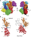
Fig. 1.
Fig. 1.
Hypothesized CA binding mode and schematic of the short-pitch cross-linking assay. (A) Hypothetical model showing proposed binding sites for the WASP family CA region on the Arp2/3 complex. The Arp2/3 complex surface representation is based on the structure of the GMFγ (Glia maturation factor gamma)-bound Arp2/3 complex (PDB ID code 4JD2) (23). (B) Ribbon diagrams of Arp2 and Arp3 in the splayed (Left) and short-pitch (Right) conformations showing the design of the engineered cross-linking assay. The structure of the GMFγ-bound Arp2/3 complex (PDB ID code 4JD2) was used to make both panels. In the right panel, the actin filament structure of Oda et al. (50) was used to move Arp2 into the short-pitch conformation. The engineered cysteines Arp3(L155C) and Arp2(R198C) are highlighted in cyan. The black arrow shows the direction of the ∼25-Å movement required to position Arp2 in the short-pitch conformation. The structure of the chemical cross-linker (BMOE) used in the short-pitch cross-linking assay is indicated. The cross-linking distance between engineered cysteines is 32.5 Å in the splayed conformation and ranges from ∼8–13 Å in different models of the short-pitch conformation. (Also see SI Appendix, Fig. S1).
Fig. 2.
Fig. 2.
Stimulation of the short-pitch conformation is a conserved feature of WASP family NPFs. (A) Single- or two-color Western blots of short-pitch cross-linking reactions containing 1 μM wild-type (wt), dual-cysteine (Cys-Arp), or single-cysteine (Arp2-Cys or Arp3-Cys) Saccharomyces cerevisiae (Sc) Arp2/3 complexes and 25 μM BMOE reacted for 0 or 5 min. (B) Single-color Western blot of 1-min cross-linking reactions containing 1 μM dual cysteine (Cys-Arp), 25 μM BMOE, and 30 μM WASP family protein, as indicated. (C) Quantification of reactions similar to those described in B, except that multiple concentrations were tested. Concentrations of WASP-CA used (expressed in micromolar) are shown in parentheses. Error bars show SE from three reactions.
Fig. 3.
Fig. 3.
CA binding to both Arp2 and Arp3 is important for stimulating the short-pitch conformation change. (A) Anti-Arp3 Western blots of short-pitch cross-linking assays containing 1 μM wild-type or CA-fusion ScArp2/3 complexes (with dual-cysteine mutations) and 25 μM BMOE reacted for 1 min in the presence or absence of 20 μM N-WASP-CA. (B) Quantification of reactions run under the conditions described in A. Error bars show SE of three reactions. (C) Cartoon summarizing the short-pitch cross-linking results for wild-type and CA fusion complexes with or without N-WASP-CA. The average percentage of short-pitch cross-linked product formed under each condition is indicated. (D) Maximum polymerization rate vs. concentration of wild-type or Arp-CA fusion complexes in 3 µM 15% pyrene actin polymerization assays. (E) Time courses of 3 µM 15% pyrene actin polymerization assays containing 100 nM wild-type or Arp2-CA fusion complex with or without 20 µM N-WASP-CA. AU, arbitrary units.
Fig. 4.
Fig. 4.
Locking the Arp2/3 complex into the short-pitch conformation bypasses the requirement for NPFs in activation. (A) Western blot of SDS/PAGE gel of cross-linking reactions containing 1 μM dual-cysteine ScArp2/3 complex (Cys-Arp) and 25 μM BMOE. Reactions were quenched with 1 mM DTT at the times indicated. (B) Quantification of data in A. Error bars show SEs from three separate reactions. (C) Time course of pyrene actin polymerization for assays containing 3 μM 15% pyrene-labeled actin and 10 nM dual-cysteine complex cross-linked for the times indicated. (D) Fold activation of the dual-cysteine complex calculated by dividing the maximum polymerization rate at a given cross-linking time by the maximum polymerization rate of the non–cross-linked complex. Data were taken from C. (E) The number of barbed ends created plotted versus time for reactions in C.
Fig. 5.
Fig. 5.
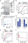
The short-pitch cross-linked complex is more active than the WASP-activated non–cross-linked complex. (A, Left) Coomassie-stained SDS/PAGE gel showing the pre–cross-linked dual-cysteine complex, the post–cross-linking reaction, and the purified cross-linked complex after passing through a MonoQ column and a GST-Dip1 affinity column. (Right) Anti-Arp3 Western blot of purified cross-linked and non–cross-linked samples. (B) Time courses of 3 μM 15% pyrene actin polymerization for reactions containing 10 nM dual cross-linked cysteine ScArp2/3 complex (“xlinked Arp2/3”) or 10 nM non–cross-linked complex with either no NPF (“dual Cys Arp2/3”), 250 nM N-WASP-VCA, or 50 nM GST-Las17-VCA, as indicated. Multiple reaction time courses are shown for the fastest reactions. (C) Maximum polymerization rate versus NPF concentration for reactions containing 10 nM non–cross-linked complex with N-WASP-VCA, GST-Las17-VCA, or LZ-dimerized N-WASP-VCA in conditions identical to those in B. The average maximum polymerization rate for purified cross-linked complex without NPFs is shown as a red circle. Error bars indicate the SD (n = 3). (D) Comparison of maximum polymerization and time to t1/2 polymerization for 10 nM cross-linked complex versus 10 nM non–cross-linked complex with 250 nM N-WASP-VCA. Error bars indicate the SD (n = 3). (E) Time course of polymerization of a reaction containing 3 μM 15% pyrene-labeled actin, 2.5 nM purified cross-linked complex, and 0–40 μM N-WASP-CA, as indicated. (F) Plot of the maximum polymerization rate versus N-WASP-CA concentration for reactions in E.
Fig. 6.
Fig. 6.
Residues in the C region are important for stimulating the short-pitch conformational change. (A) Sequence alignment of WASP family CA regions. Conserved hydrophobic (green boxes) and basic (red boxes) residues are indicated, and acidic residues in the A region are highlighted in cyan. N-WASP point mutations and deletion constructs investigated in this study are indicated. Bt, Bos taurus; Hs Homo sapiens; Sc, S. cerevisiae. (B) Maximum polymerization rate versus NPF concentration for polymerization reactions containing 3 μM 15% pyrene actin, 10 nM dual-cysteine Arp2/3 complex, and the indicated concentration of each NPF. (C) Results of 1-min short-pitch cross-linking reactions containing 1 μM dual-cysteine Arp2/3 complex, 25 μM BMOE, and the indicated concentrations (in micromolar) of mutant or wild-type N-WASP-VCA. Error bars show the SE from three reactions.
Fig. 7.
Fig. 7.
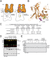
WASP-CA fused to the C terminus of Arp2 or Arp3 blocks the binding of soluble WASP-VCA to the CA-fusion subunit. (A) Sequence alignment of the C terminus of Arp2, Arp3, and actin, showing the design of Arp CA-fusion constructs. Residues conserved in Arp2 and Arp3 (cyan boxes), Arp2 only (pink boxes), or Arp3 only (green boxes) are indicated. Residues in Arp3 that mark the tip (blue line) and base (red line) of the Arp3 C-terminal tail are indicated. The majority of residues from the fused CA segments [indicated by “(CA)”] were omitted from the alignment. (B) Hypothetical model of the Arp3-CA fusion structure. The fused CA sequence is shown in magenta, and the linker is in green. The model was constructed from the crystal structure of the Arp2/3 complex with bound ATP (PDB ID code 2P9K) (22), with the WASP-C region modeled into position based on a WASP-V–bound actin structure (PDB ID code 2A3Z) (32). The A region was placed between subdomains 3 and 4 based on the structure (PDB ID code 3RSE) (27). (C) Hypothetical model of the Arp2-CA fusion structure. The model was constructed from the crystal structure of the GMF-bound Arp2/3 complex (PDB ID code 4JD2) (23), with WASP-C modeled into position as described above. (D) Schematic of the B4M cross-linking assay to probe WASP-CA binding at each site on the complex. (E) Anti-Arp3 Western blot of B4M UV cross-linking reactions containing 1.5 μM dual-cysteine (“wt”), Arp2-CA fusion, or Arp3-CA fusion complexes with 200 μM ATP and with no NPF, 4 μM N-WASP-VCA(T464C)-B4M, or 100 μM LZ-dimerized VCA (LZ-VCA), as indicated. All three complexes harbor the dual-cysteine engineered mutations. (F) Reactions from E analyzed by Western blotting with anti-Arp2 antibody. (G) Quantification of B4M cross-linking reactions as described in E and F. Error bars are SEs from three reactions.
Fig. 8.
Fig. 8.
WASP competes with the Arp3 C terminus for binding the barbed-end groove of Arp3. (A) Model for the relief of autoinhibition of the Arp2/3 complex by WASP-CA binding. B, base of the Arp3 C-terminal tail; NBC, nucleotide-binding cleft; T, tip of the Arp3 C-terminal tail. Subdomains of Arp3 and Arp3 are numbered 1–4. (B) Hypothetical model of WASP-CA (magenta) bound to Arp3 showing a clash between WASP-C and the tip of the Arp3 C-terminal tail (yellow). Cysteine 426, a residue that becomes reactive when the C terminus is deleted, is shown as green spheres. The Arp3 ribbon diagram is based on PDB ID code 1K8K, and WASP-C is modeled into barbed-end groove of Arp3 based on the WASP-V–bound actin structure (PDB ID code 2A3Z) (32). (C) Tabulated results from fluorescence anisotropy binding assays. (D) Two-color (anti-Arp3/anti-Arp2, Upper) or one-color (anti-Arp3, Lower) Western blots of cross-linking reactions containing 1.0 μM wild-type or Arp3ΔC or Arp3ΔC(C426A) fusion complexes (each with dual-cysteine mutations) with or without 20 μM N-WASP-CA showing short-pitch (sp) or non–short-pitch (non sp) products. (E) Anti-Arp3 Western blot of VCA cross-linking reactions containing 0.5 μM wild-type, Arp3ΔC, or Arp3ΔC(C426A) S. cerevisiae Arp2/3 complex, 2 μM N-WASP-VCA(V468C), 50 μM CuSO4,10 mM imidazole (pH 7.0), 50 mM KCl, 50 μM EGTA, 1 mM MgCl2, and 200 µM ATP with or without 50 μM N-WASP-CA. Note that the Arp2/3 complexes in this assay lack the engineered cysteines for short-pitch cross-linking.

Similar articles

Cited by

References

    1. Campellone KG, Welch MD. A nucleator arms race: Cellular control of actin assembly. Nat Rev Mol Cell Biol. 2010;11(4):237–251. - PMC - PubMed
    1. Skau CT, Waterman CM. Specification of Architecture and function of actin structures by actin nucleation factors. Annu Rev Biophys. 2015;44:285–310. - PMC - PubMed
    1. Amann KJ, Pollard TD. The Arp2/3 complex nucleates actin filament branches from the sides of pre-existing filaments. Nat Cell Biol. 2001;3(3):306–310. - PubMed
    1. Achard V, et al. A “primer”-based mechanism underlies branched actin filament network formation and motility. Curr Biol. 2010;20(5):423–428. - PubMed
    1. Wagner AR, Luan Q, Liu SL, Nolen BJ. Dip1 defines a class of Arp2/3 complex activators that function without preformed actin filaments. Curr Biol. 2013;23(20):1990–1998. - PMC - PubMed

Publication types

MeSH terms

Substances

LinkOut - more resources