

Bax/Bak activation in the absence of Bid, Bim, Puma, and p53
- PMID: 27310874
- PMCID: PMC5143395
- DOI: 10.1038/cddis.2016.167
Bax/Bak activation in the absence of Bid, Bim, Puma, and p53
Abstract
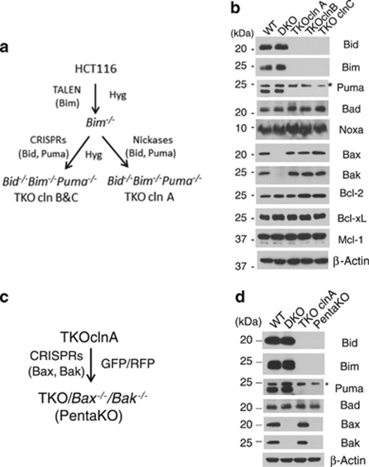
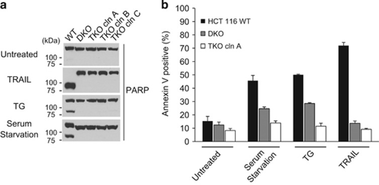
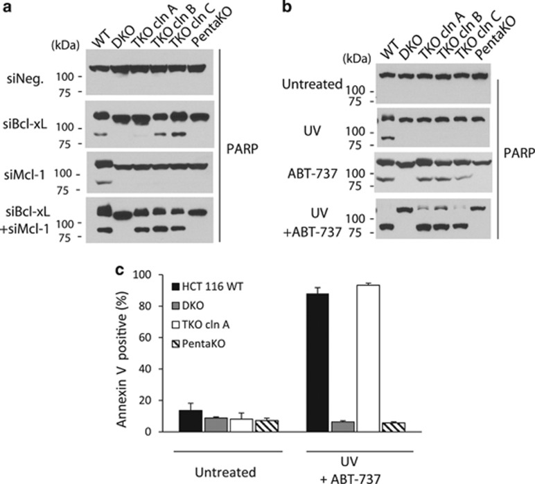
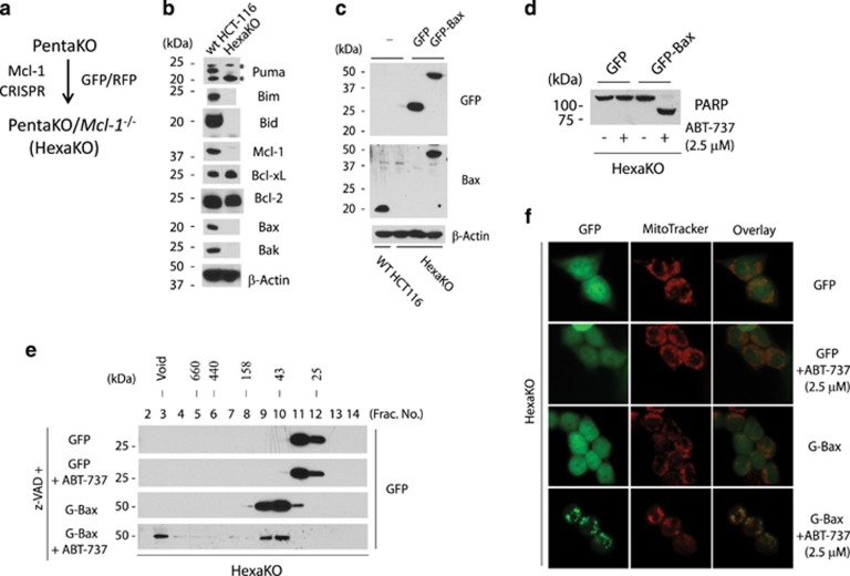
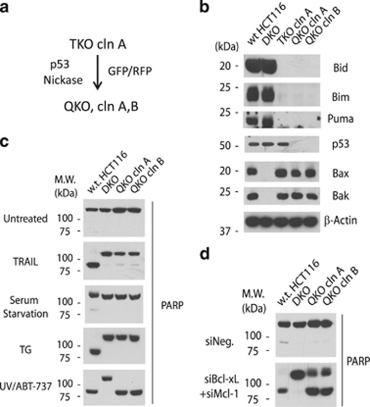
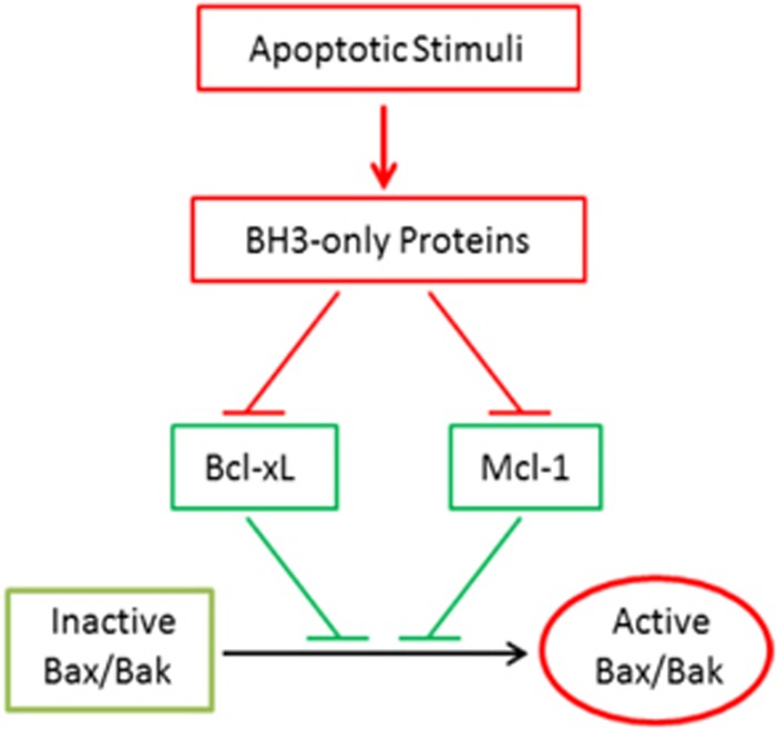
How BH3-only proteins activate Bax/Bak, the two gateway proteins of the mitochondria-dependent apoptotic pathway, remains incompletely understood. Although all pro-apoptotic BH3-only proteins are known to bind/neutralize the anti-apoptotic Bcl-2 proteins, the three most potent ones, Bid (tBid), Bim, and Puma, possess an additional activity of directly activating Bax/Bak in vitro. This latter activity has been proposed to be responsible for triggering Bax/Bak activation following apoptotic stimulation. To test this hypothesis, we generated Bid(-/)(-)Bim(-/)(-)Puma(-/)(-) (TKO), TKO/Bax(-/)(-)/Bak(-/)(-) (PentaKO), and PentaKO/Mcl-1(-/-) (HexaKO) HCT116 cells through gene editing. Surprisingly, although the TKO cells were resistant to several apoptotic stimuli, robust apoptosis was induced upon the simultaneous inactivation of Bcl-xL and Mcl-1, two anti-apoptotic Bcl-2 proteins known to suppress Bax/Bak activation and activity. Importantly, such apoptotic activity was completely abolished in the PentaKO cells. In addition, ABT-737, a BH3 mimetic that inhibits Bcl-xL/Bcl-w/Bcl-2, induced Bax activation in HexaKO cells reconstituted with endogenous level of GFP-Bax. Further, by generating TKO/p53(-/-) (QKO) cells, we demonstrated that p53, a tumor suppressor postulated to directly activate Bax, is not required for Bid/Bim/Puma-independent Bax/Bak activation. Together, these results strongly suggest that the direct activation activities of Bid (tBid), Bim, Puma, and p53 are not essential for activating Bax/Bak once the anti-apoptotic Bcl-2 proteins are neutralized.
Figures
Similar articles
-
The BH3 alpha-helical mimic BH3-M6 disrupts Bcl-X(L), Bcl-2, and MCL-1 protein-protein interactions with Bax, Bak, Bad, or Bim and induces apoptosis in a Bax- and Bim-dependent manner.J Biol Chem. 2011 Mar 18;286(11):9382-92. doi: 10.1074/jbc.M110.203638. Epub 2010 Dec 9. J Biol Chem. 2011. PMID: 21148306 Free PMC article.
-
BID, BIM, and PUMA are essential for activation of the BAX- and BAK-dependent cell death program.Science. 2010 Dec 3;330(6009):1390-3. doi: 10.1126/science.1190217. Science. 2010. PMID: 21127253 Free PMC article.
-
An interconnected hierarchical model of cell death regulation by the BCL-2 family.Nat Cell Biol. 2015 Oct;17(10):1270-81. doi: 10.1038/ncb3236. Epub 2015 Sep 7. Nat Cell Biol. 2015. PMID: 26344567 Free PMC article.
-
BCL-2 proteins and apoptosis: Recent insights and unknowns.Biochem Biophys Res Commun. 2018 May 27;500(1):26-34. doi: 10.1016/j.bbrc.2017.06.190. Epub 2017 Jul 1. Biochem Biophys Res Commun. 2018. PMID: 28676391 Review.
-
Small molecule inhibition of the Bcl-X(L)-BH3 protein-protein interaction: proof-of-concept of an in vivo chemopotentiator ABT-737.Curr Top Med Chem. 2007;7(10):961-5. doi: 10.2174/156802607780906843. Curr Top Med Chem. 2007. PMID: 17508927 Review.
Cited by
-
Cotargeting BCL-2 and PI3K Induces BAX-Dependent Mitochondrial Apoptosis in AML Cells.Cancer Res. 2018 Jun 1;78(11):3075-3086. doi: 10.1158/0008-5472.CAN-17-3024. Epub 2018 Mar 20. Cancer Res. 2018. PMID: 29559471 Free PMC article.
-
Lactobacillus raises in vitro anticancer effect of geniposide in HSC-3 human oral squamous cell carcinoma cells.Exp Ther Med. 2017 Nov;14(5):4586-4594. doi: 10.3892/etm.2017.5105. Epub 2017 Sep 5. Exp Ther Med. 2017. PMID: 29104666 Free PMC article.
-
Aberrant miRNAs Regulate the Biological Hallmarks of Glioblastoma.Neuromolecular Med. 2018 Dec;20(4):452-474. doi: 10.1007/s12017-018-8507-9. Epub 2018 Sep 4. Neuromolecular Med. 2018. PMID: 30182330 Review.
-
BH3 mimetics induce apoptosis independent of DRP-1 in melanoma.Cell Death Dis. 2018 Sep 5;9(9):907. doi: 10.1038/s41419-018-0932-z. Cell Death Dis. 2018. PMID: 30185782 Free PMC article.
-
Central role of podocytes in mediating cellular cross talk in glomerular health and disease.Am J Physiol Renal Physiol. 2024 Mar 1;326(3):F313-F325. doi: 10.1152/ajprenal.00328.2023. Epub 2024 Jan 11. Am J Physiol Renal Physiol. 2024. PMID: 38205544 Review.
References
-
- Tait SW, Green DR. Mitochondria and cell death: outer membrane permeabilization and beyond. Nat Rev Mol Cell Biol 2010; 11: 621–632. - PubMed
-
- Youle RJ, Strasser A. The BCL-2 protein family: opposing activities that mediate cell death. Nat Rev Mol Cell Biol 200; 9: 47–59. - PubMed
-
- Danial NN, Korsmeyer SJ. Cell death: critical control points. Cell 2004; 116: 205–219. - PubMed
Publication types
MeSH terms
Substances
Grants and funding
LinkOut - more resources
Full Text Sources
Other Literature Sources
Research Materials
Miscellaneous
