PA-X-associated early alleviation of the acute lung injury contributes to the attenuation of a highly pathogenic H5N1 avian influenza virus in mice
- PMID: 27289459
- PMCID: PMC7086737
- DOI: 10.1007/s00430-016-0461-2
PA-X-associated early alleviation of the acute lung injury contributes to the attenuation of a highly pathogenic H5N1 avian influenza virus in mice
Abstract
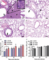
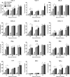
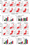
PA-X is a novel discovered accessory protein encoded by the PA mRNA. Our previous study demonstrated that PA-X decreases the virulence of a highly pathogenic H5N1 strain A/Chicken/Jiangsu/k0402/2010 in mice. However, the underlying mechanism of virulence attenuation associated with PA-X is still unknown. In this study, we compared two PA-X-deficient mutant viruses and the parental virus in terms of induction of pathology and manipulation of host response in the mouse lung, stimulation of cell death and PA nuclear accumulation. We first found that down-regulated PA-X expression markedly aggravated the acute lung injury of the infected mice early on day 1 post-infection (p.i.). We then determined that loss of PA-X expression induced higher levels of cytokines, chemokines and complement-derived peptides (C3a and C5a) in the lung, especially at early time point's p.i. In addition, in vitro assays showed that the PA-X-deficient viruses enhanced cell death and increased expression of reactive oxygen species (ROS) in mammalian cells. Moreover, we also found that PA nuclear accumulation of the PA-X-null viruses accelerated in MDCK cells. These results demonstrate that PA-X decreases the level of complement components, ROS, cell death and inflammatory response, which may together contribute to the alleviated lung injury and the attenuation of the virulence of H5N1 virus in mice.
Keywords: ALI; Highly pathogenic H5N1 AIV; Mice; PA-X; Pathogenesis.
Figures


Similar articles
-
PA-X: a key regulator of influenza A virus pathogenicity and host immune responses.Med Microbiol Immunol. 2018 Nov;207(5-6):255-269. doi: 10.1007/s00430-018-0548-z. Epub 2018 Jul 4. Med Microbiol Immunol. 2018. PMID: 29974232 Free PMC article. Review.
-
The virulence modulator PA-X protein has minor effect on the pathogenicity of the highly pathogenic H7N9 avian influenza virus in mice.Vet Microbiol. 2021 Apr;255:109019. doi: 10.1016/j.vetmic.2021.109019. Epub 2021 Feb 26. Vet Microbiol. 2021. PMID: 33676094
-
PA-X decreases the pathogenicity of highly pathogenic H5N1 influenza A virus in avian species by inhibiting virus replication and host response.J Virol. 2015 Apr;89(8):4126-42. doi: 10.1128/JVI.02132-14. Epub 2015 Jan 28. J Virol. 2015. PMID: 25631083 Free PMC article.
-
The PA-gene-mediated lethal dissemination and excessive innate immune response contribute to the high virulence of H5N1 avian influenza virus in mice.J Virol. 2013 Mar;87(5):2660-72. doi: 10.1128/JVI.02891-12. Epub 2012 Dec 19. J Virol. 2013. PMID: 23255810 Free PMC article.
-
Mammalian adaptive mutations of the PA protein of highly pathogenic avian H5N1 influenza virus.J Virol. 2015 Apr;89(8):4117-25. doi: 10.1128/JVI.03532-14. Epub 2015 Jan 28. J Virol. 2015. PMID: 25631084 Free PMC article.
Cited by
-
Host Single Nucleotide Polymorphisms Modulating Influenza A Virus Disease in Humans.Pathogens. 2019 Sep 30;8(4):168. doi: 10.3390/pathogens8040168. Pathogens. 2019. PMID: 31574965 Free PMC article. Review.
-
The Role of Viral RNA Degrading Factors in Shutoff of Host Gene Expression.Annu Rev Virol. 2022 Sep 29;9(1):213-238. doi: 10.1146/annurev-virology-100120-012345. Epub 2022 Jun 7. Annu Rev Virol. 2022. PMID: 35671567 Free PMC article. Review.
-
Prediction of potential inhibitors against SARS-CoV-2 endoribonuclease: RNA immunity sensing.J Biomol Struct Dyn. 2022 Jul;40(11):4879-4892. doi: 10.1080/07391102.2020.1863265. Epub 2020 Dec 27. J Biomol Struct Dyn. 2022. PMID: 33357040 Free PMC article.
-
N-Terminal Acetylation by NatB Is Required for the Shutoff Activity of Influenza A Virus PA-X.Cell Rep. 2018 Jul 24;24(4):851-860. doi: 10.1016/j.celrep.2018.06.078. Cell Rep. 2018. PMID: 30044982 Free PMC article.
-
PA-X: a key regulator of influenza A virus pathogenicity and host immune responses.Med Microbiol Immunol. 2018 Nov;207(5-6):255-269. doi: 10.1007/s00430-018-0548-z. Epub 2018 Jul 4. Med Microbiol Immunol. 2018. PMID: 29974232 Free PMC article. Review.
References
MeSH terms
Substances
LinkOut - more resources
Full Text Sources
Other Literature Sources
Medical
Research Materials

