

Blocking the PAH2 domain of Sin3A inhibits tumorigenesis and confers retinoid sensitivity in triple negative breast cancer
- PMID: 27286261
- PMCID: PMC5190053
- DOI: 10.18632/oncotarget.9905
Blocking the PAH2 domain of Sin3A inhibits tumorigenesis and confers retinoid sensitivity in triple negative breast cancer
Abstract
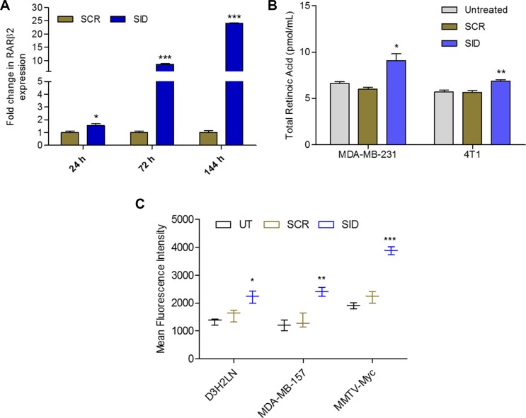
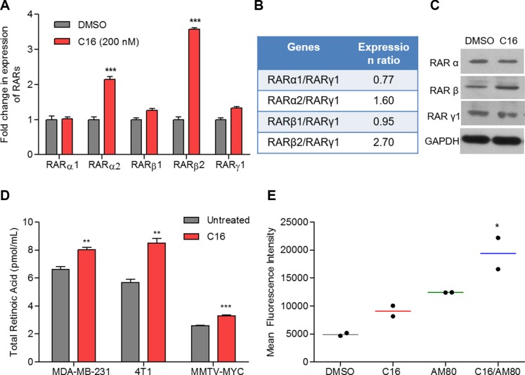
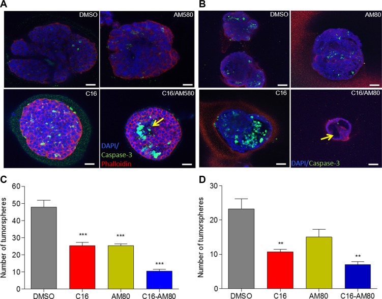
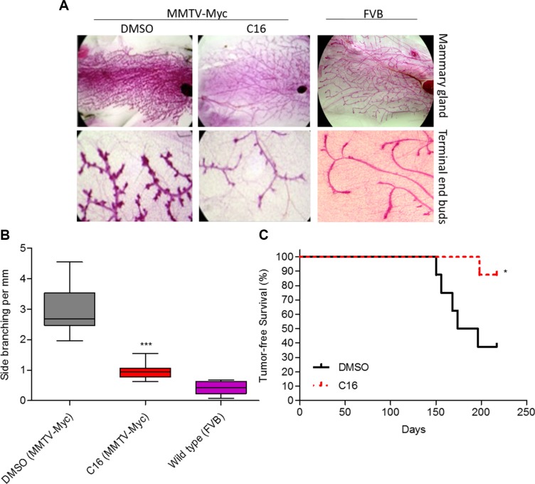
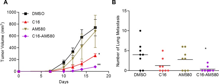
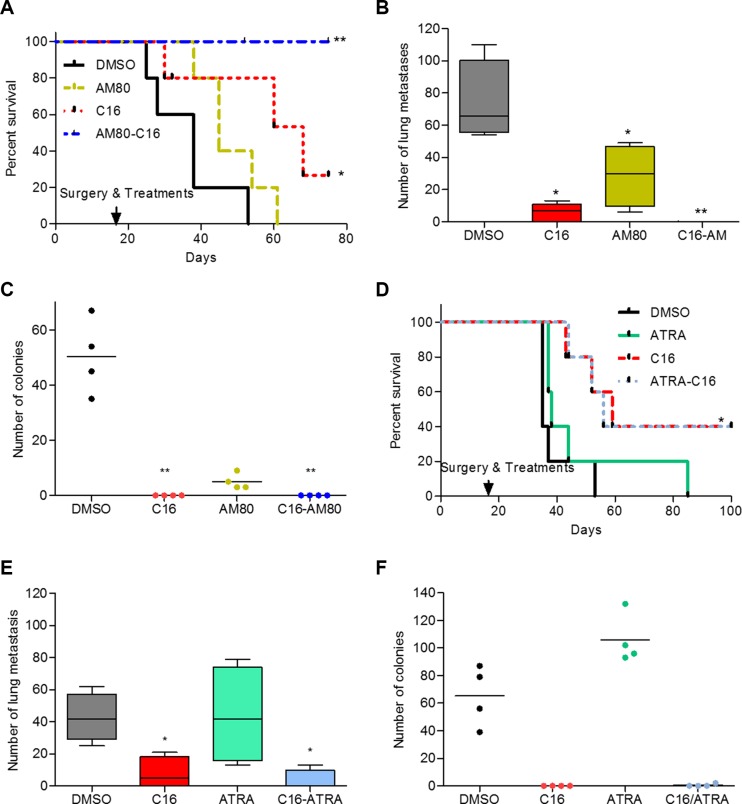
Triple negative breast cancer (TNBC) frequently relapses locally, regionally or as systemic metastases. Development of targeted therapy that offers significant survival benefit in TNBC is an unmet clinical need. We have previously reported that blocking interactions between PAH2 domain of chromatin regulator Sin3A and the Sin3 interaction domain (SID) containing proteins by SID decoys result in EMT reversal, and re-expression of genes associated with differentiation. Here we report a novel and therapeutically relevant combinatorial use of SID decoys. SID decoys activate RARα/β pathways that are enhanced in combination with RARα-selective agonist AM80 to induce morphogenesis and inhibit tumorsphere formation. These findings correlate with inhibition of mammary hyperplasia and a significant increase in tumor-free survival in MMTV-Myc oncomice treated with a small molecule mimetic of SID (C16). Further, in two well-established mouse TNBC models we show that treatment with C16-AM80 combination has marked anti-tumor effects, prevents lung metastases and seeding of tumor cells to bone marrow. This correlated to a remarkable 100% increase in disease-free survival with a possibility of "cure" in mice bearing a TNBC-like tumor. Targeting Sin3A by C16 alone or in combination with AM80 may thus be a promising adjuvant therapy for treating or preventing metastatic TNBC.
Keywords: SID decoys; Sin3A; metastases; retinoids; triple negative breast cancer.
Conflict of interest statement
The authors have declared that no conflicts of interest exists.
Figures
Similar articles
-
The Sin3A/MAD1 Complex, through Its PAH2 Domain, Acts as a Second Repressor of Retinoic Acid Receptor Beta Expression in Breast Cancer Cells.Cells. 2022 Mar 31;11(7):1179. doi: 10.3390/cells11071179. Cells. 2022. PMID: 35406744 Free PMC article.
-
Selective Inhibition of SIN3 Corepressor with Avermectins as a Novel Therapeutic Strategy in Triple-Negative Breast Cancer.Mol Cancer Ther. 2015 Aug;14(8):1824-36. doi: 10.1158/1535-7163.MCT-14-0980-T. Epub 2015 Jun 15. Mol Cancer Ther. 2015. PMID: 26078298 Free PMC article.
-
Interference with Sin3 function induces epigenetic reprogramming and differentiation in breast cancer cells.Proc Natl Acad Sci U S A. 2010 Jun 29;107(26):11811-6. doi: 10.1073/pnas.1006737107. Epub 2010 Jun 14. Proc Natl Acad Sci U S A. 2010. PMID: 20547842 Free PMC article.
-
Tamibarotene: a candidate retinoid drug for Alzheimer's disease.Biol Pharm Bull. 2012;35(8):1206-12. doi: 10.1248/bpb.b12-00314. Biol Pharm Bull. 2012. PMID: 22863914 Review.
-
Targeting triple negative breast cancer with histone deacetylase inhibitors.Expert Opin Investig Drugs. 2017 Nov;26(11):1199-1206. doi: 10.1080/13543784.2017.1386172. Epub 2017 Oct 8. Expert Opin Investig Drugs. 2017. PMID: 28952409 Review.
Cited by
-
Altered RBP1 Gene Expression Impacts Epithelial Cell Retinoic Acid, Proliferation, and Microenvironment.Cells. 2022 Feb 24;11(5):792. doi: 10.3390/cells11050792. Cells. 2022. PMID: 35269414 Free PMC article.
-
5-Azacytidine- and retinoic-acid-induced reprogramming of DCCs into dormancy suppresses metastasis via restored TGF-β-SMAD4 signaling.Cell Rep. 2023 Jun 27;42(6):112560. doi: 10.1016/j.celrep.2023.112560. Epub 2023 Jun 1. Cell Rep. 2023. PMID: 37267946 Free PMC article.
-
miR-210-3p regulates the proliferation and apoptosis of non-small cell lung cancer cells by targeting SIN3A.Exp Ther Med. 2019 Oct;18(4):2565-2573. doi: 10.3892/etm.2019.7867. Epub 2019 Aug 8. Exp Ther Med. 2019. PMID: 31555365 Free PMC article.
-
Corepressor metastasis-associated protein 3 modulates epithelial-to-mesenchymal transition and metastasis.Chin J Cancer. 2017 Mar 9;36(1):28. doi: 10.1186/s40880-017-0193-8. Chin J Cancer. 2017. PMID: 28279208 Free PMC article. Review.
-
DNA Methylation Predicts the Response of Triple-Negative Breast Cancers to All-Trans Retinoic Acid.Cancers (Basel). 2018 Oct 24;10(11):397. doi: 10.3390/cancers10110397. Cancers (Basel). 2018. PMID: 30352973 Free PMC article.
References
-
- Hudis CA, Gianni L. Triple-negative breast cancer: an unmet medical need. Oncologist. 2011;16:1–11. - PubMed
-
- Foulkes WD, Smith IE, Reis-Filho JS. Triple-negative breast cancer. N Engl J Med. 2010;363:1938–1948. - PubMed
-
- Joensuu H, Gligorov J. Adjuvant treatments for triple-negative breast cancers. Ann Oncol. 2012;23:vi40–45. - PubMed
-
- Bansal N, David G, Farias E, Waxman S. Emerging Roles of Epigenetic Regulator Sin3 in Cancer. Adv Cancer Res. 2016;130:113–135. - PubMed
MeSH terms
Substances
Grants and funding
LinkOut - more resources
Full Text Sources
Other Literature Sources
