Review
. 2016 Jul;29(3):695-747.
doi: 10.1128/CMR.00102-15.

Approved Antiviral Drugs over the Past 50 Years

Affiliations
Review

Approved Antiviral Drugs over the Past 50 Years

Erik De Clercq et al. Clin Microbiol Rev. 2016 Jul.

Abstract

Since the first antiviral drug, idoxuridine, was approved in 1963, 90 antiviral drugs categorized into 13 functional groups have been formally approved for the treatment of the following 9 human infectious diseases: (i) HIV infections (protease inhibitors, integrase inhibitors, entry inhibitors, nucleoside reverse transcriptase inhibitors, nonnucleoside reverse transcriptase inhibitors, and acyclic nucleoside phosphonate analogues), (ii) hepatitis B virus (HBV) infections (lamivudine, interferons, nucleoside analogues, and acyclic nucleoside phosphonate analogues), (iii) hepatitis C virus (HCV) infections (ribavirin, interferons, NS3/4A protease inhibitors, NS5A inhibitors, and NS5B polymerase inhibitors), (iv) herpesvirus infections (5-substituted 2'-deoxyuridine analogues, entry inhibitors, nucleoside analogues, pyrophosphate analogues, and acyclic guanosine analogues), (v) influenza virus infections (ribavirin, matrix 2 protein inhibitors, RNA polymerase inhibitors, and neuraminidase inhibitors), (vi) human cytomegalovirus infections (acyclic guanosine analogues, acyclic nucleoside phosphonate analogues, pyrophosphate analogues, and oligonucleotides), (vii) varicella-zoster virus infections (acyclic guanosine analogues, nucleoside analogues, 5-substituted 2'-deoxyuridine analogues, and antibodies), (viii) respiratory syncytial virus infections (ribavirin and antibodies), and (ix) external anogenital warts caused by human papillomavirus infections (imiquimod, sinecatechins, and podofilox). Here, we present for the first time a comprehensive overview of antiviral drugs approved over the past 50 years, shedding light on the development of effective antiviral treatments against current and emerging infectious diseases worldwide.


Figures

FIG 1
FIG 1
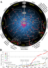
History of antiviral drugs approved between January 1959 and April 2016. (A) Approved antiviral drugs visualized in the zodiac. The gray arrow shows the dates of approval of antiviral drugs from January 1959 to April 2016. Twelve signs are positioned in a circle. Each sign indicates a drug group whose name is annotated outside the circle. In the drug group, each red star within a sign represents an approved drug, placed according to the year of approval. Yellow stars indicate approved drugs that have been discontinued or abandoned for clinical use. A total of 90 stars thus represent all approved antiviral drugs, and each drug star is positioned according to its approval date (Table 2). In this picture, every approved drug could be conceived as a “superstar,” and its contribution to human health is worthy of being remembered and respected. Therefore, this zodiac-based figure metaphorically recognizes each antiviral drug as a star in the universe, commemorating the significant contributions of antiviral drug discovery and development over the past 50 years. A list of drug abbreviations is available in Table 2. Movies and label information for approved drugs are accessible online (see http://www.virusface.com/). (B) Timeline of approval of drugs against 9 human infectious diseases (HIV, HBV, HCV, HSV, HCMV, HPV, RSV, VZV, and influenza virus). The x axis indicates the period from January 1959 to April 2016, and the y axis shows the total number of approved drugs. For each virus, a colored line demonstrates the total number of approved drugs. Moreover, years of discovery of HBV (1963), HPV (1965), HIV (1983), and HCV (1989) are indicated, while the other five viruses were discovered before 1959 (Table 1).
FIG 2
FIG 2
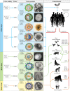
Virus family, morphology, and transmission of HIV, HBV, HCV, HSV, HCMV, HPV, RSV, VZV, and influenza virus. Nine human viruses are classified into DNA viruses (HBV, HCMV, HSV, HPV, and VZV), RNA viruses (HCV, RSV, and influenza virus), and retroviruses (HIV). These viruses are from 7 families: the Hepadnaviridae (HBV), the Papillomaviridae (HPV), the Herpesviridae (HCMV, HSV, and VZV), the Flaviviridae (HCV), the Paramyxoviridae (RSV), the Orthomyxoviridae (influenza virus), and the Retroviridae (HIV). Schematic views and electron micrograph images of viral particles are illustrated in boxes, where particle sizes measured as diameters and viral genome types (circular/linear dsDNA or linear RNA) are also indicated (Table 1). Human viruses are further characterized with the possible animal reservoirs. HIV is known to be transmitted from chimpanzees (HIV-1 groups M and N), gorillas (HIV-1 groups P and O), or sooty mangabeys (HIV-2) (11, 14, 15). Influenza viruses that infect humans originate mostly from birds, pigs, or seals (36, 401). Although the origin of HBV has yet to be clarified, bats might be a potential reservoir for HBV (47). HPV has been widely found in birds, reptiles, marsupials, and mammals, but cross-transfer between species is rare (54). Four human viruses (RSV [41], HCMV [64], HSV [74], and VZV [80]) circulate only in human populations and do not have any animal reservoir. In addition, it remains unclear whether HCV has any animal reservoir (27, 476). (The HCV electron micrograph image is republished from reference with permission of the publisher. The HPV electron micrograph image was obtained from the Laboratory of Tumor Virus Biology at the National Cancer Institute [https://visualsonline.cancer.gov/]. The electron micrograph images for HBV, HCMV [by Sylvia Whitfield], HSV [by Fred Murphy and Sylvia Whitfield], VZV [by Erskine L. Palmer and B. G. Partin], RSV [by Erskine L. Palmer], influenza virus [by Erskine L. Palmer and M. L. Martin], and HIV [by Maureen Metcalfe and Tom Hodge] were obtained from the Centers for Disease Control and Prevention [http://phil.cdc.gov/phil/home.asp].)
FIG 3
FIG 3
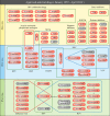
Antiviral drug groups for the treatment of 9 infectious diseases. Approved antiviral drugs are grouped for RNA viruses (HCV, RSV, and influenza virus), DNA viruses (HCMV, HBV, HPV, HSV, and VZV), and retroviruses (HIV). Names of antiviral drugs that are currently in use are enclosed in orange oblongs. Names of discontinued or abandoned antiviral drugs are enclosed in gray oblongs. Those drugs that inhibit more than one virus are shown in the overlapping regions between virus groups. For HCV drugs, a plus symbol is used to indicate the approved combination drugs (simeprevir plus sofosbuvir, sofosbuvir plus daclatasvir, daclatasvir plus asunaprevir, and ribavirin plus PegIFNα-2b).
FIG 4
FIG 4
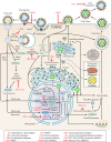
Mechanisms of drug actions during the viral life cycle. Twelve drug groups ordered by roman numerals are shown at the bottom, and their drug actions that interfere with major stages of the viral life cycle are highlighted by red arrows. Solid black arrows indicate direct biological pathways involving viral replication, and dotted black arrows indicate biological pathways with intermediate pathways inside host cells. Major viral stages are illustrated, including endocytosis, exocytosis, virus entry, reverse transcription, virus integration, viral transcription, viral translation, virus budding/release, virus maturation, and other pathways associated with cellular compartments (Golgi apparatus, mitochondria, endoplasmic reticulum [ER], ribosome, proteasome, polysome, and endosome) (for more details, see references , , and 466). Notably, replication pathways of DNA viruses (HCMV, HBV, HPV, HSV, and VZV), RNA viruses (HCV, RSV, and influenza virus), and retroviruses (HIV) diverge after entering host cells. The RNA viruses replicate in the cytoplasm, but DNA viruses and retroviruses further intrude into the nucleus for their DNA synthesis. Note that drug group XIII is not displayed because drugs in this group act mainly as immunoregulatory or antimitotic agents, and they do not directly target viral proteins. Shapes and sizes of proteins and cellular components are not to scale.
FIG 5
FIG 5
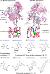
HCMV and HSV-1 DNA polymerase structures and chemical formulas of pyrophosphate analogues, 5-substituted 2′-deoxyuridine analogues, and nucleoside analogues. (A) Tertiary structures of HCMV DNA polymerase in complex with dsDNA and foscarnet (PDB accession number 3KD5). HCMV DNA polymerase is shown in pink. The dsDNA is placed in the center, where foscarnet inhibits DNA synthesis at the active site of HCMV DNA polymerase. Structural movies that demonstrate drug binding are available online (see http://www.virusface.com/). PyMOL V1.7 visualization software (http://www.pymol.org/) was used. (B) Tertiary structures of HSV-1 DNA polymerase complexed with dsDNA and ATP (PDB accession numbers 2GV9 and 4M3R). HSV-1 DNA polymerase is shown in pink. ATP near the catalytic site is displayed in the drug-binding pocket. The triphosphate form of approved antiviral inhibitors (e.g., vidarabine triphosphate) can compete with dATP to inhibit the replication activity of HSV DNA polymerase. (C) Chemical formula of foscarnet in the group of pyrophosphate analogues. (D to F) Chemical formulas of idoxuridine, trifluridine, and brivudine in the group of 5-substituted 2′-deoxyuridine analogues. (G to J) Chemical formulas of telbivudine, entecavir, vidarabine, and FV100 in the group of nucleoside analogues. Note that FV100 is an experimental inhibitor in phase 3 clinical trials.
FIG 6
FIG 6
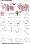
Tertiary structures of HIV-1 reverse transcriptase and chemical formulas of NRTIs and NNRTIs. (A) HIV-1 RT complexed with dsDNA and zidovudine triphosphate (left) (PDB accession number 3V4I) and nevirapine (right) (PDB accession number 4PUO). Two subunits of the HIV-1 RT heterodimer are shown in pink and orange, respectively. Zidovudine triphosphate targets the drug-binding pocket of NRTIs, known as the catalytic site, to inhibit the activity of HIV-1 RT during DNA synthesis. Nevirapine targets the drug-binding pocket of NNRTIs, known as the allosteric site, to block the activity of HIV-1 RT during DNA synthesis (see structural movies at http://www.virusface.com/). (B to H) Chemical formulas of zidovudine, stavudine, zalcitabine, emtricitabine, didanosine, lamivudine, and abacavir in the group of NRTIs. (I to M) Chemical formulas of delavirdine, nevirapine, efavirenz, rilpivirine, and etravirine in the group of NNRTIs.
FIG 7
FIG 7
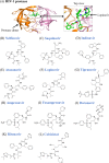
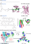
Tertiary structures of HIV-1 protease and chemical formulas of HIV protease inhibitors. (A) HIV-1 protease dimer complexed with lopinavir (PDB accession number 2Q5K). The side view (left) and top view (right) of structures are presented. (B to K) Chemical formulas of nelfinavir, saquinavir, indinavir, atazanavir, lopinavir, ritonavir, fosamprenavir, amprenavir, darunavir, and tipranavir in the group of protease inhibitors. (L) Chemical formula of cobicistat. Cobicistat is a pharmacoenhancer used with HIV protease inhibitors, but cobicistat alone shows no antiviral activity.
FIG 8
FIG 8
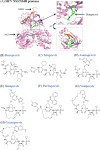
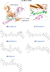
Tertiary structures of HCV NS3/NS4B protease and chemical formulas of HCV protease inhibitors. (A) HCV NS3/NS4B protease in complex with simeprevir (PDB accession numbers 3KEE and 4B76). HCV NS3 and NS4B proteins are shown in pink and orange, respectively. (B to H) Chemical formulas of boceprevir, telaprevir, asunaprevir, simeprevir, paritaprevir, vaniprevir, and grazoprevir in the group of HCV protease inhibitors.
FIG 9
FIG 9
Tertiary structures of viral integrase and chemical formulas of HIV integrase inhibitors. (A) Viral integrase of prototype foamy virus in complex with dsDNA and dolutegravir (PDB accession number 3S3N). A dimer structure of the viral integrase is shown in pink and cyan, respectively. Although the structure of HIV integrase in complex with its inhibitors is still lacking, approved antiviral inhibitors that target HIV and prototype foamy virus integrase are believed to share similar mechanisms (477). (B to D) Chemical formulas of raltegravir, elvitegravir, and dolutegravir in the group of HIV integrase inhibitors.
FIG 10
FIG 10
Chemical formulas of HIV entry inhibitors and tertiary structures of CCR5, HIV-1 GP41, and RSV glycoprotein F. (A) Chemical formula of docosanol. (B and C) Chemical formula of maraviroc and the CCR5 coreceptor in complex with maraviroc (PDB accession number 4MBS). The top and side views of the CCR5 structure are presented. (D and E) Chemical formula of enfuvirtide and tertiary structure of the HIV-1 GP41 trimer (PDB accession number 2X7R). Enfuvirtide is derived from the green region of HIV-1 GP41. The top and side views of the HIV-1 GP41 trimer are presented. Three units of the HIV-1 GP41 trimer are shown in blue, red, and pink, respectively. (F) Tertiary structure of the prefusion RSV glycoprotein F trimer in complex with the antibody motavizumab (PDB accession number 4ZYP). Motavizumab is an experimental monoclonal antibody derived from the FDA-approved drug palivizumab (478). The side views (left) and top views (right) of protein structures are presented. The heavy and light chains of motavizumab are shown in blue and green, respectively. The palivizumab-binding site (amino acid [aa] positions 254 to 277 [479]) is highlighted in red. Three units of the prefusion RSV F trimer are shown in pink, gray, and cyan, respectively.
FIG 11
FIG 11
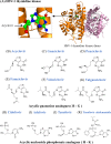
Tertiary structures of HSV-1 thymidine kinase and chemical formulas of acyclic guanosine analogues and acyclic nucleoside phosphonate analogues. (A) The HSV-1 thymidine kinase dimer in complex with acyclovir. Two units of thymidine kinase are shown in pink and orange, respectively. Acyclovir can be phosphorylated by HSV thymidine kinase and cellular enzymes (249). (B to G) Chemical formulas of acyclovir, famciclovir, valacyclovir, ganciclovir, penciclovir, and valganciclovir in the group of acyclic guanosine analogues. (H to K) Chemical formulas of cidofovir, adefovir, tenofovir, and tenofovir alafenamide in the group of acyclic nucleoside phosphonate analogues.
FIG 12
FIG 12
Tertiary structures of the HCV NS5A protein and chemical formulas of HCV NS5A inhibitors. (A) Tertiary structure of the HCV NS5A dimer in complex with daclatasvir. Two units of the HCV NS5A dimer are shown in pink and orange, respectively (PDB data were reported in reference 282). (B to F) Chemical formulas of ledipasvir, daclatasvir, ombitasvir, velpatasvir, and elbasvir in the group of HCV NS5A inhibitors. Note that velpatasvir is an experimental inhibitor currently in phase 3 clinical trials.
FIG 13
FIG 13
Tertiary structures of HCV NS5B polymerase and chemical formulas of HCV NS5B inhibitors. (A) Tertiary structure of HCV NS5B polymerase in complex with dsDNA, beclabuvir, and sofosbuvir diphosphate (PDB accession numbers 4NLD and 4WTG). Note that beclabuvir and sofosbuvir diphosphate bind to the allosteric site and the catalytic site of the HCV NS5B polymerase, respectively. (B to D) Chemical formulas of beclabuvir, dasabuvir, and sofosbuvir in the group of HCV NS5B inhibitors. Note that beclabuvir is a forthcoming inhibitor in the combination drug of daclatasvir plus asunaprevir plus beclabuvir in phase 4 clinical trials.
FIG 14
FIG 14
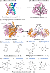
Tertiary structures of influenza virus proteins (matrix 2, neuraminidase, and RNA polymerase) and chemical formulas of influenza virus inhibitors. (A) Tertiary structure of the influenza A virus matrix 2 protein in complex with amantadine (PDB accession number 2KAD). Movies that simulate the binding of approved antiviral drugs to viral or host proteins are available online (see http://www.virusface.com/). (B) Structure of influenza A virus neuraminidase in complex with zanamivir (PDB accession number 2HTQ). (C) Tertiary structure of influenza virus RNA polymerase in complex with RNA. The RNA polymerases of influenza A virus (left) (PDB accession number 3J9B) and influenza B virus (right) (PDB accession number 4WRT) are illustrated. The PA, PB1, and PB2 subunits of RNA polymerase (see structural details in reference 480) are shown in pink, orange, and gray, respectively. Ribavirin triphosphate targets the catalytic site of the RNA polymerase to inhibit viral replication. Note that the RNA polymerase of influenza A virus is a tetramer (480), but the complete tetramer structure of influenza virus RNA polymerase in complex with its inhibitors is still lacking. (D and E) Chemical formulas of amantadine and rimantadine, which target the matrix 2 protein of influenza virus. (F and G) Chemical formulas of ribavirin and favipiravir, which target the viral RNA polymerase of influenza virus. (H to K) Chemical formulas of zanamivir, laninamivir, peramivir, and oseltamivir, which target the viral neuraminidase of influenza virus.
FIG 15
FIG 15
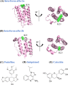
Tertiary structures of interferons and chemical formulas of podofilox, imiquimod, and catechin. (A and B) Cartoon representations of interferon alfa 2a (PDB accession number 4YPG) and interferon alfa 2b (PDB accession number 1RH2). Sequence comparison suggests that amino acid K23 in interferon alfa 2a and amino acid R23 in interferon alfa 2b mark the only sequence difference between interferon alfa 2a and interferon alfa 2b. Structural movies are available online (http://www.virusface.com/). (C to E) Chemical formulas of podofilox, imiquimod, and catechin. Note that catechin is the major ingredient of the botanical drug sinecatechin.
None
None

Similar articles

Cited by

References

    1. De Clercq E. 2004. Antivirals and antiviral strategies. Nat Rev Microbiol 2:704–720. doi:10.1038/nrmicro975. - DOI - PMC - PubMed
    1. De Clercq E. 2002. Strategies in the design of antiviral drugs. Nat Rev Drug Discov 1:13–25. doi:10.1038/nrd703. - DOI - PubMed
    1. De Clercq E. 1997. In search of a selective antiviral chemotherapy. Clin Microbiol Rev 10:674–693. - PMC - PubMed
    1. De Clercq E. 2009. The history of antiretrovirals: key discoveries over the past 25 years. Rev Med Virol 19:287–299. doi:10.1002/rmv.624. - DOI - PubMed
    1. De Clercq E. 2009. Anti-HIV drugs: 25 compounds approved within 25 years after the discovery of HIV. Int J Antimicrob Agents 33:307–320. doi:10.1016/j.ijantimicag.2008.10.010. - DOI - PubMed

Publication types

MeSH terms

Substances