. 2016 Jun 9;11(6):e0157114.
doi: 10.1371/journal.pone.0157114. eCollection 2016.

Targeted Delivery of Deoxycytidine Kinase to Her2-Positive Cells Enhances the Efficacy of the Nucleoside Analog Fludarabine

Affiliations

Targeted Delivery of Deoxycytidine Kinase to Her2-Positive Cells Enhances the Efficacy of the Nucleoside Analog Fludarabine

Sujatha P Koduvayur et al. PLoS One. .

Abstract

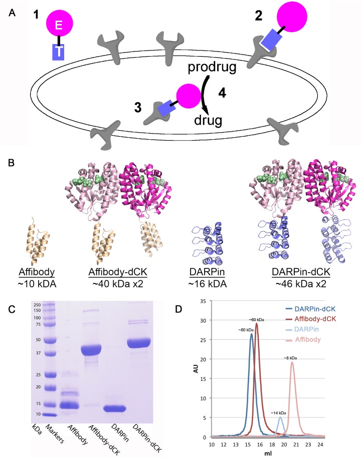
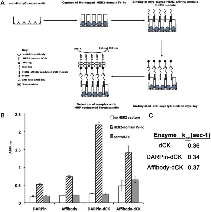
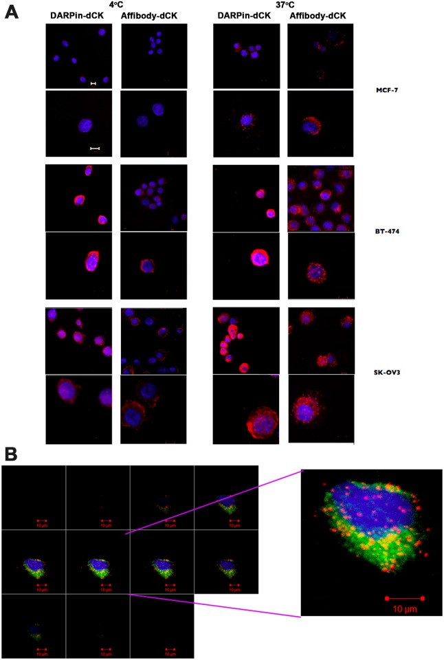
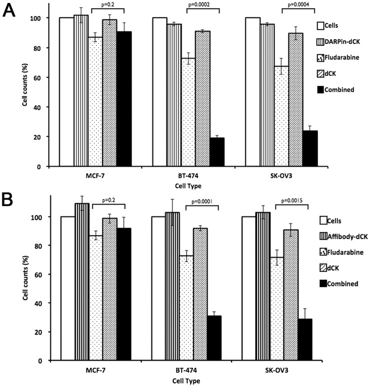
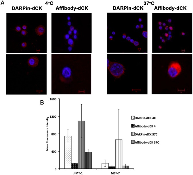
Cytotoxic drugs, such as nucleoside analogs and toxins, commonly suffer from off-target effects. One approach to mitigate this problem is to deliver the cytotoxic drug selectively to the intended site. While for toxins this can be achieved by conjugating the cell-killing moiety to a targeting moiety, it is not an option for nucleoside analogs, which rely on intracellular enzymes to convert them to their active triphosphorylated form. To overcome this limitation, and achieve site-targeted activation of nucleoside analogs, we fused the coding region of a prodrug-activating enzyme, deoxycytidine kinase (dCK), to affinity reagents that bind to the Her2 cell surface protein. We evaluated dCK fusions to an anti-Her2 affibody and Designed Ankyrin Repeat Protein (DARPin) for their ability to kill cancer cells by promoting the activation of the nucleoside analog fludarabine. Cell staining and flow cytometry experiments with three Her2 positive cancer cell lines (BT-474-JB, JIMT-1 and SK-OV-3) indicate dCK fusions binding and cellular internalization. In contrast, these reagents bind only weakly to the Her2 negative cell line, MCF-7. Cell proliferation assays indicate that SK-OV-3 and BT-474-JB cell lines exhibit significantly reduced proliferation rates when treated with targeting-module fused dCK and fludarabine, compared to fludarabine alone. These findings demonstrate that we have succeeded in delivering active dCK into the Her2-positive cells, thereby increasing the activation of fludarabine, which ultimately reduces the dose of nucleoside analog needed for cell killing. This strategy may help establish the therapeutic index required to differentiate between healthy tissues and cancer cells.


Conflict of interest statement

Competing Interests: The authors have declared that no competing interests exist.

Figures

Fig 1
Fig 1. Strategy for preferential activation of prodrugs at target cells and the Her2-affinity reagents used in this study.
(A) This strategy relies on a bi-modular fusion protein composed of a cell marker-targeting module (square labeled with T) genetically fused to an enzyme that catalyzes the activation of a prodrug (circle labeled with E). This fusion protein is administered systemically (point 1), but it accumulates at the targeted cells by binding to a specific cell surface protein (point 2). The fusion protein then enters the cell via receptor-mediated endocytosis or by membrane recycling (point 3). Subsequent administration of an appropriate prodrug results in its preferential activation in the targeted cells (point 4), thereby killing the targeted cell. (B) Ribbon diagram of the reagents with their molecular size indicated. Affibody, DARPin, and dCK models from PDB IDs 1LP1, 2JAB, and 1P5Z, respectively. The fusion proteins were modeled based on the individual structures. The dimeric nature of dCK result in molecules that contains two anti-Her2 modules, which are expected to increase its avidity to the receptor relative to the single affinity modules. Green spheres denote the substrates binding sites in dCK. (C) SDS-PAGE demonstrates the > 95% purity of the reagents. We note that the DARPin module runs as a smaller protein than the expected size. (D) Gel-filtration analysis of the reagents. The observed elution volumes correspond to the expected sizes of the reagents, with the fusion protein being dimeric, and the single affinity modules being monomeric.
Fig 2
Fig 2. Characterizing individual anti-Her2 modules and the bi-modular anti-Her2-dCK fusion proteins.
(A) Schematic of the ELISA assay used to evaluate the binding of the affinity reagents to the ectodomain IV of the Her2 receptor. (B) Binding of reagents to the ectodomain IV of Her2 receptor was measured by incubating 50 nM of reagents with 20 nM of immobilized Her2-Fc recombinant protein. Detection was performed using biotinylated anti-c-myc antibody and streptavidin conjugated to Horseradish peroxidase (HRP). Error bars represent standard deviation of triplicate measurements. We interpret the increased ELISA signal for the fusion proteins relative to the individual affinity modules resulting from the dimeric nature of the fusion proteins. (C) The observed steady state phosphorylation rate (kobs) of fludarabine by the engineered dCK (dCK-DMS74E) with, or without, the Her2-affinity modules is measured with 200 μM of fludarabine at 37°C. These measurements indicate that the affinity modules do not interfere with the dCK enzymatic activity.
Fig 3
Fig 3. Binding of bi-modular anti-Her2 -dCK fusion proteins to cancer cells.
(A) One hundred thousand cells were treated with 1 μM of Alexa Fluor® 647 dye (red in the images) conjugated reagents and incubated for 2 h at either 4°C or 37°C. The nuclei of treated cells were stained with DAPI (blue) and visualized at 40x using a Zeiss confocal laser scanning microscope. Images are representative of three independent trials. Scale bar depicts 10 μm. Note the much-increased signal for the fusion proteins with the Her2-postive cells lines (BT-474 and SK-OV3), relative to the Her2-negative MCF-7 cell line. The stronger intracellular signal at 37°C relative to that observed at 4°C indicates that at least a fraction of the fusion proteins has internalized. (B) Internalization of DARPin-dCK in BT-474 cells. Co-localization of receptor bound reagents with intracellular vesicles was detected by treating 5x10⁴ cells with 1.5x10⁶ particles each of CellLight® Reagents *BacMam 2.0* GFP markers for early and late endosomes and lysosomes, for 18 h at 37°C, according to the manufacturer’s protocol. These cells were then treated with 0.5 μM of Alexa Fluor® 647 conjugated DARPin-dCK, as in A. Image sections at the z plane at 40x resolution of a single BT-474 cell are shown on the left panel. A high power image of a single section is shown on the right panel. GFP fluorescence indicates location of endosomes and lysosomes, while red fluorescence indicates the cellular location of DARPin-dCK. Image shown is representative of 10 cells imaged from duplicate trials. Scale bar is 10 μm.
Fig 4
Fig 4. Binding of the bi-modular fusion proteins to cancer cells measured by flow cytometry.
(A) One million cells were treated with 1 μM of reagents as in Fig 2 and mean fluorescence intensity was measured using a Cyan3 fluorescent cell sorter (Beckman). Fluorescent cells were gated on cells treated with reagents that were not conjugated to dye and hence did not exhibit any fluorescence at 647 nm. Error bars correspond to standard deviations on three independent trials. (B) One million cells were treated with 1 μM dCK-AlexaFluor647 and the signal intensity was normalized to number of dye conjugations (ref 3 from Supplement). Error bars correspond to standard deviations of n = 3 trials. (C) Cell were treated with 2 μM of anti-Her2 DARPin-AlexaFluor647 and Affibody-AlexaFluor647 and measured as above. Note the much reduced mean fluorescence intensity in comparison to the fusion constructs (panel A). Error bars correspond to standard deviations of three independent trials. (D) Fluorescence intensities of cells treated with dCK-fusion protein were normalized to number of conjugated dye molecules (3), background subtracted and compared to those of cells treated with the Her2 affinity module (DARPin or affibody) alone. The resulting mean fold change in intensity was plotted for each cell type and reagent.
Fig 5
Fig 5. Effect of treatment of bi-modular fusion proteins on proliferation of cancer cells.
(A) Seven thousand cells were treated with (i) buffer as control, (ii) bi-modular fusion protein (DARPin-dCK at 5 μM, panel A or (B) Affibody-dCK at 10 μM, panel B), (iii) fludarabine (0.25 μM), (iv) dCK (10 μM) and (v) as combination of fludarabine and bi-modular fusion protein at the same concentrations when tested by themselves. After the addition of the above reagents, cells were further incubated at 37°C for 96 h in 96 well plates, upon which cell proliferation was assessed using the AlamarBlue assay. The cell proliferation signal was normalized to the buffer control signal, which was set at 100%. Results are shown for the Her2-negative MCF-7 and Her2-positive BT-474 and SK-OV3 cell lines. Error bars correspond to standard deviations of triplicate measurements. p values were calculated using the Student’s T-test macros in Microsoft Excel (tail = 1, type = 3).
Fig 6
Fig 6. Binding of fusion proteins to low Her2-expressing cancer cells.
(A) JIMT-1 cells were treated and imaged as in Fig 4. (B) Flow cytometry assay of reagents binding to Her2 expressing JIMT-1 cells. Cells were treated and counted as in Fig 5A. Mean fluorescence intensity of reagent treated JIMT-1 cells are compared to Her2- MCF-7 cells. Error bars correspond to standard deviations of triplicate measurements.

Similar articles

Cited by

References

    1. Jordheim LP, Durantel D, Zoulim F, Dumontet C. Advances in the development of nucleoside and nucleotide analogues for cancer and viral diseases. Nat Rev Drug Discov. 2013;12:447–64. 10.1038/nrd4010 - DOI - PubMed
    1. Galmarini CM, Mackey JR, Dumontet C. Nucleoside analogues and nucleobases in cancer treatment. Lancet Oncol. 2002;3:415–24. - PubMed
    1. Strebhardt K, Ullrich A. Paul Ehrlich's magic bullet concept: 100 years of progress. Nat Rev Cancer. 2008;8:473–80. 10.1038/nrc2394 - DOI - PubMed
    1. Leal M, Sapra P, Hurvitz SA, Senter P, Wahl A, Schutten M, et al. Antibody-drug conjugates: an emerging modality for the treatment of cancer. Ann N Y Acad Sci. 2014;1321:41–54. 10.1111/nyas.12499 - DOI - PubMed
    1. Lambert JM. Drug-conjugated antibodies for the treatment of cancer. Br J Clin Pharmacol. 2013;76:248–62. 10.1111/bcp.12044 - DOI - PMC - PubMed

MeSH terms