Expression of Membrane-Bound Mucins and p63 in Distinguishing Mucoepidermoid Carcinoma from Papillary Cystadenoma
- PMID: 27278378
- PMCID: PMC5082059
- DOI: 10.1007/s12105-016-0735-4
Expression of Membrane-Bound Mucins and p63 in Distinguishing Mucoepidermoid Carcinoma from Papillary Cystadenoma
Abstract
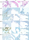
The aim of this study was to compare the immunoexpression of epithelial mucins (MUCs) in salivary duct cysts, papillary cystadenomas, and mucoepidermoid carcinomas and to evaluate if any of these markers could be useful for differentiating between mucoepidermoid carcinoma and papillary cystadenoma. We also sought to validate the p63 expression pattern found to differentiate between mucoepidermoid carcinoma and papillary cystadenoma. Immunoexpression of MUC1, MUC2, MUC4, MUC7, and p63 was studied and quantified in 22 mucoepidermoid carcinomas, 12 papillary cystadenomas, and 3 salivary duct cysts. The immunohistochemical evaluation was collectively performed by 3 oral pathologists. Scores and trends in proportions were assessed using the nonparametric Wilcoxon-Mann-Whitney rank sum test. Mucoepidermoid carcinomas, papillary cystadenomas, and salivary duct cysts demonstrated variable MUC expression patterns. All tumors were positive for p63 immunoexpression with p63 labeling in salivary duct cysts and papillary cystadenomas (15/15) limited to the basal layers of the cystic spaces, whereas in mucoepidermoid carcinomas (22/22) the p63 labeling extended throughout the suprabasal layers (p < 0.001). This study adds more confirmatory data to validate that the reactivity pattern of p63 protein can be used in distinguishing between papillary cystadenoma and low-grade mucoepidermoid carcinoma. Although positive reactivity in a tumor with MUC1 and MUC4 was inconclusive, negative reactivity suggests the diagnosis of a benign PC or SDC.
Keywords: Immunohistochemistry; Mucins; Mucoepidermoid carcinoma; Papillary cystadenoma.
Conflict of interest statement
None.
Figures
Similar articles
-
P63 expression in papillary cystadenoma and mucoepidermoid carcinoma of minor salivary glands.Oral Surg Oral Med Oral Pathol Oral Radiol. 2013 Jan;115(1):79-86. doi: 10.1016/j.oooo.2012.09.005. Oral Surg Oral Med Oral Pathol Oral Radiol. 2013. PMID: 23217538
-
Expression of membrane-bound mucins (MUC1 and MUC4) and secreted mucins (MUC2, MUC5AC, MUC5B, MUC6 and MUC7) in mucoepidermoid carcinomas of salivary glands.Am J Surg Pathol. 2005 Jun;29(6):806-13. doi: 10.1097/01.pas.0000155856.84553.c9. Am J Surg Pathol. 2005. PMID: 15897748
-
MUC1, MUC2, MUC4, and MUC5AC expression in salivary gland mucoepidermoid carcinoma: diagnostic and prognostic implications.Am J Surg Pathol. 2005 Jul;29(7):881-9. doi: 10.1097/01.pas.0000159103.95360.e8. Am J Surg Pathol. 2005. PMID: 15958852
-
Papillary cystadenoma of the lower lip exhibiting ciliated pseudostratified columnar epithelium: report of a bizarre case and review of the literature.Oral Maxillofac Surg. 2013 Sep;17(3):161-4. doi: 10.1007/s10006-012-0357-2. Epub 2012 Aug 30. Oral Maxillofac Surg. 2013. PMID: 22933035 Review.
-
Salivary gland-like tumors of the breast express basal-type immunohistochemical markers.Appl Immunohistochem Mol Morphol. 2013 Jul;21(4):283-6. doi: 10.1097/PAI.0b013e31826a277e. Appl Immunohistochem Mol Morphol. 2013. PMID: 22935826 Review.
Cited by
-
Intraoral Salivary Duct Cyst: Clinical and Histopathologic Features of 177 Cases.Head Neck Pathol. 2017 Dec;11(4):469-476. doi: 10.1007/s12105-017-0810-5. Epub 2017 Mar 27. Head Neck Pathol. 2017. PMID: 28349371 Free PMC article.
-
Analysis of the Clinical Relevance of Histological Classification of Benign Epithelial Salivary Gland Tumours.Adv Ther. 2019 Aug;36(8):1950-1974. doi: 10.1007/s12325-019-01007-3. Epub 2019 Jun 17. Adv Ther. 2019. PMID: 31209701 Free PMC article. Review.
References
-
- Ellis GL, Auclair PL. Tumors of the salivary glands. In: Silverberg SG, editor. Tumors of the salivary glands. AFIP atlas of tumor pathology. Maryland: ARP Press; 2008. pp. 173–196.
MeSH terms
Substances
Grants and funding
LinkOut - more resources
Full Text Sources
Other Literature Sources
Medical
Research Materials
Miscellaneous


