. 2016 Apr;7(2):364.
doi: 10.4172/2157-7439.1000364. Epub 2016 Apr 5.

Magnetofection Enhances Adenoviral Vector-based Gene Delivery in Skeletal Muscle Cells

Affiliations

Magnetofection Enhances Adenoviral Vector-based Gene Delivery in Skeletal Muscle Cells

Andrea Soledad Pereyra et al. J Nanomed Nanotechnol. 2016 Apr.

Abstract

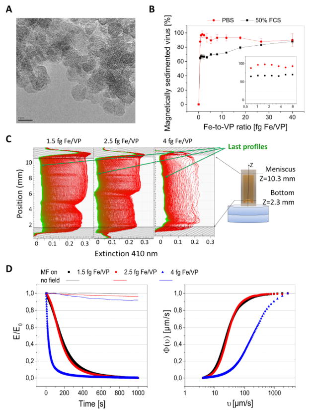
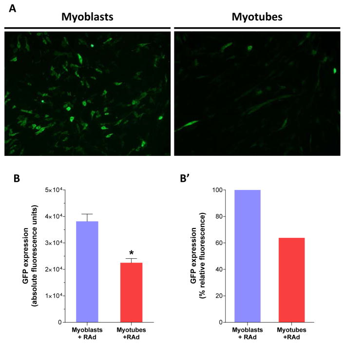
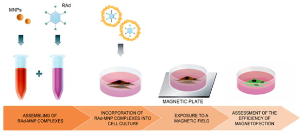
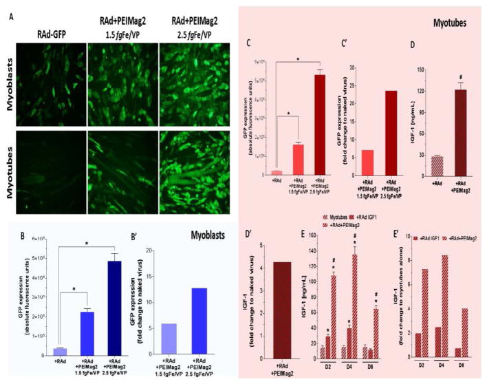
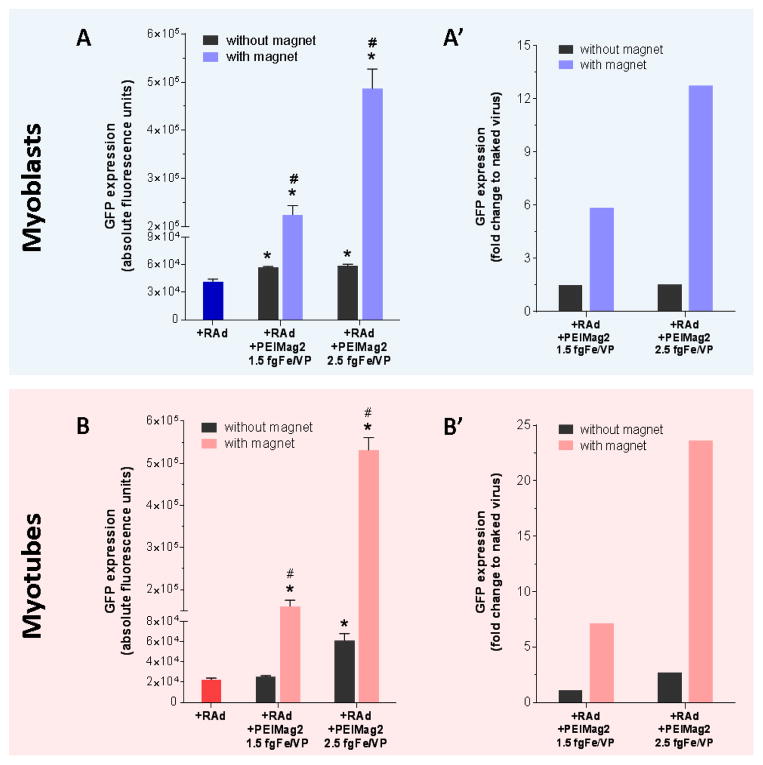
The goal of magnetic field-assisted gene transfer is to enhance internalization of exogenous nucleic acids by association with magnetic nanoparticles (MNPs). This technique named magnetofection is particularly useful in difficult-to-transfect cells. It is well known that human, mouse, and rat skeletal muscle cells suffer a maturation-dependent loss of susceptibility to Recombinant Adenoviral vector (RAd) uptake. In postnatal, fully differentiated myofibers, the expression of the primary Coxsackie and Adenoviral membrane receptor (CAR) is severely downregulated representing a main hurdle for the use of these vectors in gene transfer/therapy. Here we demonstrate that assembling of Recombinant Adenoviral vectors with suitable iron oxide MNPs into magneto-adenovectors (RAd-MNP) and further exposure to a gradient magnetic field enables to efficiently overcome transduction resistance in skeletal muscle cells. Expression of Green Fluorescent Protein and Insulin-like Growth Factor 1 was significantly enhanced after magnetofection with RAd-MNPs complexes in C2C12 myotubes in vitro and mouse skeletal muscle in vivo when compared to transduction with naked virus. These results provide evidence that magnetofection, mainly due to its membrane-receptor independent mechanism, constitutes a simple and effective alternative to current methods for gene transfer into traditionally hard-to-transfect biological models.

Keywords: Adenoviral vectors; Gene delivery; Magnetic nanoparticles; Magneto-adenovectors; Magnetofection; Skeletal muscle.


Figures

Figure 1
Figure 1
Magnetic nanoparticles and magnetic vectors: (A) Transmission Electron Microscopy image of the PEI-Mag2 nanoparticles, bar=5 nm. (B) Percent of the magnetically sedimented viral particles vs. MNP-to-Virus ratio (fg Fe/VP). Adenoviral particles labeled with a iodine-127 were assembled with PEI-Mag2 nanoparticles at different MNP-to-VP ratios in PBS for 30 min (curve “PBS”) or further 1-to-1 diluted with Fetal Calf Serum, incubated for the next 30 and then exposed at the Magnetic Plate for 30 min (curve “50%FCS”), non-sedimented radioactivity was measured in a supernatant. (C and D) Magnetophoretic mobility of the magnetic vectors characterized by kinetics of clearance of the suspensions in applied gradient magnetic fields. Figure (C) shows extinction profiles at 410 nm for the suspensions of the magnetic adenoviral complexes registered with a customized LUMiReader® device with 2 disk magnets positioned underneath optical cuvette at multiple points along the vertical axis of the cuvette (STEP-MAG measurements). Schematics at the right panel shows an optical cuvette and a set of Neodymium-Iron-Boron-Magnets positioned underneath the cuvette; resulting magnetic flux density and gradient averaged over vertical sample height were of 0.16 T and 33.5T/m, respectively. (D) Normalized integral Extinction at 410 nm, E/E0, averaged through the probe height versus time upon exposure to magnetic field (MF on) or with no filed applied (no field) and derived data on cumulative distribution functions of the effective magnetophoretic mobility F(u).
Figure 2
Figure 2
Conventional gene delivery with naked adenoviral vectors yields low efficiency in c2c12 myotubes: C2C12 myoblasts and fully differentiated myotubes were incubated for 60 minutes with a recombinant adenoviral vector, RAd-GFP, at a MOI of 60 viral particles per cell. To assess GFP expression, cell culture imaging and fluorescence quantitation were performed 48 hours after infection. A Representative microscopy images of myoblasts (left) and myotubes (right) transduced with RAd-GFP. After image acquisition, GFP quantitation was performed in cellular lysates. Comparison between both groups was plotted as absolute B and relative-to-myoblasts fluorescenceB’. Unspecific fluorescence readings from cells alone and lysis buffer were subtracted to each group previous to data analysis. * Statistically significant difference with p < 0.05 between the experimental groups. Data values represent mean ± S.E.M. n=3 for each group. Magnification, 10X.
Figure 3
Figure 3
Magnetofection in cell culture: This illustration shows the general workflow for in vitro magnetofection. The RAd-MNP complexes are pre-assembled in the test tube and later introduced to the cell culture in a dropwise manner. The culture plate is then exposed to a magnetic field created by the magnetic plate placed under it and 48 hours later.
Figure 4
Figure 4
Magnetofection enhances viral vector-mediated gene delivery in C2C12 myoblasts and myotubes: C2C12 myoblasts and fully differentiated myotubes were incubated with either naked recombinant adenoviral vectors (RAd-GFP or RAd-IGF-1) or RAd+MNPs complexes (RAd+PEIMag2) and exposed to an optimal magnetic field for 30 minutes. To assess gene delivery efficiency, GFP expression was evaluated using microscopy imaging and fluorescence quantitation 48 hours after infection for both groups. In a similar manner, IGF-1 levels were measured in the culture medium by radioimmunoassay. A Representative microscopy images of myoblasts (upper panels) and myotubes (lower panels) infected with RAd-GFP and RAd-GFP+MNPs at 1.5 and 2.5 fgFe/VP MNP-to-Viral Particle ratios. After imaging, cells lysates were used to perform quantitation of GFP expression. Comparison between conventional viral transduction and magnetofection was plotted as absolute B and C and relative-to-naked virus fluorescence levels B’ and C’. Unspecific signal from cells alone, MNPs and lysis buffer were subtracted to each group previous to data analysis. To assess magnetofection efficacy with RAd-IGF-1, culture medium from myotubes was collected 48 hs after gene transfer protocols and processed for IGF-1 extraction and quantitation. Comparison between conventional viral transduction and magnetofection was plotted as absolute D and relative-to-naked virus concentration D’. IGF-1 basal levels in myotubes alone, as well as unspecific readings from extraction buffer, were subtracted to both experimental groups in order to show only viral vector-mediated IGF-1 production. In a subset of myotubes, IGF-1 expression was analyzed in the culture medium at days 2 (D2), 4 (D4) and 6 (D6) after gene delivery and compared between non-infected, transduced with naked virus and magnetofected cells. Plots show absolute E and relative-to-myotubes alone E’ IGF-1 levels for each time point. *Statistically significant difference with p < 0.05 between non-infected myotubes and naked virus or magnetofection groups. #Statistically significant difference with p < 0.05 between naked virus and magnetofection groups. Data values represent mean ± S.E.M. n=3 for GFP groups, n=4 for IGF-1 at 48 hs and n=6 for IGF-1 longitudinal analysis. fgFe/VP=femtograms of iron per Viral Particle
Figure 5
Figure 5
The magnetic field is a critical component of magnetofection-based gene delivery: C2C12 myoblasts and fully differentiated myotubes were incubated with either naked RAd or RAd+MagneticNanoParticles complexes and 48 hours later GFP expression was evaluated as described before. With and without magnetic field comparison was performed for both, myoblasts and myotubes. Results are plotted as absolute (A and B) and relative-to-naked virus fluorescence (A’ and B’). Unspecific fluorescence readings from cells alone, MNPs and lysis buffer were subtracted to each group previous data analysis. *Statistically significant difference with p < 0.05 between naked RAd and magnetofection with and without magnet. #Statistically significant difference with p < 0.05 between magnetofection with and without magnet. Data values represent mean ± S.E.M. n=3 for each group. fgFe/VP=femtograms of iron per Viral Particle
Figure 6
Figure 6
Cellular uptake of RAd-MNPs complexes after magnetically-induced sedimentation: Magnetofection protocol was applied to fully differentiated C2C12 myotubes and Transmission Electron Microscopy (TEM) images were taken at different time points after magnetic-field exposure. (A1) 15 minutes after magnetic-field exposure. Lower-magnification image to show the highly electron-dense (black) RAd-MNPs complexes distributed over the cell surface after magnetically-induced sedimentation. (A2–A5) Red arrows indicate membrane protrusions (lamellipodia-like structures) and blue arrows indicate membrane invaginations (pits). (B) After 30 minutes, engulfment and internalization of the RAd-MNPs complexes was evident (orange arrows). (C) 60 minutes after initial magnetic-field exposure, complexes could be visualized inside the cells, loaded into cytoplasmic vesicles (orange arrows). (D) Green arrows point to adenoviral particles surrounded by PEI-Mag2 magnetic nanoparticles while being engulfed by the myotube. Inset in the lower-right corner shows isolated Recombinant Adenoviral Vectors.
Figure 7
Figure 7
Intracellular fate and localization of RAd-MNPs complexes: To allow intracellular visualization and tracking, magnetoadenovectors were assembled with fluorescent MNPs and applied to C2C12 myoblasts and myotubes. Images were taken 48 hours afterwards. (A) simultaneous visualization of Green Fluorescent Protein expression (codified by the viral genome) and Atto550PEI-Mag2 nanoparticles (red fluorescence) in myoblasts. (A’) Enlarged area focused on the punctate, perinuclear localization of the red fluorescence. (B) Fully differentiated myotubes also displaying Atto550PEI-Mag2 localization around each nuclear domain. (B’) Enlarged picture of magnetofected myotubes.
Figure 8
Figure 8
In vivo magnetofection in mouse skeletal muscle: (A) Our magnetofection protocol was evaluated in 10-month old C57BL/6 mice. Animals were anesthetized and positioned ventrally on the operating table with both hindlimbs extended and gently immobilized. Finally a 1-cm long incision was made to access the posterior muscular compartment. (B1, B2 and B3) Six days after magnetofection, freshly isolated Soleus revealed a brown area beneath the muscle surface where the RAd-MNPs were injected. (C1 and C2) Naked RAd-GFP resulted in transduction of only mononucleated cells with no GFP positive myofibers. (D1–D4) After magnetofection with RAd-MNPs complexes, GFP positive myofibers were noticeable and clearly differ from the surrounding tissue auto fluorescence. (D5 and D6) Co-localization of GFP expression and magnetic nanoparticles (Atto550PEI-Mag2 with red fluorescence) within the same myofiber.

Similar articles

Cited by

References

    1. Plank C, Zelphati O, Mykhaylyk O. Magnetically enhanced nucleic acid delivery. Ten years of magnetofection-progress and prospects. Adv Drug Deliv Rev. 2011;63:1300–1331. - PMC - PubMed
    1. Widder KJ, Senyel AE, Scarpelli GD. Magnetic microspheres: a model system of site specific drug delivery in vivo. Proc Soc Exp Biol Med. 1978;158:141–146. - PubMed
    1. Scherer F, Anton M, Schillinger U, Henke J, Bergemann C, et al. Magnetofection: enhancing and targeting gene delivery by magnetic force in vitro and in vivo. Gene Ther. 2002;9:102–109. - PubMed
    1. Gupta AK, Gupta M. Synthesis and surface engineering of iron oxide nanoparticles for biomedical applications. Biomaterials. 2005;26:3995–4021. - PubMed
    1. Tresilwised N, Pithayanukul P, Holm PS, Schillinger U, Plank C, et al. Effects of nanoparticle coatings on the activity of oncolytic adenovirus-magnetic nanoparticle complexes. Biomaterials. 2012;33:256–269. - PubMed

LinkOut - more resources