Review
. 2017 May;77(5):643-673.
doi: 10.1002/dneu.22405. Epub 2016 Jun 24.

Gap junctional signaling in pattern regulation: Physiological network connectivity instructs growth and form

Affiliations
Review

Gap junctional signaling in pattern regulation: Physiological network connectivity instructs growth and form

Juanita Mathews et al. Dev Neurobiol. 2017 May.

Abstract

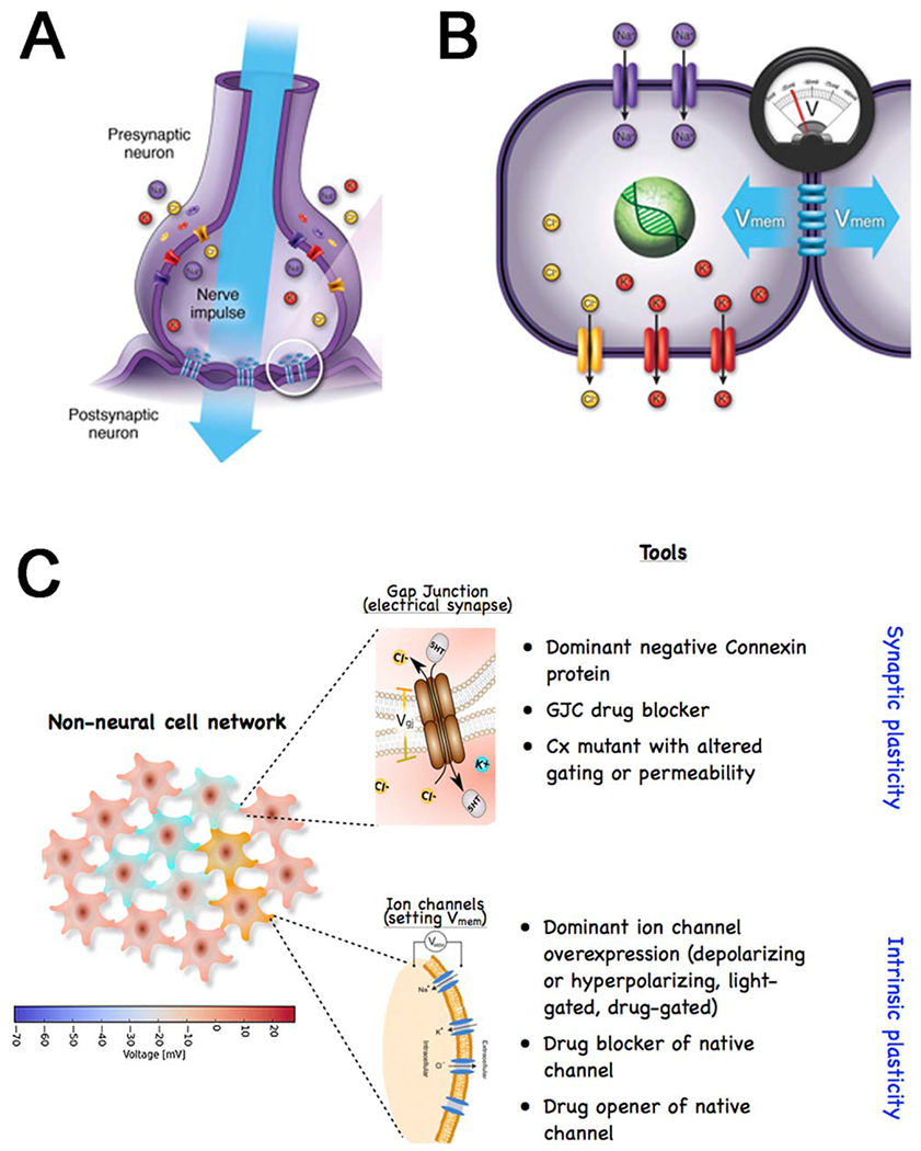
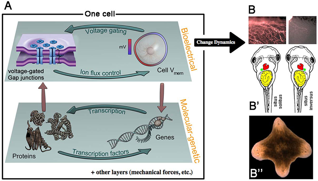
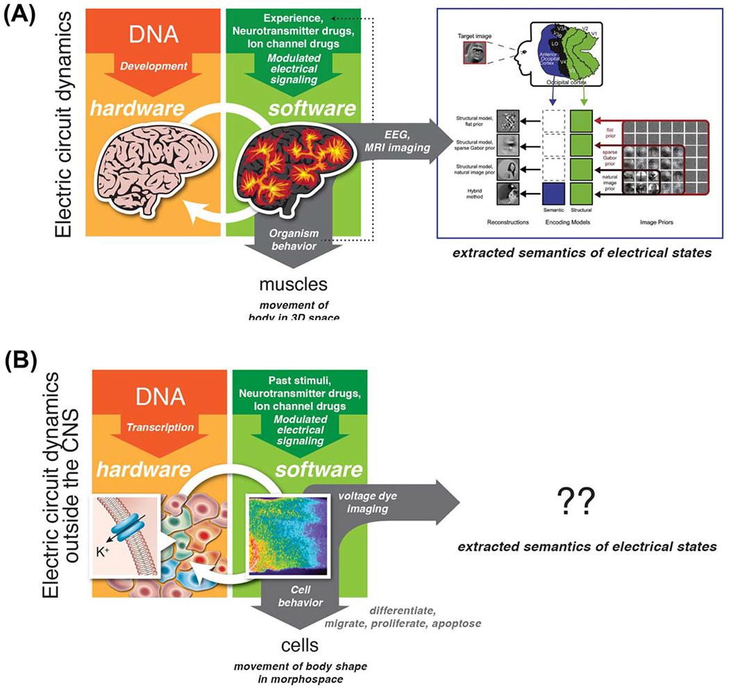
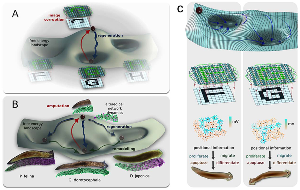
Gap junctions (GJs) are aqueous channels that allow cells to communicate via physiological signals directly. The role of gap junctional connectivity in determining single-cell functions has long been recognized. However, GJs have another important role: the regulation of large-scale anatomical pattern. GJs are not only versatile computational elements that allow cells to control which small molecule signals they receive and emit, but also establish connectivity patterns within large groups of cells. By dynamically regulating the topology of bioelectric networks in vivo, GJs underlie the ability of many tissues to implement complex morphogenesis. Here, a review of recent data on patterning roles of GJs in growth of the zebrafish fin, the establishment of left-right patterning, the developmental dysregulation known as cancer, and the control of large-scale head-tail polarity, and head shape in planarian regeneration has been reported. A perspective in which GJs are not only molecular features functioning in single cells, but also enable global neural-like dynamics in non-neural somatic tissues has been proposed. This view suggests a rich program of future work which capitalizes on the rapid advances in the biophysics of GJs to exploit GJ-mediated global dynamics for applications in birth defects, regenerative medicine, and morphogenetic bioengineering. © 2016 Wiley Periodicals, Inc. Develop Neurobiol 77: 643-673, 2017.

Keywords: bioelectric; gap junctions; morphogenesis; networks; patterning.


Conflict of interest statement

The authors declare no conflicts or competing interests.

Figures

Figure 1
Figure 1
Gap junctions form bioelectric circuits in the brain and beyond. (A) Neurons are often coupled by gap junctions, which allows electrical activity to propagate and integrate across cells. (B) The same scheme, involving ion channels to set Vmem levels and gap junctions to communicate bioelectric state to neighboring cells, is present in most somatic tissues. (C) Non-neural cells assemble into GJ-coupled networks that have many of the properties of neural networks. Manipulating the function of somatic tissues during pattern formation, by modulating GJ activity, makes use of two basic approaches, using pharmacological or genetic techniques to target connectivity (GJ gating, akin to synaptic plasticity) or resting potential (ion channels, akin to intrinsic plasticity). Graphics courtesy of Alexis Pietak and Jeremy Guay.
Figure 2
Figure 2
Information in physiological circuits instructs pattern formation. Gap junctions are key regulators of bioelectric cell state. (A) Like gene regulatory networks, which contain numerous feedback loops among gene loci, gap junctions, and ion channels both regulate and are regulated by resting potential. This establishes an autonomous layer of physiological dynamics that is coupled to transcriptional cascades, but has its own unique information and functions. Modulating bioelectric dynamics by induced changes of GJC results in large-scale alterations of pattern formation, including hyperinnervation (B), left-right organ inversions (B′), and multiple head formation in regenerating planaria (B″). Graphics courtesy of Alexis Pietak and Jeremy Guay.
Figure 3
Figure 3
Parallelism between brain and body. (A) In the brain, DNA sets the structure of the central nervous system—the hardware. Electrical circuit dynamics process information and store memories, resulting in experience-dependent (and self-organizing) patterns that control muscles resulting in the movement of the animal in three-dimensional space. Experiences (external sensory stimuli) and reagents targeting GJs, ion channels, and neurotransmitters alter the electric dynamics. Computational pipelines are beginning to be developed and applied to dynamics observed via EEG and MRI imaging, to extract the semantics—the memories represented by these bioelectric states. (B) In somatic tissues of developing or regenerating organisms, DNA sets the complement of connexins, ion channels, and neurotransmitter machinery in all cells—the hardware. Bioelectric dynamics (slow patterns of resting potential within tissues) process information and establish prepatterns for gene expression and morphogenesis, resulting in chemical signal-dependent (and self-organizing) patterns that control cell functions like migration and differentiation, resulting in anatomical changes that move the body through morphospace. Stimuli (chemical and other signals) and reagents targeting GJs, ion channels, and neurotransmitters alter the electric dynamics. Computational pipelines need to be developed and applied to these dynamics as imaged with voltage-sensitive dyes and GJ tracers, to extract the semantics—the instructive anatomical patterns represented by these bioelectric states. Graphics by Jeremy Guay of Peregrine Creative and Alexis Pietak. Neural decoding panel in (A) is reproduced with permission from Naselaris et al., 2009. Voltage pattern panel in (B right) is courtesy of Douglas J. Blackiston.
Figure 4
Figure 4
Morphogenetic memories visualized as attractors in GJ network state space. GJ dynamics may offer an opportunity to understand information processing, not only molecular biophysics, but also of pattern formation regulated by physiological cell–cell signaling. One way to visualize planarian regeneration is (A) as the function of a large network of electrically coupled cells. Some such networks have been shown (in computational neuroscience and artificial intelligence research) to have a planaria-like property of holographic memory storage: a trained network can recreate the entire pattern despite deletions of the pattern or of the network components. A well-accepted mathematical paradigm for understanding the global properties of such networks is as an energy landscape, with attractors corresponding to specific stable modes of the network. In our analogy, amputation raises the energy of the system, temporarily pulling it out of the attractor to which the system tends to return. One hypothesis is that these networks are responsible for storing the pattern of a normal planarian, and when damaged, issuing cell-level commands (differentiate, proliferate, and other instructions) that restore the anatomy (in parallel to how recall of complex geometric memories can be triggered by stimuli and induce goal-directed behavior in cognitive science studies of animal behavior). This hypothesis makes a prediction: that coherent changes in patterning will result from experimentally induced changes of the bioelectric network’s topology or dynamics. (B) It has been shown (Emmons-Bell et al., 2015) that altering the bioelectric connectivity in G. dorotocephala results in regeneration of one of four discrete head types. On this view, amputation of head and tail causes the system to move to an unable state from its basin of attraction. Partial interruption of gap-junction communication between cells (reduced connectivity and thus altered dynamics of the network) induces head wounds to regenerate (yellow arrows) new heads that resemble closely-related flatworm species that are regions of stability in the regeneration morphospace landscape (left to right: Schmidtea mediterranea, Dugesia japonica, and Philbertia felina) as well as heads of the original species in the center basin. The probability of regenerating a certain head shape is proportional to the evolutionary distance from Girardia dorotocephala. These states are non-permanent (shallow basins of attraction) and over time will remodel into their final morphological state (white dashed arrows) to the deepest and most stable basin of attraction of the original head shape. (C) The same process can be modeled as a neural-like network, with stable modes visualized as stable attractors (a.k.a. memories in neural nets), which lead to specific instructive signals regulating proliferation, migration, and differentiation that induces different but coherent patterning outcomes. Graphics by Alexis Pietak.
Figure 5
Figure 5
Stable inheritance of target morphology change after GJ network perturbation. A normal planarian has a head and tail, and regenerates each at the appropriate end of an amputated fragment (A). When cut into thirds, and the middle fragment is briefly exposed to octanol, which temporarily blocks long-range bioelectrical signaling between the wound and mature tissues, a 2-headed worm results (B). GJC, gap junctional communication. Remarkably, upon further rounds of cutting in plain water (long after the octanol has left the tissues, as confirmed by HPLC), the 2-headed form is recapitulated (C,D; images of 2-headed worms provided by Fallon Durant.). This change in the animal’s target morphology (the shape to which it regenerates upon damage) appears to be permanent, and persists across the animal’s normal reproductive mode (fissioning), despite the fact that the genomic sequence has not been altered. Chromatin modifications alone do not explain this, because the posterior wound cells, which could have been epigenetically reprogrammed to a head fate, are thrown away at each cut: the information encoding a bipolar 2-head animal is present even in the normal gut fragment—it is distributed throughout the body. We propose that this information is a kind of memory, encoded in electrical networks of somatic cells coupled by gap junctions, and is stored at the level of bioelectrical dynamics, not genetics.

Similar articles

Cited by

References

    1. Aboobaker AA. 2011. Planarian stem cells: A simple paradigm for regeneration. Trends Cell Biol 21:304–311. - PubMed
    1. Adams DS, Robinson KR, Fukumoto T, Yuan S, Albertson RC, Yelick P, Kuo L, et al. 2006. Early, H+-V-ATPase-dependent proton flux is necessary for consistent left-right patterning of non-mammalian vertebrates. Development 133:1657–1671. - PMC - PubMed
    1. Ahir BK, Pratten MK. 2014. Structure and function of gap junction proteins: Role of gap junction proteins in embryonic heart development. Int J Dev Biol 58:649–662. - PubMed
    1. Ai Z, Fischer A, Spray DC, Brown AM, Fishman GI. 2000. Wnt-1 regulation of connexin43 in cardiac myocytes. J Clin Invest 105:161–171. - PMC - PubMed
    1. Alev C, Urschel S, Sonntag S, Zoidl G. 2008. The neuronal connexin36 interacts with and is phosphorylated by CaMKII in a way similar to CaMKII interaction with glutamate receptors. Proc Natl Acad Sci U S A 105:20964–20969. - PMC - PubMed

Publication types