

Effect of mild temperature shift on poly(ADP-ribose) and γH2AX levels in cultured cells
- PMID: 27262441
- PMCID: PMC6118343
- DOI: 10.1016/j.bbrc.2016.06.001
Effect of mild temperature shift on poly(ADP-ribose) and γH2AX levels in cultured cells
Abstract
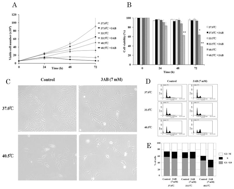
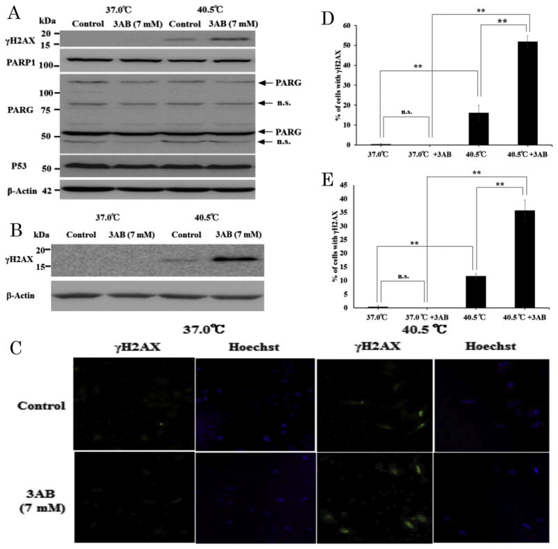
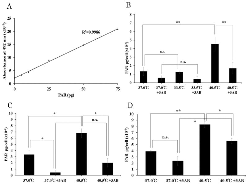
Poly (ADP-ribose) (PAR) is rapidly synthesized by PAR polymerases (PARPs) upon activation by DNA single- and double-strand breaks. In this study, we examined the quantitative amount of PAR in HeLa cells cultured within the physiological temperatures below 41 °C for verification of the effect of shifting-up or -down the temperature from 37.0 °C on the DNA breaks, whether the temperature-shift caused breaks that could be monitored by the level of PAR. While PAR level did not change significantly when HeLa cells were cultured at 33.5 °C or 37.0 °C, it was significantly increased 2- and 3-fold when cells were cultured for 12 h and 24 h, respectively, at 40.5 °C as compared to 37.0 °C. Similar to the results with HeLa cells, PAR level was increased 2-fold in CHO-K1 cells cultured at 40.5 °C for 24 h as compared to 37.0 °C. As the cellular levels of PAR polymerase1 (PARP1) and PAR glycohydrolase (PARG), a major degradation enzyme for PAR, did not seem to change significantly, this increase could be caused by activation of PARP1 by DNA strand breaks. In fact, γH2AX, claimed to be a marker of DNA double-strand breaks, was found in cell extracts of HeLa cells and CHO-K1 cells at elevated temperature vs. 37.0 °C, and these γH2AX signals were intensified in the presence of 3-aminobenzamide, a PARP inhibitor. The γH2AX immunohistochemistry results in HeLa cells were consistent with Western blot analyses. In HeLa cells, proliferation was significantly suppressed at 40.5 °C in 72 h-continuous cultures and decreased viabilities were also observed after 24-72 h at 40.5 °C. Flow cytometric analyses showed that the HeLa cells were arrested at G2/M after temperature shift-up to 40.5 °C. These physiological changes were potentiated in the presence of 3-aminobenzamide. Decrease in growth rates, increased cytotoxicity and G2/M arrest, were associated with the temperature-shift to 40.5 °C and are indirect evidence of DNA breaks. In addition to γH2AX, PAR could be a sensitive marker for DNA single- and double-strand breaks. These two molecular markers provide evidence of physiological changes occurring within cells.
Keywords: DNA double-strand break; DNA repair; DNA single-strand break; Poly(ADP-ribose); Thermal environment; γH2AX.
Copyright © 2016. Published by Elsevier Inc.
Figures
Similar articles
-
Poly(ADP-ribose) polymerases PARP1 and PARP2 modulate topoisomerase II beta (TOP2B) function during chromatin condensation in mouse spermiogenesis.Biol Reprod. 2011 May;84(5):900-9. doi: 10.1095/biolreprod.110.090035. Epub 2011 Jan 12. Biol Reprod. 2011. PMID: 21228215 Free PMC article.
-
Altered poly(ADP-ribose) metabolism impairs cellular responses to genotoxic stress in a hypomorphic mutant of poly(ADP-ribose) glycohydrolase.Exp Cell Res. 2007 Mar 10;313(5):984-96. doi: 10.1016/j.yexcr.2006.12.025. Epub 2007 Jan 10. Exp Cell Res. 2007. PMID: 17276427
-
Inhibition of poly(ADP-ribose) polymerase-1 or poly(ADP‑ribose) glycohydrolase individually, but not in combination, leads to improved chemotherapeutic efficacy in HeLa cells.Int J Oncol. 2013 Feb;42(2):749-56. doi: 10.3892/ijo.2012.1740. Epub 2012 Dec 17. Int J Oncol. 2013. PMID: 23254695 Free PMC article.
-
Functional Role of ADP-Ribosyl-Acceptor Hydrolase 3 in poly(ADP-Ribose) Polymerase-1 Response to Oxidative Stress.Curr Protein Pept Sci. 2016;17(7):633-640. doi: 10.2174/1389203717666160419144603. Curr Protein Pept Sci. 2016. PMID: 27090906 Free PMC article. Review.
-
Histone gammaH2AX and poly(ADP-ribose) as clinical pharmacodynamic biomarkers.Clin Cancer Res. 2010 Sep 15;16(18):4532-42. doi: 10.1158/1078-0432.CCR-10-0523. Epub 2010 Sep 7. Clin Cancer Res. 2010. PMID: 20823146 Free PMC article. Review.
Cited by
-
PARP Inhibitor Decreases Akt Phosphorylation and Induces Centrosome Amplification and Chromosomal Aneuploidy in CHO-K1 Cells.Int J Mol Sci. 2022 Mar 23;23(7):3484. doi: 10.3390/ijms23073484. Int J Mol Sci. 2022. PMID: 35408845 Free PMC article.
-
The clinically used PARP inhibitor olaparib improves organ function, suppresses inflammatory responses and accelerates wound healing in a murine model of third-degree burn injury.Br J Pharmacol. 2018 Jan;175(2):232-245. doi: 10.1111/bph.13735. Epub 2017 Mar 5. Br J Pharmacol. 2018. PMID: 28146604 Free PMC article.
-
Genetically Encoded Fluorescent Sensor for Poly-ADP-Ribose.Int J Mol Sci. 2020 Jul 15;21(14):5004. doi: 10.3390/ijms21145004. Int J Mol Sci. 2020. PMID: 32679873 Free PMC article.
-
Physiological levels of poly(ADP-ribose) during the cell cycle regulate HeLa cell proliferation.Exp Cell Res. 2022 Aug 1;417(1):113163. doi: 10.1016/j.yexcr.2022.113163. Epub 2022 Apr 18. Exp Cell Res. 2022. PMID: 35447104 Free PMC article.
References
-
- Lepock JR. How do cells respond to their thermal environment? Int J Hyperth. 2005;21:681–687. - PubMed
-
- Lepock JR. Cellular effects of hyperthermia: relevance to the minimum dose for thermal damage. Int J Hyperth. 2003;19:252–266. - PubMed
-
- Tansey EA, Johnson CD. Recent advances in thermoregulation. Adv Physiol Educ. 2015;39:139–148. - PubMed
-
- Rogakou EP, Pilch DR, Orr AH, Ivanova VS, Bonner WM. DNA double-stranded breaks induce histone H2AX phosphorylation on serine 139. J Biol Chem. 1998;273:5858–5868. - PubMed
MeSH terms
Substances
Grants and funding
LinkOut - more resources
Full Text Sources
Other Literature Sources
Research Materials
Miscellaneous
