. 2016 Jul 15;25(14):2997-3010.
doi: 10.1093/hmg/ddw153. Epub 2016 Jun 3.

Meckel's and condylar cartilages anomalies in achondroplasia result in defective development and growth of the mandible

Affiliations

Meckel's and condylar cartilages anomalies in achondroplasia result in defective development and growth of the mandible

Martin Biosse Duplan et al. Hum Mol Genet. .

Abstract

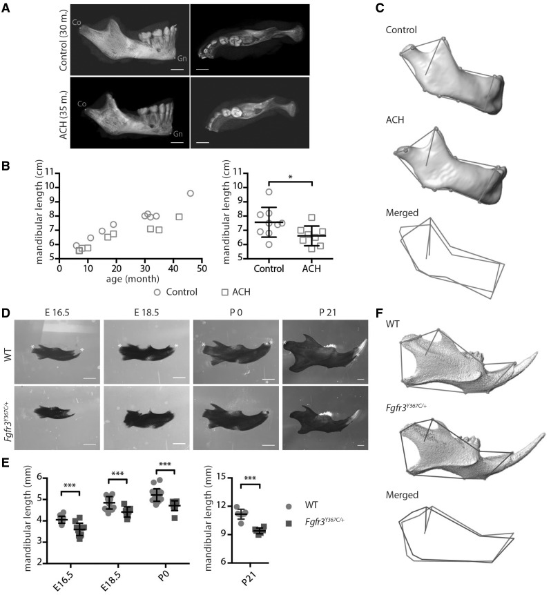
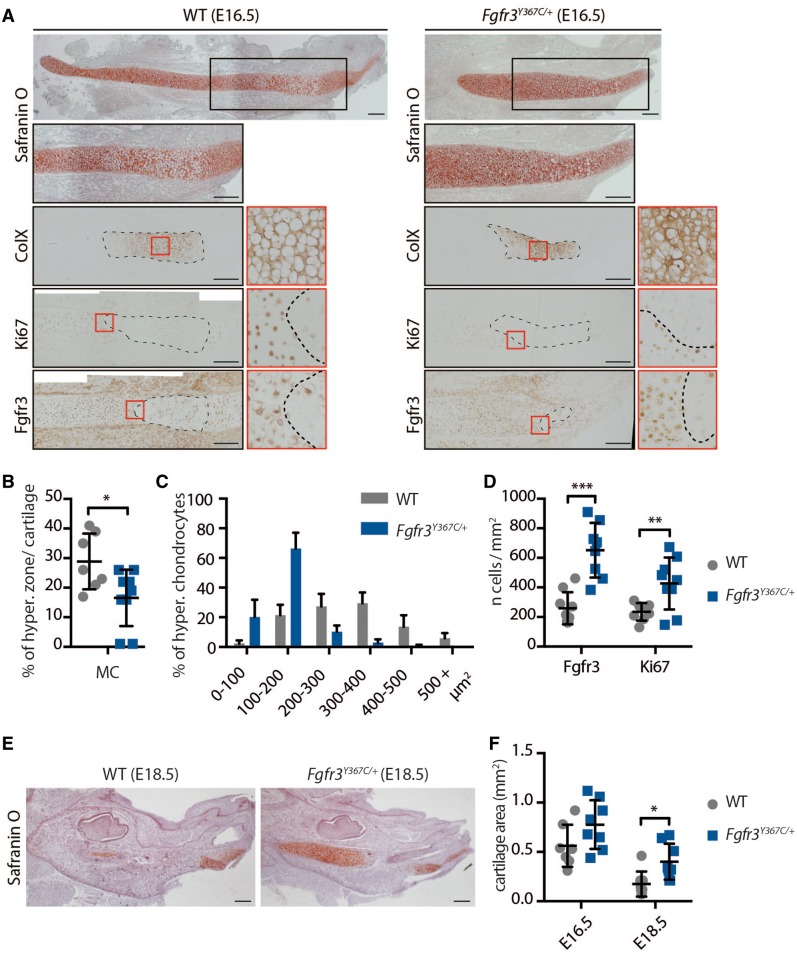
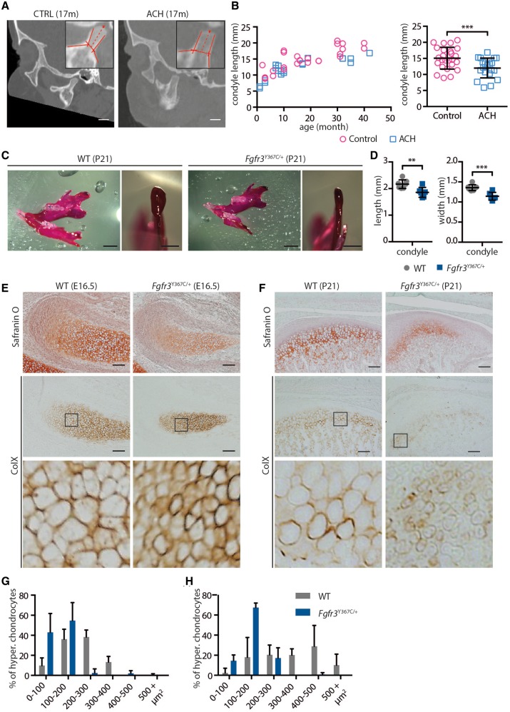
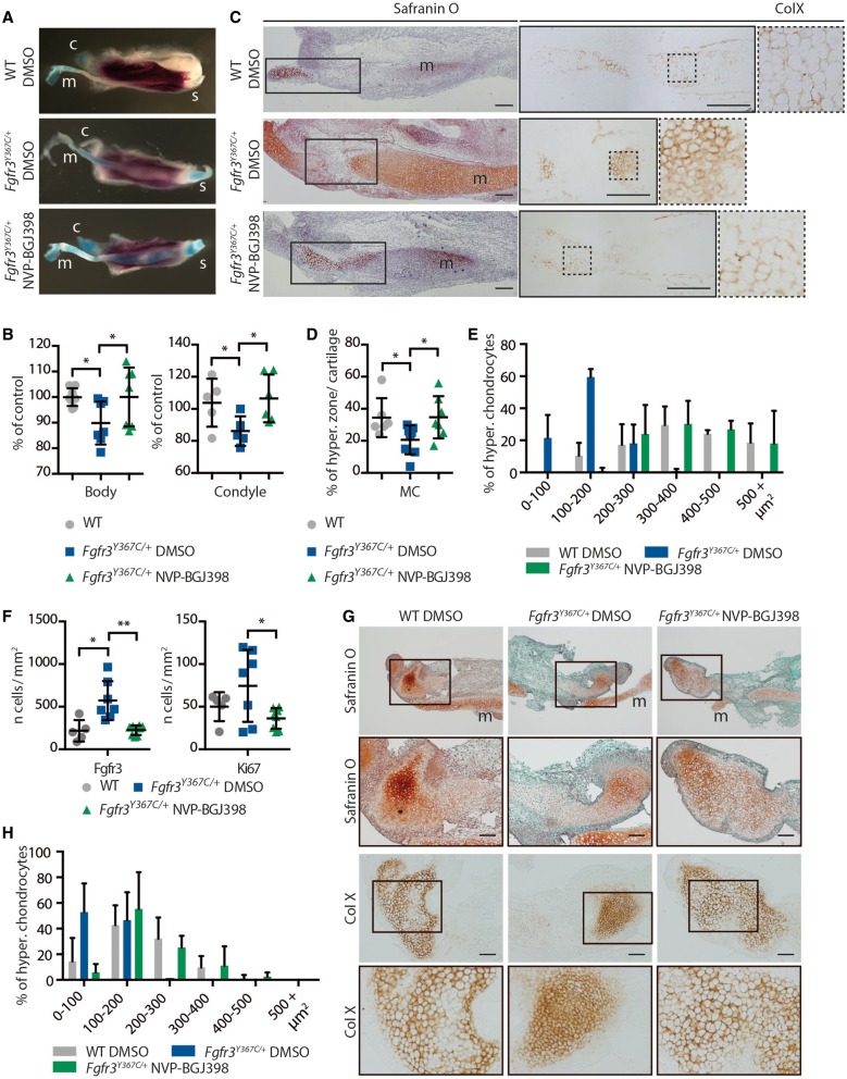
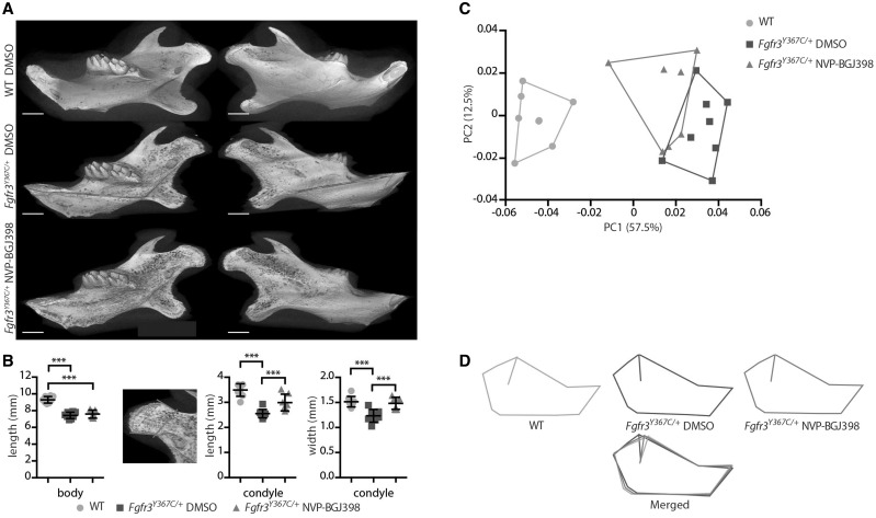
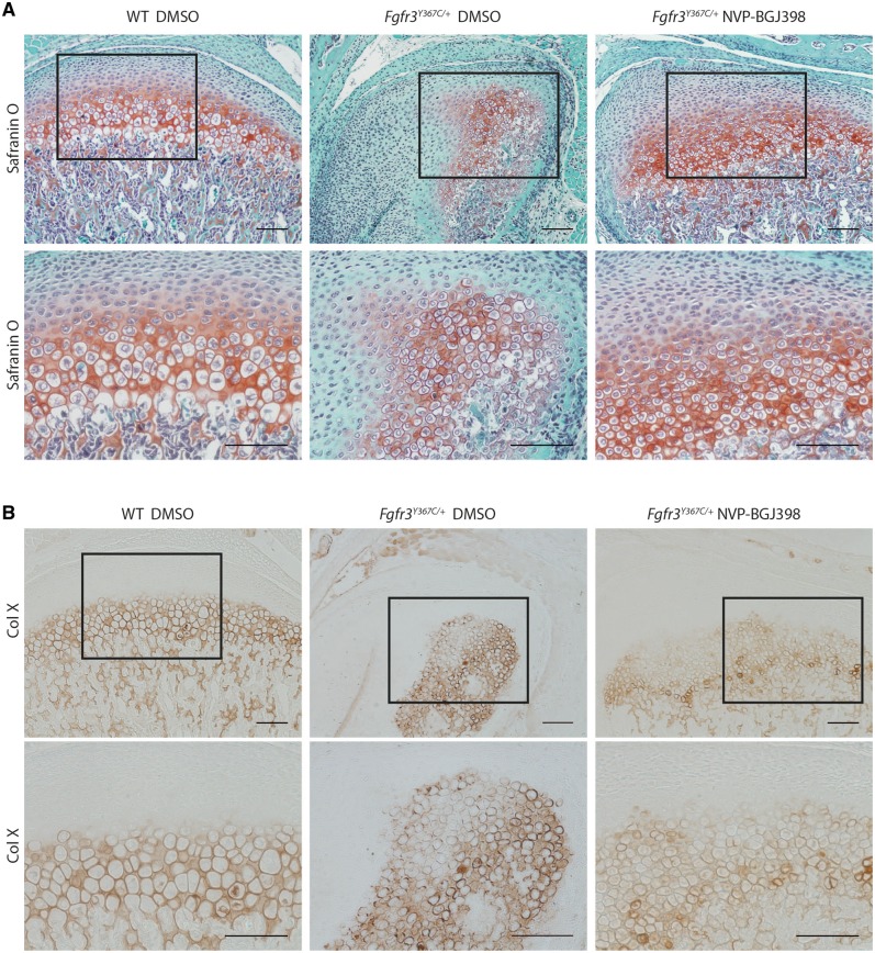
Activating FGFR3 mutations in human result in achondroplasia (ACH), the most frequent form of dwarfism, where cartilages are severely disturbed causing long bones, cranial base and vertebrae defects. Because mandibular development and growth rely on cartilages that guide or directly participate to the ossification process, we investigated the impact of FGFR3 mutations on mandibular shape, size and position. By using CT scan imaging of ACH children and by analyzing Fgfr3Y367C/+ mice, a model of ACH, we show that FGFR3 gain-of-function mutations lead to structural anomalies of primary (Meckel's) and secondary (condylar) cartilages of the mandible, resulting in mandibular hypoplasia and dysmorphogenesis. These defects are likely related to a defective chondrocyte proliferation and differentiation and pan-FGFR tyrosine kinase inhibitor NVP-BGJ398 corrects Meckel's and condylar cartilages defects ex vivo. Moreover, we show that low dose of NVP-BGJ398 improves in vivo condyle growth and corrects dysmorphologies in Fgfr3Y367C/+ mice, suggesting that postnatal treatment with NVP-BGJ398 mice might offer a new therapeutic strategy to improve mandible anomalies in ACH and others FGFR3-related disorders.


Figures

Figure 1.
Figure 1.
FGFR3 over activation in humans and mice results in mandibular hypoplasia and dysmorphogenesis. (A) 3D reconstructed CT images with volume rendering of a control and ACH patient (scale bar = 1 cm). (B) Mandible length, measured as the distance between condylion (Co) and gnathion (Gn) on sagittal sections of 3D reconstructed CT images of controls (n = 9, mean age: 24.9 months) or ACH patients (n = 8, mean age: 21.3 months) and plotted against the age of the children. (C) Landmarks and associated wireframes measured on the 3D reconstructed human mandibles and corresponding superimposition of the control and ACH wireframes computed on the basis of PC scores along PC1, the PC best separating the two groups and accounting for 47% of total shape variance. (D and E) Mandible length of embryos (E16.5 and E18.5), new born and 3-week-old WT and Fgfr3Y367C/+ mice (n ≥ 6 individuals for each age and genotype), measured following alcian blue and alizarin red staining between the condylar and symphyseal ends (marked with *) (scale bar = 1 mm). (F) Landmarks and associated wireframes measured on the 3D reconstructed mouse mandibles and corresponding superimposition of the control and Fgfr3Y367C/+ wireframes computed on the basis of PC scores along PC1, the PC best separating the two groups and accounting for 78% of total shape variance. Data shown as mean with SD; *P < 0.05, ***P < 0.005.
Figure 2.
Figure 2.
Chondrocytes homeostasis is disturbed in Meckel’s cartilage of Fgfr3Y367C/+ mice. (A) Histological staining (Safranin’O) and immunostaining for Collagen X, Ki67 (proliferation marker) and Fgfr3 of MC of WT and Fgfr3Y367C/+ E16.5 embryos. An enlargement of the ColX immunostaining (red box) highlights the modification in the size of hypertrophic chondrocytes in Fgfr3Y367C/+ embryos (scale bar = 200 μm). Area delimited with doted lines on the ColX panel corresponds to the hyperthrophic zone. Areas delimited with doted lines on the Ki67 and Fgfr3 panels correspond to immunonegative zones (respectively non-proliferative and Fgfr3-negative). Enlargements of the Ki67 and Fgfr3 immunostainings (red box) highlight the limits of the positive and negative zones. (B) Measurement of the ColX positive zone inside MC of WT and Fgfr3Y367C/+ E16.5 embryos (n ≥ 7 individuals for each genotype). (C) Mean percentage of hypertrophic chondrocytes for different size categories (expressed in μm2) inside MC of WT and Fgfr3Y367C/+ E16.5 embryos (n ≥ 50 cells from n ≥ 6 individuals for each genotype). (D) Mean number of immuno-positive cells for Fgfr3 or Ki67 inside MC of WT and Fgfr3Y367C/+ E16.5 embryos (n ≥ 7 individuals for each genotype). (E) Histological staining (Safranin’O) of WT and Fgfr3Y367C/+ E18.5 embryos (scale bar = 200 μm). (F) Mean cartilage area, measured on sagittal sections of MC in WT and Fgfr3Y367C/+ E16.5 and E18.5 embryos (n ≥ 7 individuals for each age and genotype). Data shown as mean with SD; *P < 0.05, **P < 0.01, ***P < 0.005.
Figure 3.
Figure 3.
FGFR3 over-activation in humans and mice results in condyle hypoplasia. (A) Representative sagittal sections of a control and ACH child, both 17 months of age, generated from the CT scans (scale bar = 1 cm). (B) Condylar neck length, measured on CT scans sagittal sections of controls (n = 14, mean age = 32.4 months) or ACH patients (n = 12, mean age = 32.4 months) and plotted against the age of the children. (C) Representative macroscopic views of condyles of 3-week-old WT and Fgfr3Y367C/+ mice, following alcian blue and alizarin red staining (scale bars = 2 and 1 mm). (D) Condylar neck length of 3-week-old WT and Fgfr3Y367C/+ mice, measured following alcian blue and alizarin red staining (n ≥ 6 individuals for each genotype). (E and F) Histological staining (Safranin’O) and immunostaining for Collagen X of the condylar cartilage of WT and Fgfr3Y367C/+ E16.5 embryos (E) and 3-week-old mice (F) (scale bar = 100 μm). An enlargement of the ColX immunostaining (black box) highlights the modification in the size of hypertrophic chondrocytes in Fgfr3Y367C/+ embryos and P21 mice. (G and H) Mean percentage of hypertrophic chondrocytes for different size categories (expressed in μm2) inside the condylar cartilage of WT and Fgfr3Y367C/+ E16.5 embryos (G) and 3 weeks old mice (H) (n ≥ 50 cells from n ≥ 6 individuals for each age and genotype). Data shown as mean with SD; **P < 0.01, ***P < 0.005.
Figure 4.
Figure 4.
NVP-BGJ398 corrects primary and secondary cartilages defects in ex vivo cultures of mandibles from Fgfr3Y367C/+ embryos. (A) Representative macroscopic views of hemi-mandibles from E16.5 WT or Fgfr3Y367C/+ embryos, treated with DMSO or NVP-BGJ398 and cultured for 6 days, following alcian blue and alizarin red staining. Symphyseal (s), condylar (c) and Meckel’s (m) cartilages are indicated. (B) Mandibular body and condylar neck length of cultured hemi-mandibles (n ≥ 5 individuals for each genotype and treatment). Total length of the hemi-mandible was measured as well as the length of the condylar and symphyseal cartilages (identified with the alcian blue staining). The length of the body was calculated as the total length minus the condylar and symphyseal cartilages. (C) Histological staining (Safranin’O) and immunostaining for ColX of cultured hemi-mandibles. An enlargement of the ColX immunostaining highlights the modification in the size of hypertrophic chondrocytes in MC of Fgfr3Y367C/+ embryos and the correction of the defect with NVP-BGJ398 (scale bar = 200 μm). (D) Measurement of the ColX positive zone inside MC of cultured hemi-mandibles (n ≥ 6 individuals for each genotype and treatment). (E) Mean percentage of hypertrophic chondrocytes for different size categories (expressed in μm2) inside MC of cultured hemi-mandibles (n ≥ 50 cells from n ≥ 5 individuals for each genotype and treatment). (F) Mean number of immune positive cells for Fgfr3 or Ki67 inside MC of WT, Fgfr3Y367C/+ DMSO and Fgfr3Y367C/+ NVP-BGJ398 E16.5 embryos (n ≥ 5 individuals for each genotype). (G) Histological staining (Safranin’O) and immunostaining for Collagen X of cultured hemi-mandibles condylar cartilage (scale bar = 100 μm). Meckel’s cartilage (m) is indicated. (H) Mean percentage of hypertrophic chondrocytes for different size categories (expressed in μm2) inside condylar cartilage of cultured hemi-mandibles (n ≥ 50 cells from n ≥ 5 individuals for each genotype and treatment). Data shown as mean with SD; *P < 0.05, **P < 0.01.
Figure 5.
Figure 5.
NVP-BGJ398 improves in vivo condyle growth of Fgfr3Y367C/+ mice. (A) Representative 3D reconstructions of 16-day-old WT and Fgfr3Y367C/+ mice treated with DMSO or NVP-BGJ398 for 15 days since P01 (scale bar = 1 mm). (B) Measurement of the mandible body and the condyle length and width of WT and Fgfr3Y367C/+ mice treated with DMSO or NVP-BGJ398 (n ≥ 6 individuals for each genotype and treatment). Total length of the mandible and the length of the condyle were measured . The length of the body was calculated as the total length minus the condyle. (C and D) PCA of the Procrustes shape coordinates of the landmarks measured on mandibles of WT and Fgfr3Y367C/+ mice treated with DMSO or NVP-BGJ398 and corresponding superimposition (n ≥ 6 individuals for each genotype and treatment). Data shown as mean with SD; ***P < 0.005.
Figure 6.
Figure 6.
NVP-BGJ398 improves in vivo the chondrocyte differentiation in condyle of Fgfr3Y367C/+ mice. Histological staining (Safranin’O) (A) and immunostaining for collagen X (B) of condylar cartilage of 16-day-old WT and Fgfr3Y367C/+ mice treated with DMSO or NVP-BGJ398 for 15 days since P01 (scale bar = 100 μm).

Similar articles

Cited by

References

    1. Baujat G., Legeai-Mallet L., Finidori G., Cormier-Daire V., Le Merrer M. (2008) Achondroplasia. Best Pract. Res. Clin. Rheumatol, 22, 3–18. - PubMed
    1. Horton W.A., Hall J.G., Hecht J.T. (2007) Achondroplasia. Lancet, 370, 162–172. - PubMed
    1. Di Rocco F., Biosse Duplan M., Heuzé Y., Kaci N., Komla-Ebri D., Munnich A., Mugniery E., Benoist-Lasselin C., Legeai-Mallet L. (2014) FGFR3 mutation causes abnormal membranous ossification in achondroplasia. Hum. Mol. Genet, 23, 2914–2925. - PubMed
    1. Rousseau F., Bonaventure J., Legeai-Mallet L., Pelet A., Rozet J.M., Maroteaux P., Le Merrer M., Munnich A. (1994) Mutations in the gene encoding fibroblast growth factor receptor-3 in achondroplasia. Nature, 371, 252–254. - PubMed
    1. Ornitz D.M. (2005) FGF signaling in the developing endochondral skeleton. Cytokine Growth Factor Rev., 16, 205–213. - PMC - PubMed

MeSH terms