

Low-density lipoprotein receptor-related protein 1 is a novel modulator of radial glia stem cell proliferation, survival, and differentiation
- PMID: 27258849
- PMCID: PMC5033964
- DOI: 10.1002/glia.23009
Low-density lipoprotein receptor-related protein 1 is a novel modulator of radial glia stem cell proliferation, survival, and differentiation
Abstract
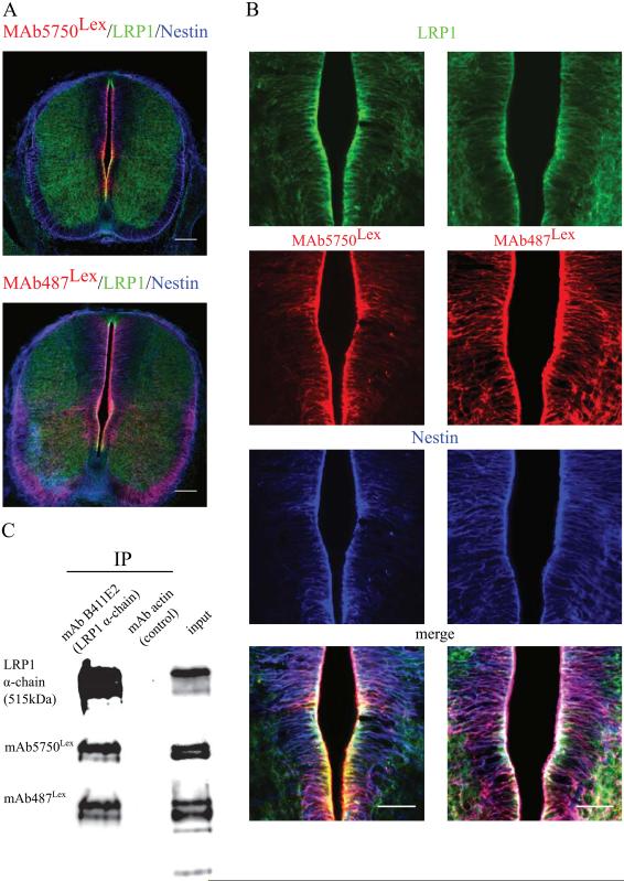
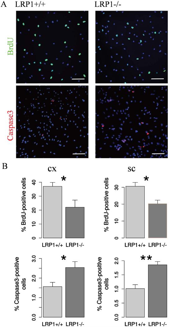
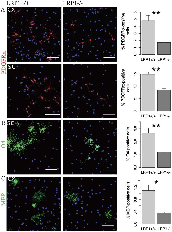
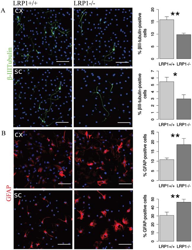
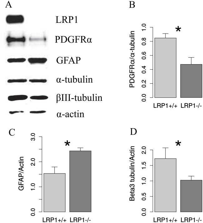
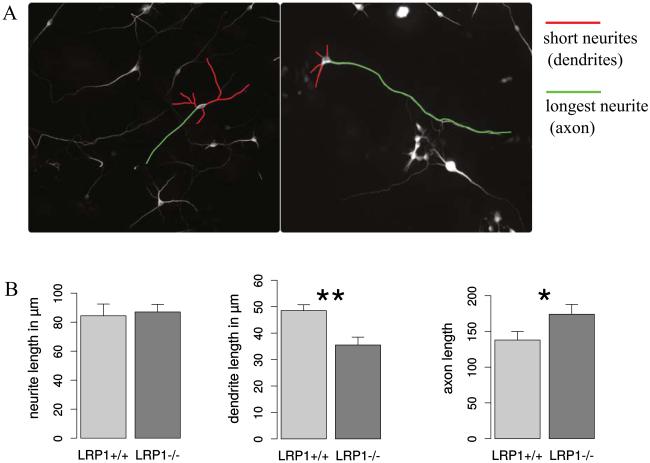
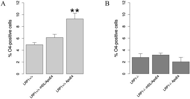
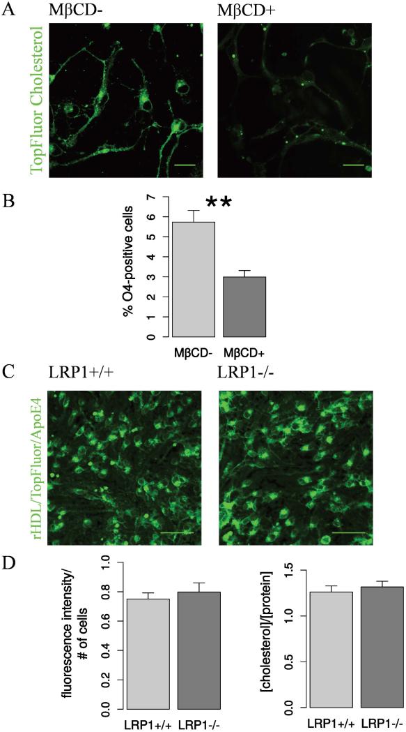
The LDL family of receptors and its member low-density lipoprotein receptor-related protein 1 (LRP1) have classically been associated with a modulation of lipoprotein metabolism. Current studies, however, indicate diverse functions for this receptor in various aspects of cellular activities, including cell proliferation, migration, differentiation, and survival. LRP1 is essential for normal neuronal function in the adult CNS, whereas the role of LRP1 in development remained unclear. Previously, we have observed an upregulation of LewisX (LeX) glycosylated LRP1 in the stem cells of the developing cortex and demonstrated its importance for oligodendrocyte differentiation. In the current study, we show that LeX-glycosylated LRP1 is also expressed in the stem cell compartment of the developing spinal cord and has broader functions in the developing CNS. We have investigated the basic properties of LRP1 conditional knockout on the neural stem/progenitor cells (NSPCs) from the cortex and the spinal cord, created by means of Cre-loxp-mediated recombination in vitro. The functional status of LRP1-deficient cells has been studied using proliferation, differentiation, and apoptosis assays. LRP1 deficient NSPCs from both CNS regions demonstrated altered differentiation profiles. Their differentiation capacity toward oligodendrocyte progenitor cells (OPCs), mature oligodendrocytes and neurons was reduced. In contrast, astrocyte differentiation was promoted. Moreover, LRP1 deletion had a negative effect on NSPCs proliferation and survival. Our observations suggest that LRP1 facilitates NSPCs differentiation via interaction with apolipoprotein E (ApoE). Upon ApoE4 stimulation wild type NSPCs generated more oligodendrocytes, but LRP1 knockout cells showed no response. The effect of ApoE seems to be independent of cholesterol uptake, but is rather mediated by downstream MAPK and Akt activation. GLIA 2016 GLIA 2016;64:1363-1380.
Keywords: LRP1; LRP1-dependent signaling; LewisX carbohydrate; membrane permeant Cre-recombinase; neural stem and progenitor cells; neural stem cell differentiation.
© 2016 Wiley Periodicals, Inc.
Figures
Similar articles
-
LRP1 regulates peroxisome biogenesis and cholesterol homeostasis in oligodendrocytes and is required for proper CNS myelin development and repair.Elife. 2017 Dec 18;6:e30498. doi: 10.7554/eLife.30498. Elife. 2017. PMID: 29251594 Free PMC article.
-
A LewisX glycoprotein screen identifies the low density lipoprotein receptor-related protein 1 (LRP1) as a modulator of oligodendrogenesis in mice.J Biol Chem. 2013 Jun 7;288(23):16538-16545. doi: 10.1074/jbc.M112.419812. Epub 2013 Apr 24. J Biol Chem. 2013. PMID: 23615909 Free PMC article.
-
Low Density Lipoprotein-Receptor Related Protein 1 Is Differentially Expressed by Neuronal and Glial Populations in the Developing and Mature Mouse Central Nervous System.PLoS One. 2016 Jun 9;11(6):e0155878. doi: 10.1371/journal.pone.0155878. eCollection 2016. PLoS One. 2016. PMID: 27280679 Free PMC article.
-
Low-density lipoprotein receptor-related protein-1 (LRP1) in the glial lineage modulates neuronal excitability.Front Netw Physiol. 2023 Jun 13;3:1190240. doi: 10.3389/fnetp.2023.1190240. eCollection 2023. Front Netw Physiol. 2023. PMID: 37383546 Free PMC article. Review.
-
The role of glutamate and its receptors in the proliferation, migration, differentiation and survival of neural progenitor cells.J Neural Transm (Vienna). 2014 Aug;121(8):819-36. doi: 10.1007/s00702-014-1174-6. Epub 2014 Feb 23. J Neural Transm (Vienna). 2014. PMID: 24562403 Review.
Cited by
-
In vitro Validation of Chimeric β-Galactosylceramidase Enzymes With Improved Enzymatic Activity and Increased Secretion.Front Mol Biosci. 2020 Jul 21;7:167. doi: 10.3389/fmolb.2020.00167. eCollection 2020. Front Mol Biosci. 2020. PMID: 32850960 Free PMC article.
-
Intestine-enriched apolipoprotein b orthologs are required for stem cell progeny differentiation and regeneration in planarians.Nat Commun. 2022 Jul 1;13(1):3803. doi: 10.1038/s41467-022-31385-2. Nat Commun. 2022. PMID: 35778403 Free PMC article.
-
LRP1 regulates peroxisome biogenesis and cholesterol homeostasis in oligodendrocytes and is required for proper CNS myelin development and repair.Elife. 2017 Dec 18;6:e30498. doi: 10.7554/eLife.30498. Elife. 2017. PMID: 29251594 Free PMC article.
-
Apolipoprotein A-I in mouse cerebrospinal fluid derives from the liver and intestine via plasma high-density lipoproteins assembled by ABCA1 and LCAT.FEBS Lett. 2021 Mar;595(6):773-788. doi: 10.1002/1873-3468.13950. Epub 2020 Oct 20. FEBS Lett. 2021. PMID: 33020907 Free PMC article.
-
The distribution of the proteoglycan FORSE-1 in the developing mouse central nervous system.J Anat. 2019 Feb;234(2):216-226. doi: 10.1111/joa.12907. Epub 2018 Nov 25. J Anat. 2019. PMID: 30474148 Free PMC article.
References
-
- Baron W, Bijlard M, Nomden A, de Jonge JC, Teunissen CE, Hoekstra D. Sulfatide-mediated control of extracellular matrix-dependent oligodendrocyte maturation. Glia. 2014;62:927–42. - PubMed
-
- Bertram B, Wiese S, von Holst A. High-efficiency transfection and survival rates of embryonic and adult mouse neural stem cells achieved by electroporation. Journal of Neuroscience Methods. 2012;209:420–427. - PubMed
-
- Blaschuk KL, Frost EE, ffrench-Constant C. The regulation of proliferation and differentiation in oligodendrocyte progenitor cells by alphaV integrins. Development. 2000;127:1961–9. - PubMed
MeSH terms
Substances
Grants and funding
LinkOut - more resources
Full Text Sources
Other Literature Sources
Molecular Biology Databases
Research Materials
Miscellaneous
