. 2016 May 31:6:26986.
doi: 10.1038/srep26986.

ALG-2 interacting protein-X (Alix) is essential for clathrin-independent endocytosis and signaling

Affiliations

ALG-2 interacting protein-X (Alix) is essential for clathrin-independent endocytosis and signaling

Vincent Mercier et al. Sci Rep. .

Abstract

The molecular mechanisms and the biological functions of clathrin independent endocytosis (CIE) remain largely elusive. Alix (ALG-2 interacting protein X), has been assigned roles in membrane deformation and fission both in endosomes and at the plasma membrane. Using Alix ko cells, we show for the first time that Alix regulates fluid phase endocytosis and internalization of cargoes entering cells via CIE, but has no apparent effect on clathrin mediated endocytosis or downstream endosomal trafficking. We show that Alix acts with endophilin-A to promote CIE of cholera toxin and to regulate cell migration. We also found that Alix is required for fast endocytosis and downstream signaling of the interleukin-2 receptor giving a first indication that CIE is necessary for activation of at least some surface receptors. In addition to characterizing a new function for Alix, our results highlight Alix ko cells as a unique tool to unravel the biological consequences of CIE.


Figures

Figure 1
Figure 1. EGFR degradation is delayed in Alix ko cells.
(a,b) Serum-starved wt or Alix ko MEFs were incubated with 100 ng/ml EGF at 37 °C for the indicated times. EGFR was revealed by immunoblotting (a) and band intensities estimated (b) using ImageJ software. EGFR intensities were normalised to actin. Graph represent the ratio between values at each time point and at t = 0 (n = 6; *P < 0.05, **P < 0.01, ***P < 0.001, two-tailed Student’s t-test). Dashed lines indicate degradation curves fitted with single exponential functions.
Figure 2
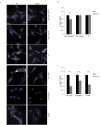
Figure 2. Endocytosis is delayed in Alix ko cells.
(a) EM quantification of BSA-gold particle endosomal trafficking. MEFs were incubated with BSA-gold for 10 min at 37 °C and fixed immediately (top panel), or washed and further incubated at 37 °C for 20 min (middle panel) or 50 min (bottom panel). 1000 gold particles were counted for each condition and were attributed to each compartment type: type 1: plasma membrane invaginations, type 2: endocytic vesicles, type 3: early endosomal compartments, type 4: multivesicular bodies (MVB), type 5: late endosomes/ lysosomes (*P < 0.05; **P < 0.01, two-tailed Student’s t-test). (b) MEFs were incubated 10 min with dextran-TRITC at 37 °C, washed and fixed. Mean fluorescent values per cell were estimated using ImageJ (Number of cells: wt, n = 123; Alix ko, n = 116 in 3 independent experiments ***P < 0.001, two-tailed Mann–Whitney U test). (c) MEFs were stained for 5 min with fluorescent WGA at 4 °C, washed and incubated 5 min at 37 °C. Mean fluorescent values per cell were estimated using ImageJ (Number of cells: wt, n = 114; Alix ko, n = 107 in 3 independent experiments ***P < 0.001, two-tailed Mann–Whitney U test).
Figure 3
Figure 3. CIE is selectively disrupted in Alix ko cells while CME is unaffected.
(a) Representative photographs of wt and Alix ko MEFs after endocytosis of EGF, Tf, GPI-GFP, β1 integrin and IL2Rβ. Serum-starved MEFs were incubated with EGF-TRITC (EGF, 2 or 100 ng/ml) or transferrin-TRITC (Tf) at 4 °C, followed by 10 min incubation at 37 °C. MEFs expressing GPI-GFP or IL2Rβ-GFP were incubated with anti-GFP antibodies at 4 °C, followed by 5 min incubation at 37 °C. To follow β1-integrin endocytosis, MEFs were incubated at 4 °C with anti-active-β1 integrin antibodies followed by 10 min incubation at 37 °C. In both latter cases, cell surface bound antibodies were removed by acid treatment. Hoechst stained nuclei appear in blue. (b,c) Mean fluorescence values per cell were estimated using ImageJ. Number of cells in 3 independent experiments: EGF 100 ng/ml, wt, n = 161; Alix ko, n = 128; two-tailed Student’s t test; EGF 2 ng/ml, wt, n = 95; Alix ko, n = 85; Tf, wt, n = 87; Alix ko, n = 96; GPI-GFP, wt, n = 59; Alix ko, n = 82, two-tailed Mann–Whitney U test; β1-integrin, wt, n = 81; Alix ko, n = 76; two-tailed Student’s t-test; IL2Rβ, wt, n = 100; Alix ko, n = 102; two-tailed Mann–Whitney U test; ***P < 0.001).
Figure 4
Figure 4. CIE but not CME of CTxB is impaired in Alix ko cells.
(a) Representative images of 5 min CTxB-TRITC uptake by wt and Alix ko MEFs. The reduced endocytosis of CTxB-TRITC seen in Alix ko cells is further decreased by chlorpromazine (chlorpro) but not by filipin. Ø: no treatment. Hoechst stained nuclei appear in blue. (b) Quantification of the experiment shown in (a). Mean fluorescence values per cell were calculated using ImageJ (Number of cells in 3 independent experiments: wt Ø, n = 107; wt filipin, n = 90; wt chlorpro, n = 72; Alix ko Ø, n = 145; Alix ko filipin, n = 100; Alix ko chlorpro, n = 95; *P < 0.05; **P < 0.01; ***P < 0.001, Dunn’s multiple comparison test). (c) Quantification of 5 min CTxB uptake by wt and Alix ko MEFs expressing GFP or GFP-Eps15DN. Mean fluorescence values per cell were calculated using ImageJ (Number of cells in 5 independent experiments: wt GFP, n = 147; wt GFP-Eps15DN, n = 93; Alix ko GFP, n = 147; Alix ko Eps15DN, n = 105; **P < 0.01; ***P < 0.001, Dunn’s multiple comparison test). (d) Quantification of CTxB uptake by wt and Alix ko MEFs expressing sh-clathrin-GFP. Wt and Alix ko MEF cells were transfected with an sh vector directed against clathrin (sh-clathrin-GFP) or a control vector (sh-scramble-GFP). 72 h later, cells were incubated with CTxB for 20 min at 4 °C, followed by 5 min at 37 °C and internalized CTxB was quantified. (Number of cells in 4 independent experiments: wt sh-scramble-GFP, n = 62; wt sh-clathrin-GFP, n = 68; Alix ko sh-scramble-GFP, n = 60; Alix ko sh-clathrin-GFP, n = 63; *P < 0.05; ***P < 0.001, Dunn’s multiple comparison test). Western blot analyses using anti-clathrin antibody demonstrate the efficacy of the sh-clathrin plasmid in downregulating clathrin expression in N2a cells 48 h after transfection.
Figure 5
Figure 5. Alix and endophilin A2 relocalise to CTxB labeled membrane patches.
(a) Images from time-lapse video microscopy, acquired by spinning-disk confocal microscopy of an Alix ko MEF expressing GFP-Alix 5 min after addition of CTxB-TRITC. Arrows indicate GFP-Alix (green) recruitment at the level of preexisting (right) or appearing (left) CTxB labeled patches (magenta). Both arrows show disappearance of CTxB and GFP-Alix. (b) Recruitment of Alix (green) to CTxB patches (magenta). Alix ko MEFs expressing GFP-Alix were left untreated, or incubated for 20 min with CTxB-TRITC at 4 °C prior to fixation. (c) Alix (green) is not recruited to membrane patches containing TfR (magenta). Alix ko MEFs expressing GFP-Alix were incubated for 20 min with Tf-TRITCat 4 °C prior to fixation. (d) Upper panels: Endophilin A2 recruitment to CTxB patches in both wt and Alix ko cells. wt MEFs and Alix ko MEFs expressing Myc-endophilin-A2 (endo A2), were left untreated, or incubated with CTxB-TRITC for 20 min at 4 °C before fixation and immunostaining with anti-Myc antibodies. Lower panel: Endophilin A2 (blue) and Alix (green) colocalize to CTxB patches (magenta). Alix ko MEFs expressing GFP-Alix and Myc-endophilin were incubated with CTxB-TRITC for 20 min at 4 °C before immunolabeling with anti-Myc antibodies. The image on the right shows the 3-color merge. Arrows indicate representative patches containing all 3 proteins. (e) Graph showing the percentage of CTxB labeled patches colocalizing with GFP, GFP-Alix, Flag-AlixΔendo or Myc-endophilin-A2 in wt (black) or Alix ko cells (gray). Quantification was performed using ImageJ (Number of cells in 3 independent experiments: GFP, n = 31; Alix, n = 53; AlixΔendo, n = 43; endoA2, n = 57 for wt and n = 56 for Alix ko. ***P < 0.001, one way ANOVA and Dunett’s test).
Figure 6
Figure 6. Alix and endophilin A2 mediate CIE of CTxB.
(a) Alix mediated CIE of CTxB requires binding to endophilins. CTxB-TRITC uptake by wt and Alix ko MEFs (upper panel) or by Alix ko cells transduced with viruses (lower panel) encoding for Alix (Alix ko + Alix), or for a mutant of Alix unable to bind to endophilin-A (Alix ko + AlixΔendo). Cells were incubated with CTxB during 20 min at 4 °C, followed by 5 min at 37 °C and acid stripping. (b) Quantification of CTxB uptake using the same type of experiments as shown in (a). Uptake of CTxB was quantified in wt or Alix ko cells or in Alix ko cells transduced with Alix or Alix deletion mutants lacking several amino-acids of the PRD corresponding to binding sites for Tsg101 (AlixΔTsg101: AlixΔP717–P720), endophilin-A (AlixΔendo: AlixΔP748–P761), or Cin85 (AlixΔCin85: AlixΔP739–R745). Mean fluorescence values per cell were estimated using ImageJ. (Number of cells in 4 independent experiments: wt, n = 104; Alix ko, n = 98; rescue Alix, n = 112; AlixΔTsg101, n = 117; AlixΔendo, n = 147; AlixΔCin85, n = 133. ***P < 0.001, Dunn’s multiple comparison test). (c) Endophilin-A2 and Alix act in a common pathway in CIE of CTxB. wt and Alix ko MEFs were transfected with plasmids containing sh-endophilin and coding for GFP (sh-endo-GFP) or a scrambled version of the same shRNA sequence (sh-scramble-GFP), and cultured for 72 h. Cells were then incubated with CTxB-TRITC (magenta) for 20 min at 4 °C, followed by 5 min at 37 °C. Surface bound toxin was removed by acid treatment prior to fixation and processing. Asterisks indicate the nuclei of transfected cells. (d) Quantification of the experiments shown in (c). CTxB mean fluorescence values per transfected cell were calculated using ImageJ software (Number of cells in 4 independent experiments: wt sh-scramble-GFP, n = 93; wt sh-endo-GFP, n = 70; Alix ko sh-scramble-GFP, n = 76; Alix ko sh-endo-GFP, n = 67. ***P < 0.001, one way ANOVA and Dunnett’s test).
Figure 7
Figure 7. Physiological consequences of loss of Alix expression.
(a) Decreased motility of Alix ko cells in a wound healing assay. Cells migrating into the gap were observed for 20 h and migration rates were measured using Metamorph (Number of cells in 2 independent experiments: wt and ko, n = 200; ***P < 0.001, two-tailed Student’s t-test). Representative recordings of migration tracks are shown in the lower panel. (b) Phenotypic rescue of Alix ko cells transduced with Alix (Alix ko + Alix) or AlixΔendophilin (Alix ko + AlixΔendo). Velocities were measured as in (a) (Number of cells in 2 independent experiments: Alix ko + Alix and Alix ko + AlixΔendo, n = 200; ***P < 0.001, one-way ANOVA and Tukey HSD). (c) Alix knock-down in EBV-B cells decreases IL2-induced phosphorylation of STAT5. Cells were transfected with pSuper-empty or pSuper-shAlix and stimulated with IL2 for 10 min. Immunoblots were performed to analyze the effect on tyrosine phosphorylation of STAT5. Band intensities were estimated using ImageJ. P-Stat5 and Stat5 intensities were normalized to α-tubulin intensities. Graph shows the ratio between P-Stat5 and Stat5 values (n = 3; ***P < 0.001, two-tailed Student’s t-test).

Similar articles

Cited by

References

    1. Doherty G. J. & McMahon H. T. Mechanisms of endocytosis. Annu Rev Biochem 78, 857–902, 10.1146/annurev.biochem.78.081307.110540 (2009). - DOI - PubMed
    1. Mayor S., Parton R. G. & Donaldson J. G. Clathrin-independent pathways of endocytosis. Cold Spring Harbor perspectives in biology 6, 10.1101/cshperspect.a016758 (2014). - DOI - PMC - PubMed
    1. Boucrot E. et al. Endophilin marks and controls a clathrin-independent endocytic pathway. Nature, 10.1038/nature14067 (2014). - DOI - PubMed
    1. Renard H. et al. Endophilin-A2 functions in membrane scission in clathrin-independent endocytosis. Nature, 10.1038/nature14064 (2014). - DOI - PMC - PubMed
    1. Ringstad N., Nemoto Y. & De Camilli P. The SH3p4/Sh3p8/SH3p13 protein family: binding partners for synaptojanin and dynamin via a Grb2-like Src homology 3 domain. Proc Natl Acad Sci USA 94, 8569–8574, 10.1073/pnas.94.16.8569 (1997). - DOI - PMC - PubMed

Publication types

MeSH terms