. 2016 May 31;11(5):e0156139.
doi: 10.1371/journal.pone.0156139. eCollection 2016.

Multi-Compartmentalisation in the MAPK Signalling Pathway Contributes to the Emergence of Oscillatory Behaviour and to Ultrasensitivity

Affiliations

Multi-Compartmentalisation in the MAPK Signalling Pathway Contributes to the Emergence of Oscillatory Behaviour and to Ultrasensitivity

Aban Shuaib et al. PLoS One. .

Abstract

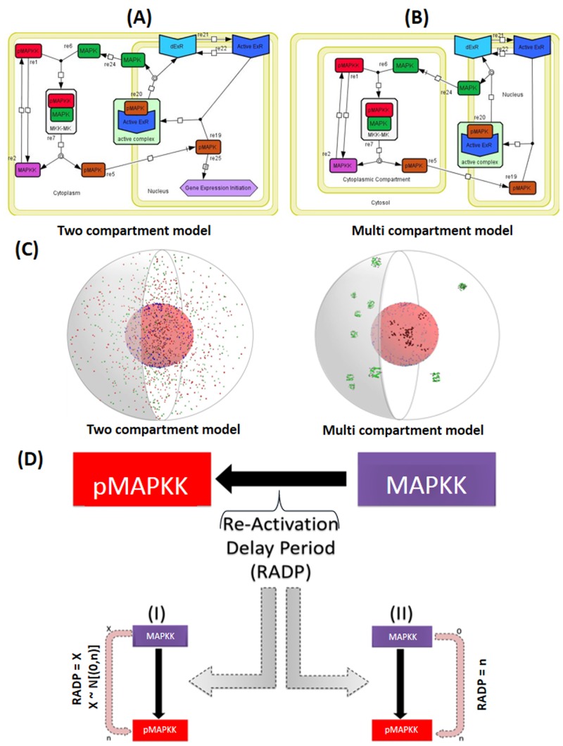
Signal transduction through the Mitogen Activated Protein Kinase (MAPK) pathways is evolutionarily highly conserved. Many cells use these pathways to interpret changes to their environment and respond accordingly. The pathways are central to triggering diverse cellular responses such as survival, apoptosis, differentiation and proliferation. Though the interactions between the different MAPK pathways are complex, nevertheless, they maintain a high level of fidelity and specificity to the original signal. There are numerous theories explaining how fidelity and specificity arise within this complex context; spatio-temporal regulation of the pathways and feedback loops are thought to be very important. This paper presents an agent based computational model addressing multi-compartmentalisation and how this influences the dynamics of MAPK cascade activation. The model suggests that multi-compartmentalisation coupled with periodic MAPK kinase (MAPKK) activation may be critical factors for the emergence of oscillation and ultrasensitivity in the system. Finally, the model also establishes a link between the spatial arrangements of the cascade components and temporal activation mechanisms, and how both contribute to fidelity and specificity of MAPK mediated signalling.


Conflict of interest statement

Competing Interests: The authors have declared that no competing interests exist.

Figures

Fig 1
Fig 1. A schematic representation of the MAPK cascade and its activation mechanisms.
The MAPK pathway is composed of three levels. The signal is transduced through phosphorylation events where mitogen activated protein kinase kinase kinase (MAP3K, also known as MAPKKK) phosphorylates mitogen activated protein kinase kinase (MAP2K, also known as MAPKK) leading to its activation and thus the phosphorylation and activation of the mitogen activated protein kinase (MAPK). Active MAPK phosphorylates protein targets in the cytoplasm and the nucleus. For mediating nuclear events MAPK translocates to the nucleus where it phosphorylates many proteins, which control gene expression.
Fig 2
Fig 2. Graphical representation of cytoplasmic and nuclear events in the two-compartment and multi-compartment agent-based models (ABMs).
The basic two dimensional design of the two and multi-compartment models of the two tier MAPK pathway represented using Systems Biology Graphical Notation (SBGN) standard annotations. (A) Illustrates the design of the two-compartment ABM whilst (B) describes the design of the multi-compartment ABM. Details of the two model design, structure and functionality are provided in the Materials and Methods section. (C) A three-dimensional (3D) visualisation of both the two-compartment vs. the multi-compartment model. The right hand side of both 3D representations is a 3D cross section of the “cell”. The cytoplasm is represented by the grey space around the nucleus. Inside the cytoplasm green spheres are MAPK, red spheres are pMAPKK, violet spheres are MAPKK, within the nuclear space, black spheres are pMAPK agents, dark blue are ExRs and light blue are dExRs. (D) Modelling the Re-Activation Delay Period (RADP) in the ABM: once pMAPKK agents change state into MAPKKs, they become dormant for period of time, and once this dormancy period is passed MAPKKs are re-activated. RADP was modelled either stochastically (I) or deterministically/periodically (II). In the stochastic model (I), RADP (X) was generated randomly for every individual pMAPKK agent, where X was a value between 0 and the chosen maximum value n (X ~ N ([0, n])). Periodic RADP was always identical for every MAPKK formed (RADP = n).
Fig 3
Fig 3. Analysis of MAPK activation dynamics observed in vitro in the published literature.
84 MAPK activation dynamics values were collected from published literature and the time to achieve Emax were plotted. A whisker plot with the median values are presented. (A) 63 in vitro data points from the analysed data were selected, plotted and analysis for normality was conducted. D'Agostino & Pearson omnibus normality test showed that the in vitro data for MAPK activation dynamics were not normally distributed (**: p<0.01). (B) 21 In silico data-points were extracted from the above literature and normality analysis was performed as for (A), demonstrating that the in silico data was normally distributed.
Fig 4
Fig 4. Robustness and sensitivity analysis of the ABM models.
The basic two compartment (A) and multi-compartment models (B) were run multiple times (n = 3). (A) The graph shows a run of the complete two compartment model in the presence of constitutively active MAPKK agents and the emergent kinetic behaviour of pMAPK and MAPK agents. The graph shows the interaction between pMAPKKs and MAPKs until the level of pMAPKs and MAPKs plateau as the interaction reaches equilibrium. The pattern emerging is a graded ultrasensitive response (whereby Emax ≥ 10 min). The model shows rapid activation of MAPK and formation of pMAPK but does not show ultrasensitive behaviour. (B) A graph generated from the multi-compartment model without a constitutively active MAPKK, this model shows that the multi-compartment system is capable of generating high level of pMAPK within a short period of time and with a gradual activation of MAPKK agents in addition to demonstrating an ultrasensitive behaviour. Individual data points for each run and the mean of the values are plotted. (C-H) Sensitivity analysis of the multi-compartment ABM to examine model sensitivity to manipulation of initial agent numbers. The number of each agent was altered by ±20%, compared to the control model. The number of pMAPK (E, F) and pMAPKK (G, H) agents were plotted. Time to achieve both Emax and EC50 were determined under each condition and the analysis of variance (ANOVA) was used to test for statistically significant changes. The analyses showed no significant difference between the different ABM conditions.
Fig 5
Fig 5. The effect of delaying MAPKK re-activation on the dynamics of MAPK activation and MAPKK levels.
Once pMAPKK agents bind and activate MAPKs to pMAPKs, pMAPKKs convert to a dormant state (MAPKK). The length of this dormancy period was set and its effects on the levels of pMAPK, MAPK, pMAPKK and MAPKK were monitored. In (A) and (B) the re-activation delay period (RADP) was set at a short period (0 ≤ RADP ≤ 90 s), while in (C) and (D) RADP was set to an intermediate period (0 ≤ RADP ≤ 4.53 min); in (E) and (F) RADP was set to a the highest range of the intermediate period (0 ≤ RADP ≤ 7.55 min); while in (G) and (H) RADP was set to long periods (0 ≤ RADP ≤ 22.6 min). The figures on the left hand side were stochastic (where the RADP was set stochastically within the specified delay period every time pMAPKK switched state to MAPKK); while models on the right hand side were deterministic (where MAPKK returns to the active pMAPKK state after a fixed period.
Fig 6
Fig 6. The effect of MAPKK re-activation delays on the dynamics of pMAPK formation and pMAPKK levels in two-compartment system.
The re-activation delay characteristics of pMAPKK (red) were applied to the two-compartment ABM and the effects were monitored. Initially the effect of a deterministic versus a stochastic model were looked at. In (A) and (B) short RADPs (0 ≤ RADP ≤ 90 s) were tested, (A) was the model with stochastic RADP while (B) was the model with deterministic/periodic RADP. There was no significant difference between the graphs generated by either ABMs when the analysis of variance (ANOVA) was used. However, both of the models had generated lower activation rate and formation of pMAPK (brown) and pMAPKK (violet) in comparison to the multi-compartment system. The graphs in (C) and (D) were generated with long RADPs (0 ≤ RADP ≤ 22.6 min), pMAPK formation, pMAPKK and MAPK (green) activations patterns were similar to those with short RADP seen in (A) and (B). Unlike multi-compartment models, deterministic models with intermediate or long RADPs did not generate any oscillatory pattern.
Fig 7
Fig 7. pMAPKK and pMAPK levels and rate of activation are significantly enhanced in the two compartmental model in the presence of signalosome clusters though with no significant difference between deterministic and stochastic re-activation delay (RADP) models.
Deterministic and stochastic models of MAPKK RADPs were tested again in the two-compartment ABM, in the context of assembly and disassembly of pMAPKK-MAPK signalsome clusters. In both models the presence of the clusters caused a rapid rate for pMAPKK (red) activation and pMAPK formation (green). This observation shares similarity with the multi-compartment system; however, only at the initial MAPK activation stage. Yet, these cluster models differ with the multi-compartment model in three aspects; (1) the cluster model exhibits a two phase response (activation [turn on] and deactivation [turn off/recovery] phases); (2) the recovery of MAPK (seen in the post-activation phase of the signalosome cluster model) and (3) that high levels of active pMAPKK are incapable of re-establishing high levels of pMAPK. In (A) and (B) short RADPs (0 ≤ RADP ≤ 90 s) were tested, (A) was the model with stochastic RADP while (B) was the model with deterministic RADP (RADP = 90 s). The graphs in (C) and (D) were generated with long RADPs (0 ≤ RADP ≤ 22.6 min), where (C) stochastic RADP was employed while (D) deterministic RADP was utilised (RADP = 22.6 min). The dynamics of pMAPK formation, MAPKK and MAPK activations in the long RADP models were similar to those noted in the short RADP models. Student t-test no significant difference in the responses generated by stochastic and deterministic models of RADPs at long periods, except for the slightly higher pMAPKK levels in the deterministic model once the steady state was reached. This also applies to the models with short RADPs, though the stochastic models generate higher levels of pMAPK in the initial phase.
Fig 8
Fig 8. The effect of changeable input-output dynamics at the level of MAPKK on phosphorylated MAPK (pMAPK) formation characteristics in a multi-compartment system.
Using the multi-compartment model, the MAPK pathway was run with different re-activation delay period (RADP) configurations to assess how switching between different MAPKK dormancy periods affect the formation of pMAPK. This was done to resemble a cellular system where a cell is initially faced with a strong, yet short activating signal, followed by the take-over of the inhibitory mechanisms, which is subsequently succeeded by a moderate and persistent activating signal. This simulation is similar to what cells are exposed to during somatogenesis. In the initial phase, a highly stochastic model of MAPKK RADP (0 ≤ RADP ≤ 90 s) was used (green solid line), once pMAPK level reached its maximum and was at equilibrium, the simulation was switched to deterministic-intermediate RADP model (RADP = 7.55 min, solid blue line). Once the level of pMAPK reached its lowest and was at equilibrium, the re-activation delay was switched to a model with stochastic-intermediate RADP (0 ≤ RADP ≤ 7.55 min; solid black line). This combination of the different modes of the MAPKK re-activation shows that once strong activation inputs of MAPKK are substantially reduced, inhibitory inputs which cause the deactivation of pMAPKK for long periods are capable of rapidly reducing the levels of pMAPK. However, they are still not capable of re-establishing the initial levels of MAPK seen at t0 as only 58.7% of t0 MAPK level was re-established. The final stage of the simulation (solid blue lines), reflects that in a multi-compartment system, even with a high stochasticity for MAPKK activation, a low number of active pMAPKK is sufficient to fundamentally increase and maintain high pMAPK levels.

Similar articles

Cited by

References

    1. Seger R, Krebs EG. The MAPK signaling cascade. FASEB journal: official publication of the Federation of American Societies for Experimental Biology. 1995;9(9):726–35. Epub 1995/06/01. . - PubMed
    1. Brunet A, Roux D, Lenormand P, Dowd S, Keyse S, Pouyssegur J. Nuclear translocation of p42/p44 mitogen-activated protein kinase is required for growth factor-induced gene expression and cell cycle entry. EMBO J. 1999;18(3):664–74. - PMC - PubMed
    1. Khokhlatchev AV, Canagarajah B, Wilsbacher J, Robinson M, Atkinson M, Goldsmith E, et al. Phosphorylation of the MAP Kinase ERK2 Promotes Its Homodimerization and Nuclear Translocation. Cell. 1998;93(4):605–15. - PubMed
    1. Chambard J-C, Lefloch R, Pouysségur J, Lenormand P. ERK implication in cell cycle regulation. Biochimica et Biophysica Acta (BBA)—Molecular Cell Research. 2007;1773(8):1299–310. - PubMed
    1. Choi TG, Lee J, Ha J, Kim SS. Apoptosis signal-regulating kinase 1 is an intracellular inducer of p38 MAPK-mediated myogenic signalling in cardiac myoblasts. Biochimica et Biophysica Acta (BBA)—Molecular Cell Research. 2011;1813(8):1412–21. - PubMed

Publication types

MeSH terms

Substances