. 2016 Jul 29;291(31):16208-20.
doi: 10.1074/jbc.M115.706747. Epub 2016 May 19.

Role of Conserved Disulfide Bridges and Aromatic Residues in Extracellular Loop 2 of Chemokine Receptor CCR8 for Chemokine and Small Molecule Binding

Affiliations

Role of Conserved Disulfide Bridges and Aromatic Residues in Extracellular Loop 2 of Chemokine Receptor CCR8 for Chemokine and Small Molecule Binding

Line Barington et al. J Biol Chem. .

Abstract

Chemokine receptors play important roles in the immune system and are linked to several human diseases. The initial contact of chemokines with their receptors depends on highly specified extracellular receptor features. Here we investigate the importance of conserved extracellular disulfide bridges and aromatic residues in extracellular loop 2 (ECL-2) for ligand binding and activation in the chemokine receptor CCR8. We used inositol 1,4,5-trisphosphate accumulation and radioligand binding experiments to determine the impact of receptor mutagenesis on both chemokine and small molecule agonist and antagonist binding and action in CCR8. We find that the seven-transmembrane (TM) receptor conserved disulfide bridge (7TM bridge) linking transmembrane helix III (TMIII) and ECL-2 is crucial for chemokine and small molecule action, whereas the chemokine receptor conserved disulfide bridge between the N terminus and TMVII is needed only for chemokines. Furthermore, we find that two distinct aromatic residues in ECL-2, Tyr(184) (Cys + 1) and Tyr(187) (Cys + 4), are crucial for binding of the CC chemokines CCL1 (agonist) and MC148 (antagonist), respectively, but not for small molecule binding. Finally, using in silico modeling, we predict an aromatic cluster of interaction partners for Tyr(187) in TMIV (Phe(171)) and TMV (Trp(194)). We show in vitro that these residues are crucial for the binding and action of MC148, thus supporting their participation in an aromatic cluster with Tyr(187) This aromatic cluster appears to be present in a large number of CC chemokine receptors and thereby could play a more general role to be exploited in future drug development targeting these receptors.

Keywords: CCR8; G protein-coupled receptor (GPCR); chemokine receptor; metal ion-protein interaction; molecular pharmacology; mutagenesis in vitro; protein-protein interaction; receptor structure-function.


Figures

FIGURE 1.
FIGURE 1.
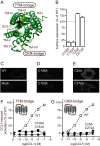
Importance of conserved disulfide bridges for CCR8 cell surface expression and CCL1-induced activation. A, crystal structure of CCR5 (top view, Protein Data Bank code 4MBS), highlighting the two disulfide bridges in yellow. The figure was made using PyMOL software. B, surface expression of the four mutant receptors, as tested by ELISA (n = 3–4). The data were normalized to the expression level of CCR8 WT (100%, mean A450 = 0.415 ± 0.039) and empty vector (0%, mean A450 = 0.135 ± 0.02). C–E, representative confocal microscopy images of COS-7 cells transiently transfected with the indicated constructs. C, CCR8 WT and pcDNA empty vector (mock). D, C106A and C183A. E, C25A and C272A. F and G, IP3 accumulation experiments in transiently transfected COS-7 cells that show activation with CCL1 of CCR8 mutants lacking the 7TM receptor conserved disulfide bridge (F) or the chemokine receptor conserved disulfide bridge (G). A schematic representation of the relevant bridge is shown as an inset in each panel. The data were normalized to CCR8 WT activation (dotted line). The average maximal WT count (100%) was 5093 ± 885 cpm, and the average empty vector count (0%) was 639 ± 136 cpm. The error bars (barely visible) represent S.E. (n = 4–5 for the mutants and n = 33 for the WT).
FIGURE 2.
FIGURE 2.
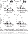
Importance of conserved disulfide bridges for chemokine binding to CCR8. A–D, homologous competition binding experiments in transiently transfected COS-7 cells. Binding of 125I-CCL1 (A and B) or 125I-MC148 (C and D) to CCR8 mutants with disrupted 7TM receptor conserved bridge (A and C) or chemokine receptor conserved bridge (B and D) is shown. The data were normalized to WT binding, which is represented by a dotted line. In CCL1 experiments, the average maximal count for the WT (100%) was 1965 ± 105 cpm, and the average count for the empty vector (0%) was 788 ± 46 cpm. In MC148 experiments, the average maximal count for the WT was 605 ± 62 cpm, and the average count for the empty vector was 63 ± 10 cpm. Error bars represent S.E. (n = 3–5). Schematic representations of the relevant bridge are shown as insets in A and B. E, alignment using CLUSTALW 1.7 of the sequences of the agonist CCL1 and the antagonist MC148 (25). The CC motif in both chemokines is highlighted with a box.
FIGURE 3.
FIGURE 3.
Importance of disulfide bridges for small molecule agonist and antagonist actions in CCR8. IP3 accumulation experiments in transiently transfected COS-7 cells. A and B, CuPhe-induced activation of mutant receptors lacking the 7TM receptor conserved disulfide bridge (A) or the chemokine receptor conserved disulfide bridge (B). C, effect of the small molecule antagonist LMD-A on CuPhe-induced activation of mutant receptors lacking the chemokine receptor conserved disulfide bridge. The data were normalized to WT activation. The average maximal count for the CuPhe-induced activation of the WT (100%) was 4042 ± 42 cpm, and the average count for the empty vector (0%) was 882 ± 49 cpm. The error bars represent S.E. (n = 3–5 for the mutants and n = 20 for the WT). The molecular structure of the relevant ligand, either CuPhe (39) or LMD-A (28), is shown below each panel.
FIGURE 4.
FIGURE 4.
Aromatic amino acids in the ECL-2 region of human CC-chemokine receptors. Alignment of amino acid sequences of ECL-2 and flanking regions in human CC-chemokine receptors is shown. The alignment was made in ICM (Molsoft), and the zero end-gap global alignment algorithm was used (58). Black background highlights 100% conservation of a single amino acid. Gray background denotes ≥70% conservation of similar amino acids (in this case Tyr, Phe, and Trp). The three aromatic amino acids just C-terminal to the conserved cysteine in CCR8 are highlighted with black boxes. The schematic illustration in the left panel illustrates the area included in the alignment.
FIGURE 5.
FIGURE 5.
Importance of selected aromatic amino acids in extracellular loop 2 for chemokine- and small molecule-induced activation of CCR8. IP3 accumulation experiments in transiently transfected COS-7 cells are shown. A–F, CCL1-induced (A–C) or CuPhe-induced (D–F) activation of CCR8 mutants Y184A (A and D), F186A (B and E), or Y187A (C and F). The data were normalized to WT activation (shown with a dotted line). The average maximal count for the CCL1-induced activation of the WT (100%) was 4151 ± 1705 cpm, and the average count for the empty vector (0%) was 1247 ± 335 cpm. For the CuPhe-induced activation, the average maximal count for the WT was 4527 ± 153 cpm, and the average count for the empty vector was 867 ± 198 cpm. The error bars represent S.E. (n = 3–4 for the mutants and n = 20–28 for the WT). In A–C, vertical dotted lines indicate the approximate EC50 values.
FIGURE 6.
FIGURE 6.
Importance of selected aromatic amino acids in extracellular loop 2 for chemokine- and small molecule-mediated antagonism on CCR8. IP3 accumulation experiments in transiently transfected COS-7 cells are shown. A–F, MC148-mediated (A–C) or LMD-A-mediated (D–F) antagonism of CCL1-induced activation of CCR8 mutants Y184A (A and D), F186A (B and E), or Y187A (C and F). WT activation is illustrated with a dotted line. The data were normalized to WT activation. The average maximal count for the CCL1-induced activation of the WT (100%) was 4151 ± 1705 cpm, and the average count for the empty vector (0%) was 1247 ± 335 cpm. For the CuPhe-induced activation, the average maximal count for the WT was 4527 ± 153 cpm, and the average count for the empty vector was 867 ± 198 cpm. The error bars represent S.E. (n = 3 for the mutants and n = 4–7 for the WT).
FIGURE 7.
FIGURE 7.
Dependence of CC-chemokine binding on selected aromatic residues in extracellular loop 2 of CCR8. A–F, homologous competition binding using 125I-CCL1 (A–C) and 125I-MC148 (D–F) in transiently transfected COS-7 cells expressing Y184A (A and D), F186A (B and E), or Y187A (C and F). The data were normalized to WT binding, which is illustrated with a dotted line. In 125I-CCL1 binding experiments, the maximal average value for the WT receptor was 1970 ± 103 cpm, and the average count for the empty vector was 625 ± 109 cpm. In 125I-MC148 binding experiments, the maximal average value for the WT receptor was 463 ± 127 cpm, and the average count for the empty vector was 31 ± 10 cpm. The error bars represent S.E. (n = 3–9).
FIGURE 8.
FIGURE 8.
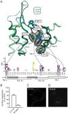
Involvement of aromatic residues in the top of transmembrane helices IV and V in a putative aromatic cluster in CCR8. A, structural conservation of the aromatic residues at the TMIV/TMV interface in the CC-chemokine receptor subfamily. The CCR5-based homology model of CCR8 (blue) was superimposed onto the high resolution crystal structure of CCR5 (green; Protein Data Bank code 4MBS). Important side chains are shown as stick representations. The van der Waals surface is shown for aromatic ring systems of conserved residues. The amino acid conservation of the extracellular sides of TMIV/TMV and ECL-2 is shown as sequence logo for the human CC receptor subfamily (CCR1–10). The logo displays the frequencies of amino acids on each position as the relative heights of letters, along with the degree of sequence conservation as the total height of a stack of letters, measured in bits of information. Secondary structure elements are shown as white boxes (α-helix) and gray arrows (β-sheet). Two of the positions where aromatic residues are conserved, 4.63 (Phe171 in CCR8) and 5.34 (Trp194 in CCR8), are highlighted with black boxes. The figure was made using ICM (59). B–D, surface expression of aromatic amino acid mutants F171A and W194A. B, ELISA on COS-7 cells transiently transfected with each mutant receptor (n = 7–11). The surface expression is shown as a percentage of WT receptor surface expression (100%, average A450 = 0.214 ± 0.02). The average empty vector value (0%) was 0.168 ± 0.021. C and D, representative confocal microscopy pictures of COS-7 cells transiently transfected with F171A (C) or W194A (D).
FIGURE 9.
FIGURE 9.
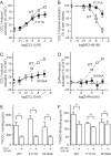
Role of selected aromatic amino acids in the transmembrane regions flanking extracellular loop 2 in receptor activation and binding in CCR8. A–D, IP3 accumulation experiments in transiently transfected COS-7 cells. A, CCL1-induced activation of F171A. B, MC148-mediated antagonism of CCL1-induced activation of F171A. C and D, CCL1-induced (C) or ZnPhe-induced (D) activation of W194A. The data were normalized to WT values (A, C, and D) or to own values (B). WT graphs are shown as dotted lines for comparison. For CCL1-mediated activation, the average maximal WT value was 1268 ± 99 cpm. For ZnPhe-mediated activation, the average maximal WT value was 655 ± 64 cpm. In B, the maximal value of CCL1-mediated activation of F171A, which was 849 ± 44 cpm, was used for the normalization. Error bars represent S.E., n = 6–7, except for CCL1 on the WT, where n = 33. E and F, homologous competition binding assays in transiently transfected COS-7 cells. The cpm values for the binding of 125I-CCL1 (E) or 125I-MC148 (F) to CCR8 WT, F171A, and W194A are given. Binding in the presence (+) or absence (−) of 100 nm competing cold ligand (CCL1 in E and MC148 in F) is shown for all receptor constructs (n = 4–5). An asterisk denotes a statistically significant (p < 0.05) difference. ns means that there is no statistically significant difference.
FIGURE 10.
FIGURE 10.
Structure of the TMIV/TMV aromatic interface in available crystal structures of class A 7TM receptors. A, structural arrangement of the aromatic amino acid at position Cys + 4 (in red) in nine crystal structures of class A 7TM receptors. The conserved cysteine is shown in yellow. The names of the nine receptors are listed in the top list to the right. The receptors written below in italics either do not have an aromatic residue in Cys + 4 (10 receptors) or their aromatic residue is differently arranged (GPR40 and rhodopsin). B, the human receptor CCR5 (Protein Data Bank code 4MBS). C, the human receptor CXCR4 (Protein Data Bank code 4RWS). D, the cytomegalovirus receptor US28 (Protein Data Bank code 4XT1). Important side chains are shown as stick representation. The van der Waals surface is shown for aromatic ring systems of selected receptor residues. A was made in PyMOL, and B–D were made using ICM (59).

Similar articles

Cited by

References

    1. Gerard C., and Rollins B. J. (2001) Chemokines and disease. Nat. Immunol. 2, 108–115 - PubMed
    1. Rosenkilde M. M., and Schwartz T. W. (2004) The chemokine system: a major regulator of angiogenesis in health and disease. APMIS 112, 481–495 - PubMed
    1. Viola A., and Luster A. D. (2008) Chemokines and their receptors: drug targets in immunity and inflammation. Annu. Rev. Pharmacol. Toxicol. 48, 171–197 - PubMed
    1. Schwarz M. K., and Wells T. N. (2002) New therapeutics that modulate chemokine networks. Nat. Rev. Drug Discov. 1, 347–358 - PubMed
    1. Allen S. J., Crown S. E., and Handel T. M. (2007) Chemokine: receptor structure, interactions, and antagonism. Annu. Rev. Immunol. 25, 787–820 - PubMed

MeSH terms