. 2016 Jul 1;143(13):2325-33.
doi: 10.1242/dev.136499. Epub 2016 May 25.

The tyrosine kinase FER is responsible for the capacitation-associated increase in tyrosine phosphorylation in murine sperm

Affiliations

The tyrosine kinase FER is responsible for the capacitation-associated increase in tyrosine phosphorylation in murine sperm

Antonio Alvau et al. Development. .

Abstract

Sperm capacitation is required for fertilization. At the molecular level, this process is associated with fast activation of protein kinase A. Downstream of this event, capacitating conditions lead to an increase in tyrosine phosphorylation. The identity of the tyrosine kinase(s) mediating this process has not been conclusively demonstrated. Recent experiments using stallion and human sperm have suggested a role for PYK2 based on the use of small molecule inhibitors directed against this kinase. However, crucially, loss-of-function experiments have not been reported. Here, we used both pharmacological inhibitors and genetically modified mice models to investigate the identity of the tyrosine kinase(s) mediating the increase in tyrosine phosphorylation in mouse sperm. Similar to stallion and human, PF431396 blocks the capacitation-associated increase in tyrosine phosphorylation. Yet, sperm from Pyk2(-/-) mice displayed a normal increase in tyrosine phosphorylation, implying that PYK2 is not responsible for this phosphorylation process. Here, we show that PF431396 can also inhibit FER, a tyrosine kinase known to be present in sperm. Sperm from mice targeted with a kinase-inactivating mutation in Fer failed to undergo capacitation-associated increases in tyrosine phosphorylation. Although these mice are fertile, their sperm displayed a reduced ability to fertilize metaphase II-arrested eggs in vitro.

Keywords: Capacitation; FER; Tyrosine phosphorylation.


Conflict of interest statement

Competing interests

The authors declare no competing or financial interests.

Figures
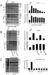
Fig. 1.
Fig. 1.
PF431396 inhibits the capacitation-associated increase in tyrosine phosphorylation. Sperm suspensions (106) were diluted in media supplemented (CAP) or not (NON) with NaHCO3 and BSA and in the presence of different compounds as detailed. After 1 h, sperm were analyzed for protein phosphorylation with anti-phosphotyrosine (anti-PY) and anti-phospho PKA substrates (anti-pPKAs). For each experiment (n=3), western blots were analyzed using ImageJ. For comparison between blots, pixels for each lane contained in the region marked by # (proteins in the 70-100 kDa range for PY and 80-200 kDa for pPKAs) were quantified and normalized using the CAP lane as reference (100%). The comparison between groups was performed using two-tailed analysis of variance (ANOVA) in one-way blocks. Multiple comparisons were performed by Tukey's test. Bar graphs represent mean±s.e.m. of the normalized values (**P<0.01). (A) PF431396 concentration curve. Sperm were incubated either in NON or CAP media with increasing concentrations of PF431396, which was added from the beginning of the 60 min incubation. Anti-PY and anti-pPKAs western blot analyses (left panels) and ImageJ quantification (right panels) were conducted. (B) PF431396 blocks cAMP-induced tyrosine phosphorylation. Sperm were incubated either in NON or CAP media in the presence or absence of 1 mM dbcAMP and 100 µM IBMX for 1 h in the presence or in the absence of 10 µM PF431396. Quantification was conducted as described. (C) Okadaic acid (OA) does not overcome inhibition by PF431396. Sperm were incubated for 1 h either in NON or in CAP media in the absence or in the presence of 10 µM PF431396 and in the presence of increasing concentrations of OA. Analyses were carried out as described.
Fig. 2.
Fig. 2.
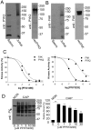
Presence of FAK subfamily members in sperm and analysis of in vitro activity. (A,B) Recombinant proteins (recPYK2, recFAK) or lysates from sperm or Jurkat cells were subjected to western blotting with antibodies for PYK2 (A) or FAK (B). (C) In vitro kinase activity of recombinant PYK2 and FAK. Recombinant PYK2 and FAK (60 ng/reaction) were assayed using poly Glu:Tyr as substrate as described. Each of the kinases (tested in triplicate) was assayed in the presence of increasing concentrations of PF431396 (left) and PF573228 (right). Numbers on the x-axis are expressed as logarithm of molar concentration. Error bars are in the order of 0.002-0.01 and do not appear in the graph. (D) PF573228 concentration curve. Sperm were incubated for 1 h in NON or in CAP media with increasing concentrations of PF573228. Western blotting using anti-PY antibodies (left) and ImageJ quantification (right) were conducted from independent experiments (n=3). Bars represent the mean±s.e.m. [**P<0.01 versus both CAP (0 μM PF573228) and NON CAP] of the normalized values.
Fig. 3.
Fig. 3.
Sperm from Pyk2−/− mice undergo normal tyrosine phosphorylation. (A) Tyrosine phosphorylation analysis. Sperm from either wild-type (Pyk2+/+) or knockout (Pyk2−/−) mice were incubated either under NON or CAP conditions. After 1 h incubation, tyrosine phosphorylation was assessed by anti-PY western blotting. Images are representative of three independent experiments (n=3) using three different animals. (B,C) PF431396 and PF573228 concentration curves. The effect of PF431396 (B) and of PF573228 (C) on tyrosine phosphorylation was analyzed as described for Fig. 1A. (D) Anti-PYK2 and anti-FAK western blot analyses of Pyk2+/+ and Pyk2−/− sperm. Cauda epididymal sperm from either Pyk2+/+ or Pyk2−/− mice were obtained in TYH-HEPES media as described above, and sperm cell lysates prepared. The presence of PYK2 and FAK was analyzed by independent western blotting run in parallel. (E) Sperm from Pyk2−/− animals are fertile in vitro. Cauda epididymal sperm from either Pyk2+/+ or Pyk2−/− mice were collected in TYH media as described in Materials and Methods and capacitated for 1 h, and rates of in vitro fertilization (IVF) evaluated as described in Materials and Methods. Bar graphs represent the mean±s.e.m. of the normalized values of three independent experimental replicates (n=3), using three different animals. IVF data were analyzed by the simple χ2 test.
Fig. 4.
Fig. 4.
Presence of FER in mouse sperm. (A) Presence of FERT in mouse sperm. Sperm (or NIH3T3 cell lysates for control) were analyzed by western blotting using anti-FER antibodies. (B) FER in vitro kinase assay. Recombinant FER (60 ng) in vitro kinase activity was assayed as described for PYK2 and FAK in Fig. 2C. Values represent mean±s.e.m. (n=3). Error bars are in the order of 0.01-0.08 and do not appear in the graph. (C-F) Distribution of PYK2, FAK, FER and PY substrates after solubilization with Triton X-100 (1%). Cauda epididymal sperm obtained as described in Fig. 1 were incubated in conditions that support capacitation for 1 h. Sperm were then washed in PBS, and Triton X-100-treated samples prepared as described in Materials and Methods. Triton X-100-soluble (SOL) and insoluble (IN) fractions were separated, boiled in SDS-sample buffer for 5 min and analyzed by western blotting using anti-PY (C), anti -PYK2 (D), anti -FAK (E) and anti -FER (F). Results are representative of three independent experiments (n=3) using different animals.
Fig. 5.
Fig. 5.
Sperm from FerDR/DR mice do not display capacitation-associated increases in tyrosine phosphorylation. (A,B) Sperm from either wild-type (+/+) (A) or FerDR/DR (DR/DR) mice (B) were obtained as described for Fig. 1 and incubated under NON or CAP conditions. Time-dependent response by phosphorylation of PKA substrates and by tyrosine phosphorylation was assessed using anti-PY (upper panels) and anti-pPKAs (lower panels) by western blotting. Images shown are representative of at least three independent experiments (n=3). (C) Sperm from FerDR/DR mice are subfertile in vitro. Cauda epididymal sperm from either wild-type (+/+) or FerDR/DR (DR/DR) mice were obtained in TYH media and capacitated for 1 h, and IVF rates were evaluated as described in Materials and Methods. Bar graphs represent the mean±s.e.m. of the normalized values (**P<0.01) (n=3). IVF data were analyzed by the simple χ2 test.
Fig. 6.
Fig. 6.
Localization of FER and tyrosine-phosphorylated proteins in mouse sperm by STORM. (A) 3D STORM images of FER in xy projection. The color represents the relative distance from the focal plane along the z-axis. (B) FER localization in the midpiece (MP), yz projection. (C) FER localization in the principal piece (PP), yz projection. (D) 3D STORM images of tyrosine phosphorylation (pY) in xy projection. The color represents the relative distance from the focal plane along the z-axis. (E) Tyrosine-phosphorylated protein (PY) localization in the MP, yz projection. (F) Tyrosine-phosphorylated protein (PY) localization in the PP, yz projection. (G) Radial distribution of FER and PY in the MP. (H) Radial distribution of FER and PY in the PP. Images are representative of 15 cells from three different animals for PY analysis and of nine cells from three different animals for FER analysis.

Similar articles

Cited by

References

    1. Austin C. R. (1951). Observations on the penetration of the sperm in the mammalian egg. Aust. J. Sci. Res. Ser. B Biol. Sci. 4, 581-596. - PubMed
    1. Baker M. A., Hetherington L. and Aitken R. J. (2006). Identification of SRC as a key PKA-stimulated tyrosine kinase involved in the capacitation-associated hyperactivation of murine spermatozoa. J. Cell Sci. 119, 3182-3192. 10.1242/jcs.03055 - DOI - PubMed
    1. Baker M. A., Hetherington L., Curry B. and Aitken R. J. (2009). Phosphorylation and consequent stimulation of the tyrosine kinase c-Abl by PKA in mouse spermatozoa; its implications during capacitation. Dev. Biol. 333, 57-66. 10.1016/j.ydbio.2009.06.022 - DOI - PubMed
    1. Baldi E., Casano R., Falsetti C., Krausz C., Maggi M. and Forti G. (1991). Intracellular calcium accumulation and responsiveness to progesterone in capacitating human spermatozoa. J. Androl. 12, 323-330. - PubMed
    1. Baldi E., Luconi M., Bonaccorsi L. and Forti G. (2002). Signal transduction pathways in human spermatozoa. J. Reprod. Immunol. 53, 121-131. 10.1016/S0165-0378(01)00089-4 - DOI - PubMed

Publication types