. 2016 Jun 28;7(26):39980-39995.
doi: 10.18632/oncotarget.9556.

Suppression of progranulin expression inhibits bladder cancer growth and sensitizes cancer cells to cisplatin

Affiliations

Suppression of progranulin expression inhibits bladder cancer growth and sensitizes cancer cells to cisplatin

Simone Buraschi et al. Oncotarget. .

Erratum in

Abstract

We have recently demonstrated a critical role for progranulin in bladder cancer. Progranulin contributes, as an autocrine growth factor, to the transformed phenotype by modulating Akt-and MAPK-driven motility, invasion and anchorage-independent growth. Progranulin also induces F-actin remodeling by interacting with the F-actin binding protein drebrin. In addition, progranulin is overexpressed in invasive bladder cancer compared to normal tissue controls, suggesting that progranulin might play a key role in driving the transition to the invasive phenotype of urothelial cancer. However, it is not established whether targeting progranulin could have therapeutic effects on bladder cancer. In this study, we stably depleted urothelial cancer cells of endogenous progranulin by shRNA approaches and determined that progranulin depletion severely inhibited the ability of tumorigenic urothelial cancer cells to migrate, invade and grow in anchorage-independency. We further demonstrate that progranulin expression is critical for tumor growth in vivo, in both xenograft and orthotopic tumor models. Notably, progranulin levels correlated with response to cisplatin treatment and were upregulated in bladder tumors. Our data indicate that progranulin may constitute a novel target for therapeutic intervention in bladder tumors. In addition, progranulin may serve as a novel biomarker for bladder cancer.

Keywords: anchorage-independent growth; bladder cancer; motility; progranulin; tumor formation in vivo.


Conflict of interest statement

The authors declare no conflicts of interest.

Figures
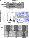
Figure 1
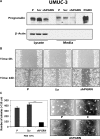
Figure 1. Progranulin depletion inhibits UMUC-3 urothelial cancer cell motility
(A) The generation of UMUC-3/shScr (scramble control) and UMUC-3/shPGRN cells has been described in Materials and Methods. The progranulin-specific shRNA was TI350373 (Origine). Progranulin expression in lysates and conditioned media from parental (P), control (Scr) or shRNA progranulin-transfected (shPGRN) UMUC-3 cells was detected by immunoblot with anti-progranulin polyclonal antibodies. (B) Migration of the various UMUC-3 cell lines was performed using transwells as described in detail in Materials and Methods. Data are the average of three independent experiments run in duplicates ± SD. ***P < 0.001. Recombinant human progranulin was supplemented at 80 nM (C) The in vitro “wound healing” motility assay in UMUC-3 cells in either SFM or SFM supplemented with 1% FBS was performed as described in Materials and methods. Cells were analyzed with a cell live microscope using the Metamorph Image Acquisition and Analysis software (Universal Imaging) (×100). 10 fields/plate were examined. Pictures from representative fields are shown.
Figure 2
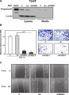
Figure 2. Progranulin depletion inhibits T24T urothelial cancer cell motility
(A) The generation of T24T/shScr (scramble control) and T24T/shPGRN cells has been described in Materials and Methods. The progranulin-specific shRNA was TI350373 (Origine). Progranulin expression in lysates and conditioned media from parental (P), control (Scr) or shRNA progranulin-transfected (shPGRN) T24T cells was detected by immunoblot with anti-progranulin polyclonal antibodies. (B) Migration of the various T24T cell lines was performed using transwells as described in detail in Materials and Methods. Data are the average of three independent experiments run in duplicates ± SD. ***P < 0.001. (C) The in vitro “wound healing” motility assay in T24T cells in either SFM or SFM supplemented with 1% FBS was performed as described in Materials and methods. Cells were analyzed with a cell live microscope using the Metamorph Image Acquisition and Analysis software (Universal Imaging) (×100). 10 fields/plate were examined. Pictures from representative fields are shown.
Figure 3
Figure 3. Progranulin targeting modulates invasion and anchorage-independent growth of UMUC-3 urothelial cancer cells
(A) Parental (P), shScr-transfected (Scr) control and Progranulin-depleted (shPGRN) UMUC-3 cells were assessed for invasive ability through Matrigel-coated transwells as described in Materials and Methods. Data are the average of three independent experiments ± SD. ***P < 0.001. Recombinant human progranulin was supplemented at 80 nM. (B) Anchorage-independent growth was measured by colony formation in soft-agar as previously described [18, 19, 49]. Colonies > 150 μM were counted. The experiment is the average of three independent experiments run in duplicates ± SD. ***P < 0.001.
Figure 4
Figure 4. Progranulin targeting modulates invasion and anchorage-independent growth of T24T urothelial cancer cells
(A) Parental (P), shScr-transfected (Scr) control and Progranulin-depleted (shPGRN) T24T cells were assessed for invasive ability through Matrigel-coated transwells as described in Materials and Methods. Data are the average of three independent experiments ± SD. ***P < 0.001. (B) Anchorage-independent growth was measured by colony formation in soft-agar as described IN Materials and Methods. Colonies >150 μM were counted. The experiment is the average of three independent experiments run in duplicates ± SD. ***P < 0.001.
Figure 5
Figure 5. Progranulin depletion inhibits UMUC-3 urothelial cancer cell motility and anchorage-independent growth
(A) The generation of UMUC-3/shScr (scramble control) and UMUC-3/shPGRN cells has been described in Materials and Methods. The progranulin-specific shRNA used here was TI350374 (Origine). Progranulin expression in lysates and conditioned media from parental (P), control (Scr) or shRNA progranulin-transfected (shPGRN) UMUC-3 cells was detected by immunoblot with anti-progranulin polyclonal antibodies. (B) The in vitro “wound healing” motility assay in UMUC-3 cells in either SFM or SFM supplemented with 1% FBS was performed as described in Materials and Methods. Cells were analyzed with a cell live microscope using the Metamorph Image Acquisition and Analysis software (Universal Imaging) (×100). 10 fields/plate were examined. Pictures from representative fields are shown. (C) Anchorage-independent growth was measured by colony formation in soft-agar as described in Materials and Methods. Colonies > 150 μM were counted. The experiment is the average of three independent experiments run in duplicates ± SD. ***P < 0.001.
Figure 6
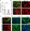
Figure 6. Stable depletion of endogenous progranulin in UMUC-3 urothelial cancer cells inhibits tumor xenograft growth
(A) Xenograft volume at day 52 of mice injected with UMUC-3/shScr (shScr) and UMUC-3/shPGRN (shPGRN). ***P < 0.001. (B) Representative macroscopic photograph of a mouse 53 days post injection with UMUC-3/shScr in the left flank and UMUC3-shPGRN cell in the right flank. (C) Tumors extracted from UMUC-3/shScr and UMUC-3/sh-PGRN injected mice were tested for progranulin expression by immunofluorescence analysis. Three-dimensional surface plots are indicative of progranulin expression levels of the relative fluorescence images. Bar = 5 μm.
Figure 7
Figure 7. Progranulin regulates orthotopic bladder tumor formation
(A) Tumor volume at day 20 of mice (n = 22) injected orthotopically, under ultrasound guidance, with UMUC-3/shScr (shScr) and UMUC-3/shPGRN (shPGRN) cells. ***P < 0.001. (B) Immunofluorescence staining of progranulin (green signal) and actin (red signal) in frozen sections of UMUC-3/shScr and UMUC-3/shPGRN orthotopic xenografts. The two insets represent relative magnified areas of the immunofluorescence pictures. White arrows point at the cortical actin fibers, which are disrupted in the presence of progranulin in the ShScr orthotopic tumors. (CE) Immunostaining of Ki67, actin, E-cadherin and vimentin in frozen sections of the orthotopic tumors. Nuclei: blue. Bar = 10 μm.
Figure 8
Figure 8. Progranulin depletion sensitized UMUC-3 urothelial cancer cells to cisplatin treatment
(A–B) UMUC-3/shScr and UMUC-3/shPGRN were plated onto six-well plates at a density of 3.5 × 10⁵ cells/well in 10% FBS-containing media. After 24 hours, cells were washed and transfer to 1% FBS-containing media with or without cisplatin at the concentrations of 1 μM and 10 μM. Cells were then counted after 24 hours as previously described [31, 49, 50]. Data are the average of 5 independent experiments run in duplicates ± SD. Groups were compared by ANOVA. *P < 0.05. **P < 0.01. ***P < 0.001. (B) Cell survival was assessed in 1% serum-containing media by MTS assays at cisplatin concentration of 1, 10 and 20 μM. ***P < 0.001.
Figure 9
Figure 9. Progranulin is upregulated in bladder cancer tissues
(A) Human bladder tumor microarray containing 69 validated cases of various types and stages of bladder cancers and normal bladder tissue controls. The panels show light micrographs outlining the expression of progranulin in different tumors as indicated. Progranulin is expressed at high levels in all cancers analyzed compared to the normal bladder except for the mucinous carcinoma. The arrows show areas of high staining for progranulin in the epithelial compartments of all cancer studied. (B) Quantification of progranulin staining levels was assessed using ImageJ software. All cases in the tissue microarray were analyzed and quantified. Briefly, the threshold of each image was adjusted in order to show only the specific staining. The background was removed and the data were quantified and plotted using SigmaPlot software. Student's t-tests were run to determine statistical significance of each class of cancers compared to normal bladder. *P < 0.05; ***P < 0.001. Bars = 100 μm. (C) Additional progranulin IHC on archived bladder cancer tissue samples, which include low and high grade superficial papillary tumors (Ta) and CIS (Ti). These results complement the TMA analysis with a broader spectrum of various urothelial carcinoma stages (D) Progranulin expression in archived tissue samples was analyzed and quantified as described above. *P < 0.05; ***P < 0.001.

Similar articles

Cited by

References

    1. Siegel RL, Miller KD, Jemal A. Cancer statistics, 2015. CA Cancer J Clin. 2015;65:5–29. - PubMed
    1. Mitra AP, Cote RJ. Molecular Pathogenesis and Diagnostics of Bladder Cancer. Annu Rev Pathol. 2009;4:251–285. - PubMed
    1. Knowles MA. Molecular pathogenesis of bladder cancer. Int J Clin Oncol. 2008;13:287–297. - PubMed
    1. Botteman MF, Pashos CL, Redaelli A, Laskin B, Hauser R. The health economics of bladder cancer: a comprehensive review of the published literature. Pharmacoeconomics. 2003;21:1315–1330. - PubMed
    1. He Z, Bateman A. Progranulin (granulin-epithelin precursor, PC-cell-derived growth factor, acrogranin) mediates tissue repair and tumorigenesis. J Mol Med. 2003;81:600–612. - PubMed

MeSH terms