. 2016 May 23:6:26509.
doi: 10.1038/srep26509.

SUMO5, a Novel Poly-SUMO Isoform, Regulates PML Nuclear Bodies

Affiliations

SUMO5, a Novel Poly-SUMO Isoform, Regulates PML Nuclear Bodies

Ya-Chen Liang et al. Sci Rep. .

Abstract

Promyelocytic leukemia nuclear bodies (PML-NBs) are PML-based nuclear structures that regulate various cellular processes. SUMOylation, the process of covalently conjugating small ubiquitin-like modifiers (SUMOs), is required for both the formation and the disruption of PML-NBs. However, detailed mechanisms of how SUMOylation regulates these processes remain unknown. Here we report that SUMO5, a novel SUMO variant, mediates the growth and disruption of PML-NBs. PolySUMO5 conjugation of PML at lysine 160 facilitates recruitment of PML-NB components, which enlarges PML-NBs. SUMO5 also increases polySUMO2/3 conjugation of PML, resulting in RNF4-mediated disruption of PML-NBs. The acute promyelocytic leukemia oncoprotein PML-RARα blocks SUMO5 conjugation of PML, causing cytoplasmic displacement of PML and disruption of PML-NBs. Our work not only identifies a new member of the SUMO family but also reveals the mechanistic basis of the PML-NB life cycle in human cells.


Figures
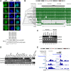
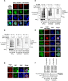
Figure 1
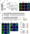
Figure 1. A primate- and tissue-specific SUMO variant, SUMO5, forms NBs.
(A) SUMO5 forms NBs. (Top panel) localization of GFP-tagged SUMO proteins and their associated subcellular structures in 293 cells were assessed by confocal microscopy. DNA (blue) was stained with DAPI. Scale bar, 10 μm. (Bottom panel) whole cell lysates were immunoblotted with anti-GFP or anti-β-actin antibodies. (B) UCSC Genome Browser display of Multiz Alignments of 20 mammals (17 primates) at the SUMO5 gene locus (chr20:53, 874, 198-53, 876, 011), indicating SUMO5 is conserved among primate species. The height of species track (green histogram) reflects the value of normalized MULTIZ score. Organisms (species) included are: Human (Homo sapiens), Chimp (Pan troglodytes), Bonobo (Pan paniscus), Gorilla (Gorilla gorilla gorilla), Orangutan (Pongo pygmaeus abelii), Gibbon (Nomascus leucogenys), Proboscis monkey (Nasalis larvatus), Golden snub-nosed monkey (Rhinopithecus roxellana), Green monkey (Chlorocebus sabaeus), Crab-eating macaque (Macaca fascicularis), Rhesus (Macaca mulatta), Baboon (Papio anubis), Squirrel monkey (Saimiri boliviensis), Marmoset (Callithrix jacchus), Tarsier (Tarsius syrichta), Mouse lemur (Microcebus murinus), Bushbaby (Otolemur garnettii), Tree shrew (Tupaia belangeri), Dog (Canis lupus familiaris), and Mouse (Mus musculus). Their phylogenetic relationship is listed at the left of the panel. (C) SUMO5 is expressed in human cell lines. Expression of SUMO5 mRNA was analyzed by RT-PCR. (D) SUMO5 expression is tissue-specific. mRNA levels of SUMO5 and SUMO1 were analyzed by RT-PCR. SM, skeletal muscle. SI, small intestine. PBL, peripheral blood leukocyte. (E) UCSC Genome Browser display of translation initiation sites and histone H3 K27 acetylation marks at the putative SUMO5 promoter region. (Ribosome profile) Translation initiation sites were indicated by ribosome profiles of THP-1 cells treated with puromycin (blue) and with cycloheximide (purple) as control. The ribosome profiles data and track were generated as described. The height of the histogram at each location corresponds to the number of reads. A vertical, gray block indicates the coding region of SUMO5. (Bottom three tracks) SUMO5 is transcribed in the lung and the spleen. H3 K27 acetylation marks, which indicate actively transcribed genes, were mapped using ChIP-Seq by the Roadmap Epigenomics Project for the lung (GEO Accession: GSM906395), spleen (GSM906398), and small intestine (GSM915330).
Figure 2
Figure 2. SUMO5 forms NBs through SUMO conjugation.
(A) SAE2, a SUMO E1 enzyme, is a member of the SUMO5 protein complex. Lysates from 293 cells expressing Flag-SUMO5 were purified by an immunoaffinity column and analyzed by 8% and 12% SDS-PAGE. Protein bands specific to the SUMO5 complex were identified by mass spectrometry. (B) Ubc9 is a SUMO E2 enzyme for SUMO5. 293 cells were transfected with expression plasmids as indicated. Anti-Flag immunoprecipitates of cell lysates were immunoblotted with anti-Flag or anti-HA antibodies. Arrowhead indicates SUMO-conjugated Ubc9. (C) Ubc(DN) and SENP1 block the formation of SUMO5 NBs. NBs in 293 cells co-expressing GFP-SUMO5 and Flag-tagged Ubc9, Ubc9(DN), or SENP1 were visualized by confocal microscopy. (D) Alignment of amino acid sequences of SUMO5, SUMO1, SUMO2, SUMO3, and SUMO4. Conserved SUMO conjugation motifs are boxed by solid lines. The di-glycine motif whose removal is required for SUMO activation is boxed by dashed lines. *indicates amino acid residues in SUMO5 that were point-mutated in this study. Identical amino acids between SUMO5 and SUMO1 are shaded. Sequence alignment was performed using ClustalW. Diagram at lower right shows the conserved SUMO modification motif and the Gly-Gly conjugation site of SUMO5. (E) SUMO5 forms NBs through polySUMOylation at K18. 293 cells were transfected with expression plasmids as indicated and analyzed by confocal microscopy. The locations of mutations are listed in the right panel. Arrowheads indicate the positions of small PML-NBs. Scale bars, 10 μm.
Figure 3
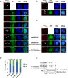
Figure 3. SUMO5 facilities the formation of PML-NBs.
(A) The conjugation site and the SUMO modification motif of SUMO5 are required for the formation and the enlargement of PML-NBs, respectively. GFP-tagged SUMO5, SUMO5(AA), SUMO5(sm), SUMO1, SUMO2, SUMO3, and SUMO4 were transfected into 293 cells. The formation of endogenous PML-NBs (red) was assessed by confocal microscopy using an anti-PML antibody. Arrowheads indicate the positions of small PML-NBs. All of the cells expressing GFP-SUMO5, -SUMO5(sm), -SUMO1, -SUMO2, or -SUMO3 contained NBs, and representative images are shown. None of the cells expressing GFP-SUMO4 contained NBs. 8% of the cells expressing GFP-SUMO5(AA) contained detectable NBs. (B) SUMO5(K18R) forms small yet detectable PML-NBs. 293 cells were transfected with GFP-tagged SUMO5(K18R) and analyzed as in (A). Arrowheads indicate the positions of PML-NBs. (C) SUMO5 restores PML-NBs that had been demolished by SUMO shRNA. 293 cells were transfected with an shRNA construct targeting both SUMO1 and SUMO5 (shSUMO1/5) and then re-transfected with HA-SUMO5 or HA-SUMO1 24 hours later. Restoration of endogenous PML-NBs (red) was assessed by immunofluorescence microscopy with anti-PML antibody and indicated by arrowheads. Expression of SUMO1/5 shRNA was verified by GFP fluorescence. (D) The percentages of GFP-expressing cells from (C) with or without PML-NBs were calculated. “n” represents the total number of cells counted. In untransfected cells, 73% had detectable NBs (NB+). In shRNA-treated (shRNA) cells, 54% were NB+. In SUMO5-rescued cells (shRNA + S5), 82% were NB+. In SUMO1-rescued cells (shRNA+S1), 40% were NB+. (E) Adding back SUMO1 or SUMO5 restores expression after shRNA knockdown. 293 cells were treated as in (C). Whole cell lysates were immunoblotted with anti-HA or anti-γ-tubulin antibodies. Scale bars, 10 μm.
Figure 4
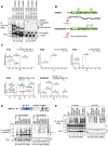
Figure 4. SUMO5 conjugates PML as polymeric chains.
(A) SUMO5 can conjugate PML. GFP-tagged SUMO5 was co-expressed with Flag-tagged PML-I, PML-IV, or PML-VI in 293 cells. Anti-Flag immunoprecipitates were immunoblotted with anti-Flag or anti-GFP antibodies as indicated. (B) SUMO5(I95R), which retains the di-glycine motif (G96G97) after tryptic digest, is used to identify peptide fragments conjugated by SUMO5 in LC-MS/MS analyses. (C) MS/MS confirms that SUMO5 conjugates PML at K160, K380, K400, K490, K497, and K490/K497. Anti-Flag immunoprecipitates from 293 cell lysates expressing Flag-PML-IV and GFP-SUMO5(I95R) were separated by SDS-PAGE and analyzed by LC-MS/MS. (D) SUMO5 conjugates PML at K65, K160, and K490. Flag-tagged SUMO5 was co-expressed with GFP-tagged wild-type or mutant PML. Anti-Flag immunoprecipitates were immunoblotted with anti-GFP or anti-Flag antibodies. PML(3K), mutant PML with lysines 65, 160, and 490 mutated to arginines. Bracket, SUMO-modified PML. Arrowhead, unmodified PML. +1 next to an arrowhead indicates the position of PML conjugated by one SUMO5 molecule. (E) SUMO5 poly-SUMOylates PML. GFP-tagged SUMO5 or mutants were co-expressed with HA-tagged PML in 293 cells. Anti-HA immunoprecipitates were immunoblotted with anti-HA or anti-GFP antibodies as indicated. Bracket, SUMO-modified PML. Arrowhead, unmodified PML.
Figure 5
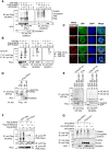
Figure 5. SUMO5 conjugation facilitates interaction among PML-NB components.
(A) SUMO1 enhances SUMO5 conjugation of PML. 293 cells were transfected with GFP-PML, Flag-SUMO5, and increasing amounts of HA-SUMO1 expression plasmids. Anti-Flag immunoprecipitates were immunoblotted with anti-HA, anti-Flag, and anti-GFP antibodies as indicated. (B) SUMO1 and SUMO5 sequentially conjugate PML. 293 cells were transfected with HA-PML, Flag-SUMO5 and GFP-SUMO1 expression plasmids. Serial immunoprecipitation (serial IP) was performed using an anti-Flag antibody first and then anti-HA antibodies. Final immunoprecipitates were immunoblotted with anti-HA, anti-Flag, and anti-GFP antibodies as indicated. * : SUMO5-conjugated PML (PML*S5). ∙ : SUMO1-conjugated PML (PML*S1). ▴: SUMO5- and SUMO1-conjugated PML (PML*S5*S1). (C) SUMO5 enlarges nuclear domains of SP100 and HIPK2. 293 cells were transfected with Flag-tagged SP100 or HIPK2 together with GFP-SUMO5 or GFP vector. The formation of nuclear bodies containing SP100 or HIPK2 was assessed by confocal microscopy. Scale bar, 10 μm. (D,E) SUMO5 conjugates HIPK2 and SP100. Flag-tagged HIPK2 or SP100 were co-expressed with GFP-tagged SUMO5 or SUMO5(K18R). Anti-Flag immunoprecipitates were immunoblotted with anti-GFP or anti-Flag antibodies. HIPK2*S5 indicates SUMO5-conjugated HIPK2. SP100*S5 indicates different species of SP100 reflecting different degrees of SUMO5 conjugation. (F) SUMO5 interacts with PML and Daxx. Interaction of SUMO5 with PML or Daxx was analyzed by co-immunoprecipitation from lysates of 293 cells transfected with plasmids indicated. (G) Daxx and HIPK2 associate with SUMO5-conjugated proteins. 293 cells were transfected with expression plasmids indicated. SUMO-conjugated proteins associated with Daxx and HIPK2 were visualized by co-immunoprecipitation.
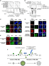
Figure 6
Figure 6. SUMO5 mediates the normal turnover of PML-NBs.
(A) SUMO5 recruits SUMO2. GFP-tagged SUMO2 was transfected into 293 cells alone or with DsRed-tagged SUMO5. PML-NBs were assessed by confocal microscopy. (B) SUMO5 enhances poly-SUMO2/3 conjugation of PML. 293 cells were transfected with HA-PML, Flag-SUMO3, HA-Ubc9, and increasing amounts of GFP-SUMO5 expression plasmids. Immunoprecipitates with anti-Flag antibodies were immunoblotted with anti-HA, anti-Flag, and anti-GFP antibodies as indicated. (C) SUMO3 promotes deconjugation of SUMO5 from PML. 293 cells were transfected with HA-PML, Flag-SUMO5, and increasing amounts of GFP-SUMO3 expression plasmids. Cell lysates were analyzed as in (B). (D) SUMO5 induces disruption of PML-NBs in an RNF4-dependent manner. 293 cells were transfected with combinations of GFP-SUMO5, Flag-RNF4, Flag-RNF4(CS), or vector controls. Images were obtained by confocal microscopy. (E) Ubiquitin is recruited into SUMO5-induced PML-NBs. 293 cells expressing Flag-ubiquitin were cotransfected with GFP-SUMO5 or GFP vector alone. Cell images were obtained by confocal microscopy. (F) RNF4 interacts with SUMO2/3 only. Interaction of RNF4 with different SUMOs was analyzed by co-immunoprecipitation from cell lysates of 293 cells transfected with plasmids indicated. Scale bars, 10 μm.
Figure 7
Figure 7. Competition for SUMO5 conjugation from PML-RARα causes cytoplasmic displacement of PML.
(A) PML-RARα can be conjugated by SUMO5, SUMO1, SUMO2, SUMO3, but not by SUMO4. 293 cells were transfected with plasmids indicated. Anti-HA immunoprecipitates from cell lysates were immunoblotted with anti-HA or anti-GFP antibodies. (B) PML-RARα outcompetes PML for SUMO5 conjugation. 293 cells were transfected with expression plasmids indicated. Cell lysates were immunoblotted with anti-GFP, anti-HA, or anti-Flag antibodies. o, cross-reacting bands of anti-GFP antibody. (C) SUMO5-conjugated PML-RARα moves PML into cytoplasmic aggregates. 293 cells were transfected with GFP-SUMO5 alone (SUMO5 alone) or co-transfected with GFP-SUMO5 and HA-PML-RARα (SUMO5 and PML-RARα). An anti-PML antibody (anti-PML staining) was used in confocal microscopy to obtain cell images. (D) SP100 remains largely localized to PML-null SUMO5 NBs after PML is recruited into the cytoplasm by PML-RARα. 293 cells expressing Flag-SP100 were cotransfected with GFP-SUMO5 (Flag-SP100/GFP-SUMO5), HA-PML-RARα (Flag-SP100/HA-PML-RARα), or GFP-SUMO5 and HA-PML-RARα (Flag-SP100/GFP-SUMO5/HA-PML-RARα). (E) Without SUMO5, PML-RARα fails to form cytoplasmic aggregates. 293 cells were co-transfected with HA-PML-RARα and SUMO5 shRNA targeting endogenous SUMO5 or control shRNA (shNC). The shRNA constructs contained the GFP gene, and green fluorescence indicated expression of shRNAs. Localization of HA-PML-RARα was revealed by anti-HA staining in confocal microscopy. Scale bars, 10 μm. (F) PolySUMO5 conjugation regulates the formation and disruption of PML-NBs. Enlargement of PML-NBs is facilitated by polySUMO5 conjugation of PML, which recruits other proteins into PML-NBs. PolySUMO5 conjugation also promotes polySUMO2/3 conjugation, which replaces SUMO5 conjugation on PML. De-conjugation of SUMO5 and polySUMO2/3 conjugation then initiate RNF4-dependent disruption of PML-NBs.

Similar articles

Cited by

References

    1. Lallemand-Breitenbach V. & de The H. PML nuclear bodies. Cold Spring Harb Perspect Biol 2, a000661 (2010). - PMC - PubMed
    1. Ishov A. M. et al. PML is critical for ND10 formation and recruits the PML-interacting protein daxx to this nuclear structure when modified by SUMO-1. J Cell Biol 147, 221–234 (1999). - PMC - PubMed
    1. D’Orazi G. et al. Homeodomain-interacting protein kinase-2 phosphorylates p53 at Ser 46 and mediates apoptosis. Nat Cell Biol 4, 11–19 (2002). - PubMed
    1. Szostecki C., Guldner H. H., Netter H. J. & Will H. Isolation and characterization of cDNA encoding a human nuclear antigen predominantly recognized by autoantibodies from patients with primary biliary cirrhosis. J Immunol 145, 4338–4347 (1990). - PubMed
    1. Skinner P. J. et al. Ataxin-1 with an expanded glutamine tract alters nuclear matrix-associated structures. Nature 389, 971–974 (1997). - PubMed

Publication types

MeSH terms