. 2016 Jul 1;129(13):2673-83.
doi: 10.1242/jcs.183103. Epub 2016 May 20.

Chromatibody, a novel non-invasive molecular tool to explore and manipulate chromatin in living cells

Affiliations

Chromatibody, a novel non-invasive molecular tool to explore and manipulate chromatin in living cells

Denis Jullien et al. J Cell Sci. .

Abstract

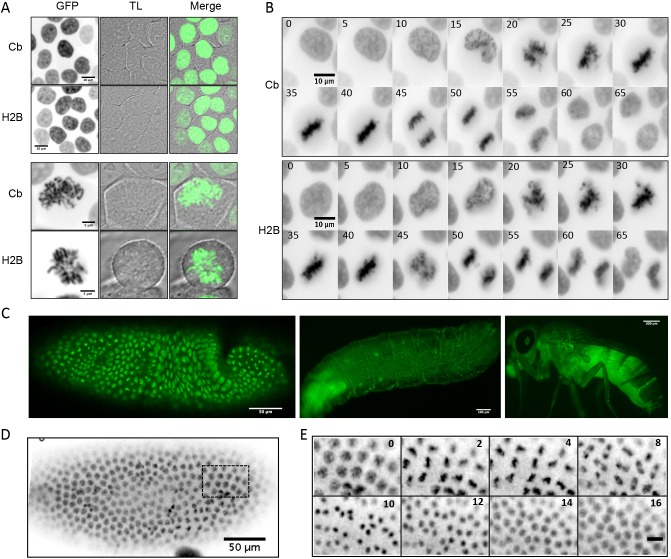
Chromatin function is involved in many cellular processes, its visualization or modification being essential in many developmental or cellular studies. Here, we present the characterization of chromatibody, a chromatin-binding single-domain, and explore its use in living cells. This non-intercalating tool specifically binds the heterodimer of H2A-H2B histones and displays a versatile reactivity, specifically labeling chromatin from yeast to mammals. We show that this genetically encoded probe, when fused to fluorescent proteins, allows non-invasive real-time chromatin imaging. Chromatibody is a dynamic chromatin probe that can be modulated. Finally, chromatibody is an efficient tool to target an enzymatic activity to the nucleosome, such as the DNA damage-dependent H2A ubiquitylation, which can modify this epigenetic mark at the scale of the genome and result in DNA damage signaling and repair defects. Taken together, these results identify chromatibody as a universal non-invasive tool for either in vivo chromatin imaging or to manipulate the chromatin landscape.

Keywords: Chromatin; Chromatin function; Epigenetic; Real-time imaging; Single-domain antibody.


Conflict of interest statement

Competing interests

D.J., J.V., B.S. and G.M. are co-inventors on the patent WO2014202745 A1, concerning the chromatibody discovery and applications. The other authors declare no conflict of interest.

Figures

Fig. 1.
Fig. 1.
Specificity of the chromatibody binding. (A) Purified histones H2A or H2B were transferred after SDS-PAGE. The left panel shows the H2A and H2B histones upon Ponceau staining. The overlay was performed with the purified H2A or H2B histones, followed by the incubation with chromatibody revealed by anti-HA and the HRP-conjugated secondary antibody (right panel). (B) The chromatibody (Cb) binding specificity to purified core histones (H2A, H2B, H3 or H4), the H2A–H2B dimer, the H3–H4 tetramer or nucleosomes (Nucl.) was assessed by ELISA assays. A control VHH was used as a negative control (right panel). Histone concentrations coated on the plates are indicated (antigen [Ag] concentrations are in µg/ml). Three independent experiments were performed (n=3), each point as triplicates. Results are mean±s.e.m. $$$$, P<0.0001 [for the statistical significance between the chromatibody binding to nucleosomes, H2A–H2B or the other antigens indicated (Ag concentration of 10 µg/ml), and compared to control VHH]. (C–E) Modeling of the interaction between chromatibody and the H2A–H2B dimer. (C) Secondary-structure-guided sequence alignment between the anti-cholera toxin VHH structural template (PDB code: 4IDL) and the chromatibody chromatibody. The three complementary determining regions (CDRs) are delineated under the alignment (turquoise, green and red, respectively) and are similarly color-coded in the optimized view of the 3D models. (D) Prediction of the complex between chromatibody and the H2A–H2B dimer. Three acidic residues from the H2A second helix (displayed in CPK mode), E1056 (brown), E1061 (purple) and E1064 (red), form hydrogen-bonds with chromatibody R29 (green), S108 (pink) and R111 (red), respectively. Hydrogen bonds between chromatibody and H2B (marked by black arrowheads, Fig. 1C) strengthen the interaction. (E) Modeled structure resulting from docking the chromatibody CDR3 hairpin into the H2A–H2B acidic cavity at the surface of the nucleosome core.
Fig. 2.
Fig. 2.
Chromatin staining in different model systems. Chromatibody (Cb) allows the immunostaining of chromatin in a wide interspecies system. In the merged images, VHH and DAPI signals are shown in green and red, respectively. Control VHH was used as a negative control. (A) HCT116 cells were immunostained with chromatibody or control VHH, and DNA was labeled with DAPI. The right panels (merge) show both DAPI and VHH stainings. (B) Fluorescence pictures (inverted gray scale) of blastoderm Drosophila embryos immunostained with chromatibody or control VHH. Scale bars: 50 µm. (C) Higher magnification of the stained embryos and overlap between VHH and DAPI (merge). Scale bars: 10 µm. (D) Budding yeast cells stained with chromatibody or control VHH and DAPI. Scale bars: 5 µm.
Fig. 3.
Fig. 3.
Chromatibody–GFP fusion expressed in living cells. (A) Confocal images of HCT116 cells expressing chromatibody (Cb)–GFP or H2B–GFP at interphase (upper panels, scale bars: 10 µm) or mitosis (lower panels, scale bars: 5 µm). Left, middle and right panels show the GFP fluorescence (inverted gray scale), transmitted light (TL) and merged signals, respectively. (B) Time-lapse fluorescence imaging (inverted gray scale) of HCT116 cells stably expressing the chromatibody–GFP or H2B–GFP. The time sequence is indicated in minutes. (C) Confocal imaging of a Drosophila blastoderm (scale bar: 50 µm), living larvae (scale bar: 100 µm) and adult (scale bar: 300 µm) expressing the chromatibody–GFP under the control of the tubulin promoter. (D) Fluorescence image (inverted gray scale) of a living Drosophila embryo expressing chromatibody–GFP. (E) Higher magnification and time-lapse fluorescence microscopy of the embryo shown in D. Scale bar: 10 µm. The time sequence is indicated in minutes.
Fig. 4.
Fig. 4.
Chromatibody dynamic properties. (A) Parental HCT116 cells (NT, not transfected) or HCT116 cells stably expressing chromatibody–GFP (Cb) or the bivalent chromatibody–chromatibody–GFP (Cb-Cb) were subjected to biochemical fractionation. The soluble (s) and crude chromatin (cc) fractions were analyzed by western blotting with antibodies against the HA tag or H2B. (B) Images showing the fluorescence recovery after photobleaching (FRAP) kinetics in the nucleus of HCT116 cells expressing the chromatibody–GFP, chromatibody–chromatibody–GFP, GFP or H2B–GFP. The photobleached areas are indicated by the dashed circle. GFP and H2B–GFP were used as highly mobile and low-mobile controls, respectively. The time sequence is indicated. Scale bars: 5 µm. (C) FRAP curves established from HCT116 cells expressing the GFP constructs mentioned on the right. Mean±s.d. intensities in the photobleached area relative to pre-bleaching intensities are plotted as a function of time after bleaching (n=9). (D) Histogram showing the mean±s.d. recovery half time (t1/2) calculated for chromatibody–GFP and bivalent chromatibody–chromatibody–GFP from the FRAP experiments shown in C.
Fig. 5.
Fig. 5.
Chromatibody-driven DDR alteration. (A) DNA-damage-dependent RNF8 and UbH2A foci. HeLa cells were transfected with the indicated forms of RNF8 24 h prior treatment with calicheamicin (10 pM) and immunostained for ubiquitylated H2A (UbH2A). Cb, chromatibody. Scale bar: 20 µm. (B) RNF8-dependent H2A di-ubiquitylation. HT1080 cells were transfected with the indicated constructs for 24 h. Whole-cell extracts were analyzed with an anti-ubiquitinylated H2A antibody (UbH2A). (C) 53BP1 recruitment to DSBs. HT1080 cells were transfected with the indicated forms of RNF8 for 24 h, treated (10 pM calicheamicin) and immunostained for 53BP1. (D) BRCA1 foci formation. HT1080 cells were transfected with the indicated forms of RNF8 for 24 h and fixed before being immunostained for BRCA1. (E) Accumulation of spontaneous unrepaired DSBs. HT1080 cells were transfected with the indicated constructs for 24 or 42 h and immunostained for γH2AX without any genotoxic treatment. The graphs are mean±s.d. from three independent experiments (n=3) in which at least 100 cells for each category were scored. *P<0.05, **P<0.01, ***P<0.001 (Student's t-test).

Similar articles

Cited by

References

    1. Arbabi Ghahroudi M., Desmyter A., Wyns L., Hamers R. and Muyldermans S. (1997). Selection and identification of single domain antibody fragments from camel heavy-chain antibodies. FEBS Lett. 414, 521-526. 10.1016/S0014-5793(97)01062-4 - DOI - PubMed
    1. Bannister A. J. and Kouzarides T. (2011). Regulation of chromatin by histone modifications. Cell Res. 21, 381-395. 10.1038/cr.2011.22 - DOI - PMC - PubMed
    1. Barbera A. J., Chodaparambil J. V., Kelley-Clarke B., Joukov V., Walter J. C., Luger K. and Kaye K. M. (2006). The nucleosomal surface as a docking station for Kaposi's sarcoma herpesvirus LANA. Science 311, 856-861. 10.1126/science.1120541 - DOI - PubMed
    1. Bhaumik S. R., Smith E. and Shilatifard A. (2007). Covalent modifications of histones during development and disease pathogenesis. Nat. Struct. Mol. Biol. 14, 1008-1016. 10.1038/nsmb1337 - DOI - PubMed
    1. Bischof J., Maeda R. K., Hediger M., Karch F. and Basler K. (2007). An optimized transgenesis system for Drosophila using germ-line-specific phiC31 integrases. Proc. Natl. Acad. Sci. USA 104, 3312-3317. 10.1073/pnas.0611511104 - DOI - PMC - PubMed

Publication types