

doi: 10.1007/s00285-016-1027-z.
Epub 2016 May 20.
Existence of and decay to equilibrium of the filament end density along the leading edge of the lamellipodium
Affiliations
- PMID: 27206776
- PMCID: PMC5206285
- DOI: 10.1007/s00285-016-1027-z
Item in Clipboard
Existence of and decay to equilibrium of the filament end density along the leading edge of the lamellipodium
J Math Biol.
2017 Jan.
Abstract
A model for the dynamics of actin filament ends along the leading edge of the lamellipodium is analyzed. It contains accounts of nucleation by branching, of deactivation by capping, and of lateral flow along the leading edge by polymerization. A nonlinearity arises from a Michaelis-Menten type modeling of the branching process. For branching rates large enough compared to capping rates, the existence and stability of nontrivial steady states is investigated. The main result is exponential convergence to nontrivial steady states, proven by investigating the decay of an appropriate Lyapunov functional.
Keywords: Actin; Lamellipodium; Lyapunov function.
Figures
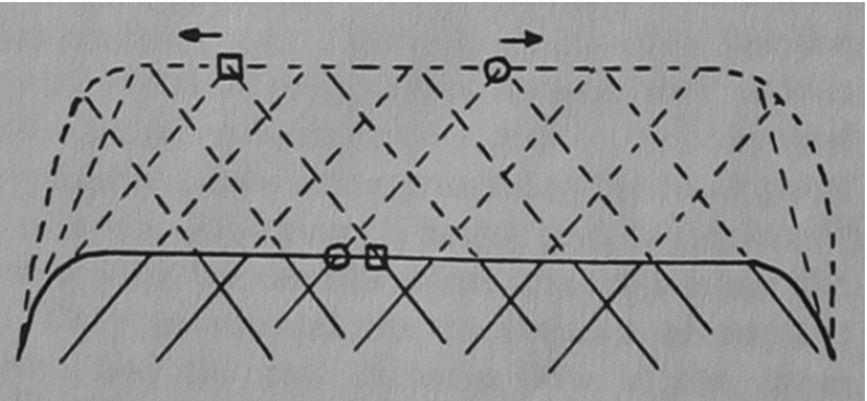
Lateral flow. Solid and dashed lines represent the present and, respectively, a future state of filaments of the leading edge (drawing courtesy of J. Vic Small)
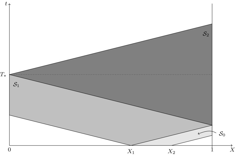
Illustration of the proof of Proposition 2
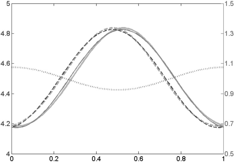
Approximate and numerical solution for and with c(x) given by (26). The end density of the right moving filaments u is depicted in dark and light blue (dashed) and that of the left moving filaments v (solid) in red and orange. Thin lines (red and dark blue) are the asymptotic approximations, thick lines (orange and light blue) are the numerical solution of the time dependent problem after and the left y-axis applies. In green (dotted) the lateral flow c(x) is depicted, the values are in relation to the right y-axis
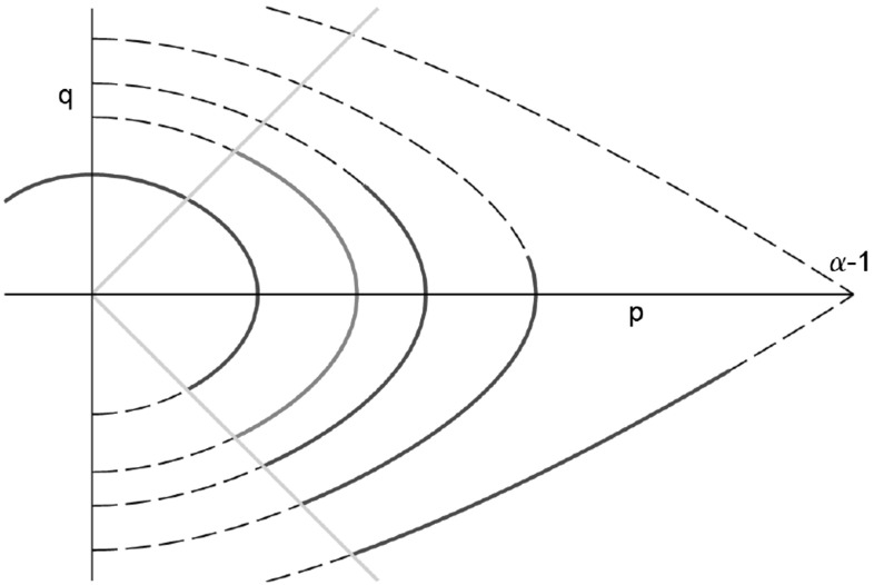
This figure shows the trajectories in (p, q)-phase space of the stationary equation with constant c and (DBC). The orange, diagonal lines represent the boundary conditions at and at . Dashed curves represent (29) for different values of and the blue lines the corresponding solutions curves fulfilling . The red line is desired the solution satisfying both boundary conditions

Density distribution of filaments along the leading edge for Dirichlet boundary conditions as given in Eq. (2) using the equation prior to non-dimensionalization. Red-dotted and blue-dashed lines refer to right and left moving filaments respectively; the black-solid line shows the total filament end density. Parameters (not scaled): , , , ,
Similar articles
-
An extended Filament Based Lamellipodium Model produces various moving cell shapes in the presence of chemotactic signals.J Theor Biol. 2015 Oct 7;382:244-58. doi: 10.1016/j.jtbi.2015.06.044. Epub 2015 Jul 17. J Theor Biol. 2015. PMID: 26192155
-
Model of turnover kinetics in the lamellipodium: implications of slow- and fast- diffusing capping protein and Arp2/3 complex.Phys Biol. 2016 Dec 6;13(6):066009. doi: 10.1088/1478-3975/13/6/066009. Phys Biol. 2016. PMID: 27922825 Free PMC article.
-
Leading edge maintenance in migrating cells is an emergent property of branched actin network growth.Elife. 2022 Mar 11;11:e74389. doi: 10.7554/eLife.74389. Elife. 2022. PMID: 35275060 Free PMC article.
-
Actin dynamics: growth from dendritic branches.Curr Biol. 2005 May 10;15(9):R346-57. doi: 10.1016/j.cub.2005.04.029. Curr Biol. 2005. PMID: 15886095 Review.
-
The comings and goings of actin: coupling protrusion and retraction in cell motility.Curr Opin Cell Biol. 2005 Oct;17(5):517-23. doi: 10.1016/j.ceb.2005.08.004. Curr Opin Cell Biol. 2005. PMID: 16099152 Review.
Cited by
-
Computational estimates of mechanical constraints on cell migration through the extracellular matrix.PLoS Comput Biol. 2020 Aug 27;16(8):e1008160. doi: 10.1371/journal.pcbi.1008160. eCollection 2020 Aug. PLoS Comput Biol. 2020. PMID: 32853248 Free PMC article.
References
-
- Crandall MG, Rabinowitz PH. Bifurcation from simple eigenvalues. J Funct Anal. 1971;8(2):321–340. doi: 10.1016/0022-1236(71)90015-2. - DOI
MeSH terms
LinkOut - more resources
Full Text Sources
Other Literature Sources
