. 2016 Jul;36(7):1328-37.
doi: 10.1161/ATVBAHA.115.306670. Epub 2016 May 19.

Deficiency of ATP-Binding Cassette Transporters A1 and G1 in Endothelial Cells Accelerates Atherosclerosis in Mice

Affiliations

Deficiency of ATP-Binding Cassette Transporters A1 and G1 in Endothelial Cells Accelerates Atherosclerosis in Mice

Marit Westerterp et al. Arterioscler Thromb Vasc Biol. 2016 Jul.

Abstract

Objective: Plasma high-density lipoproteins have several putative antiatherogenic effects, including preservation of endothelial functions. This is thought to be mediated, in part, by the ability of high-density lipoproteins to promote cholesterol efflux from endothelial cells (ECs). The ATP-binding cassette transporters A1 and G1 (ABCA1 and ABCG1) interact with high-density lipoproteins to promote cholesterol efflux from ECs. To determine the impact of endothelial cholesterol efflux pathways on atherogenesis, we prepared mice with endothelium-specific knockout of Abca1 and Abcg1.

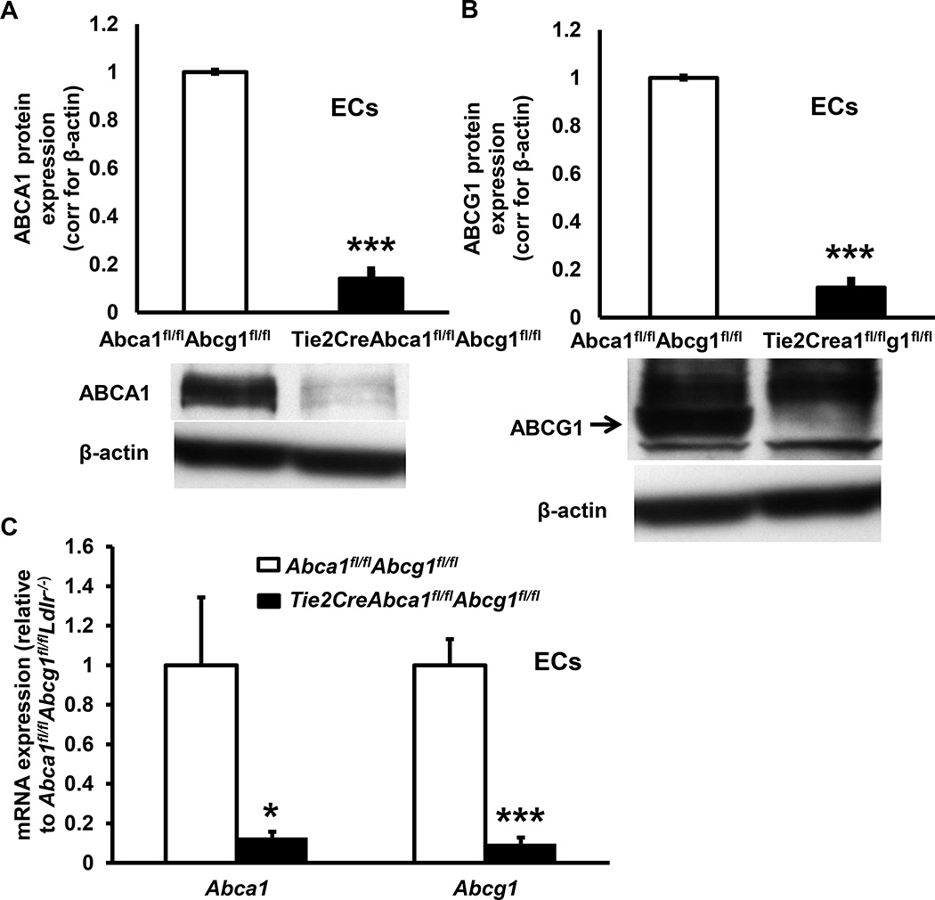
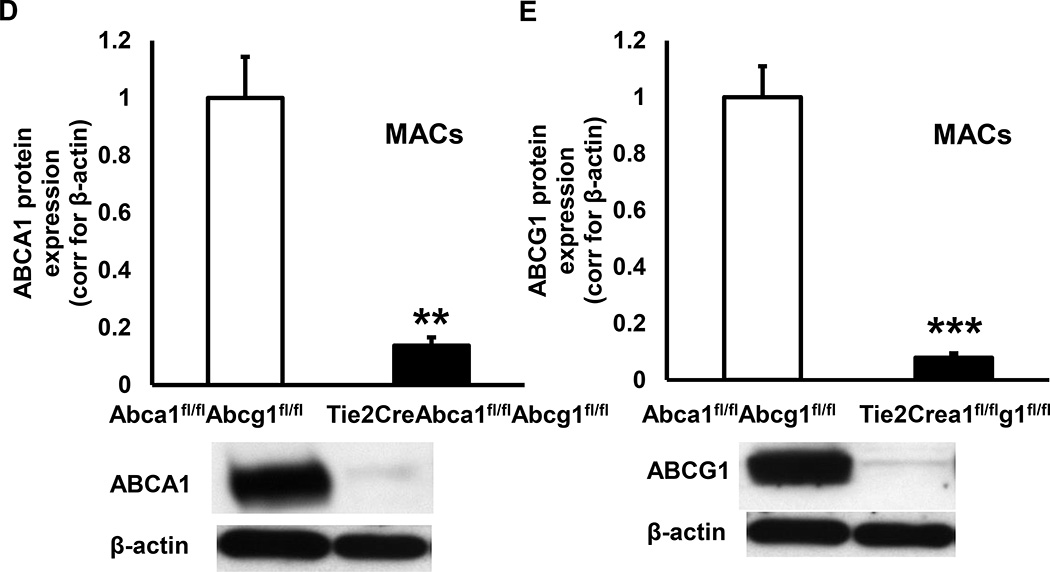
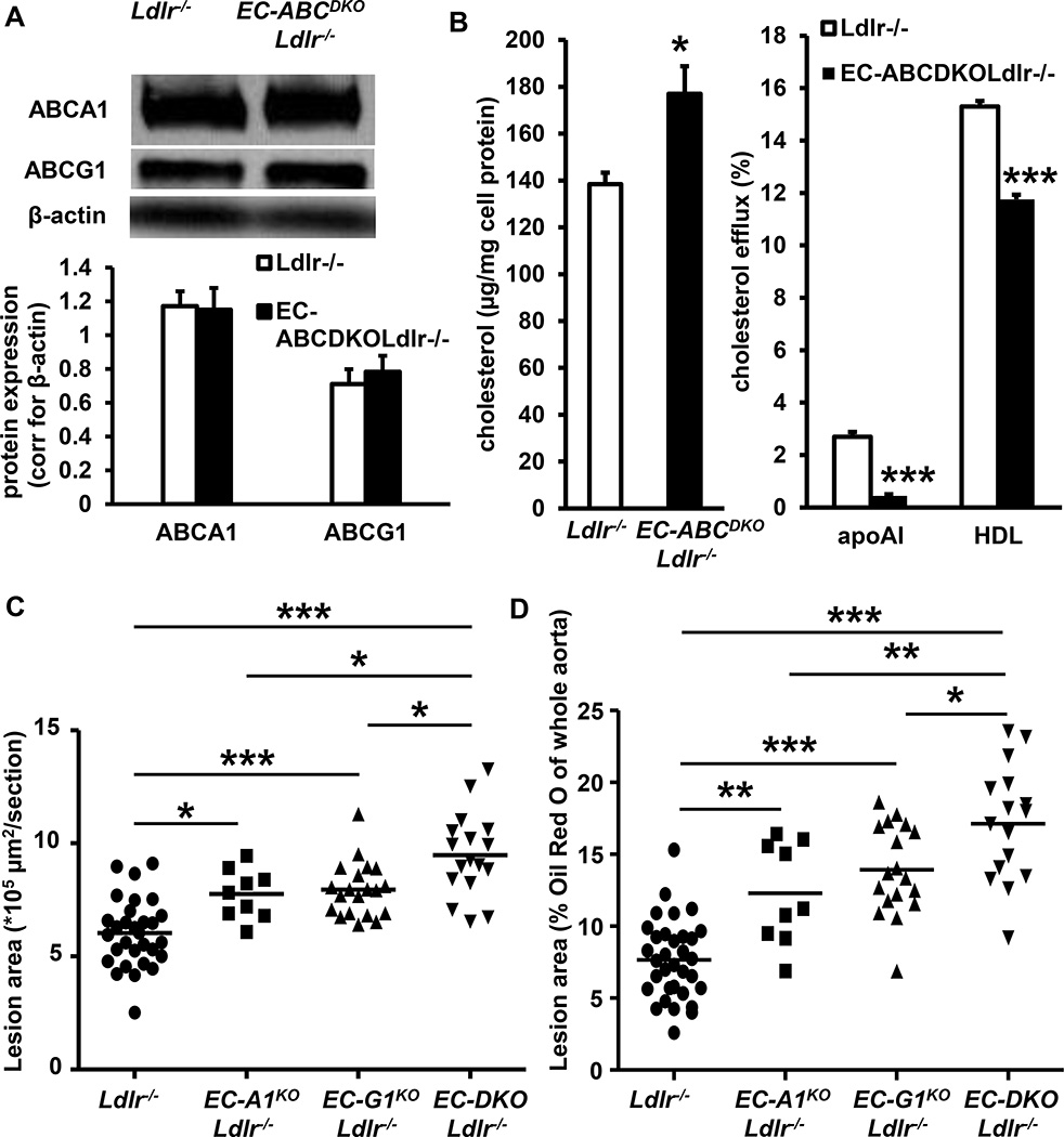
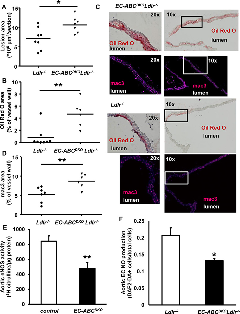
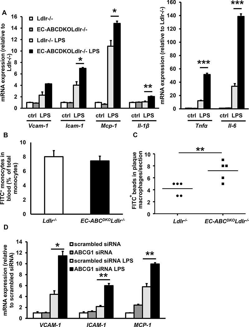
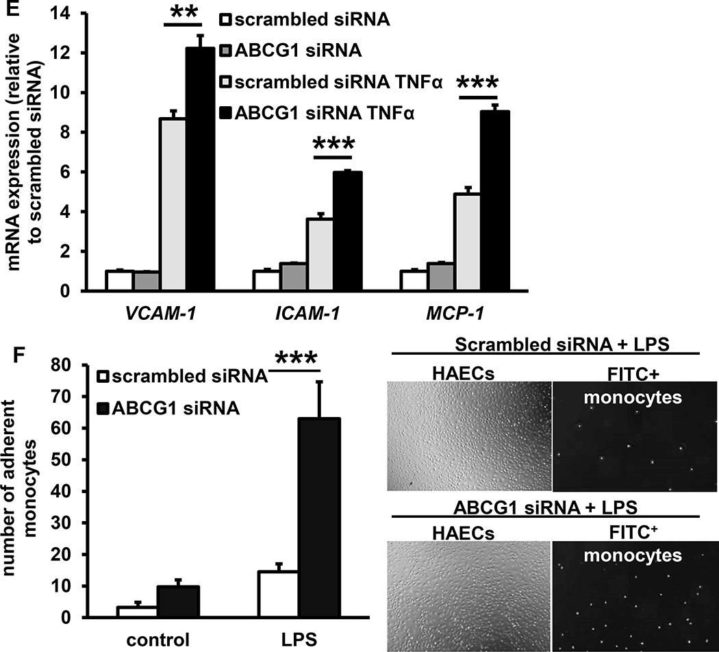
Approach and results: Generation of mice with EC-ABCA1 and ABCG1 deficiency required crossbreeding Abca1(fl/fl)Abcg1(fl/fl)Ldlr(-/-) mice with the Tie2Cre strain, followed by irradiation and transplantation of Abca1(fl/fl)Abcg1(fl/fl) bone marrow to abrogate the effects of macrophage ABCA1 and ABCG1 deficiency induced by Tie2Cre. After 20 to 22 weeks of Western-type diet, both single EC-Abca1 and Abcg1 deficiency increased atherosclerosis in the aortic root and whole aorta. Combined EC-Abca1/g1 deficiency caused a significant further increase in lesion area at both sites. EC-Abca1/g1 deficiency dramatically enhanced macrophage lipid accumulation in the branches of the aorta that are exposed to disturbed blood flow, decreased aortic endothelial NO synthase activity, and increased monocyte infiltration into the atherosclerotic plaque. Abca1/g1 deficiency enhanced lipopolysaccharide-induced inflammatory gene expression in mouse aortic ECs, which was recapitulated by ABCG1 deficiency in human aortic ECs.

Conclusions: These studies provide direct evidence that endothelial cholesterol efflux pathways mediated by ABCA1 and ABCG1 are nonredundant and atheroprotective, reflecting preservation of endothelial NO synthase activity and suppression of endothelial inflammation, especially in regions of disturbed arterial blood flow.

Keywords: ATP-binding cassette transporters; atherosclerosis; endothelium; hypercholesterolemia.


Conflict of interest statement

Disclosures

A.R. Tall is a consultant to Amgen, Arisaph, and CSL. The other authors report no conflicts.

Figures

Figure 1
Figure 1. Tie2CreAbca1fl/flAbcg1fl/fl mice show >90% decreased ABCA1 and ABCG1 protein expression in endothelial cells (ECs) and macrophages (MACs) compared to Abca1fl/flAbcg1fl/fl controls
A and B. Lung ECs were isolated using ICAM-2 positive beads. After two purifications, cells were incubated with the liver X receptor (LXR) ligand T0901317 (3 µM, 24 h), and ABCA1 (A), and ABCG1 (B) protein expression was assessed using Western blot, quantified, and corrected for β-actin. C. Aortic ECs were isolated and Abca1 and Abcg1 mRNA expression determined and corrected for the housekeeping gene m36B4. D and E. Thioglycollate-elicited MACs were isolated, incubated with T0901317 (3 µM, 24 h), and ABCA1 (D), and ABCG1 (E) protein expression was assessed as indicated in A–B. n=5 per genotype. **P<0.01, ***P<0.001, by t-test.
Figure 2
Figure 2. Deficiency of ABCA1 and ABCG1 in ECs increases cholesterol accumulation and accelerates atherosclerosis
A. Tie2CreAbca1fl/flAbcg1fl/flLdlr−/− and Abca1fl/flAbcg1fl/flLdlr−/− mice were transplanted with Abca1fl/flAbcg1fl/fl BM. These mice are referred to as EC-ABCDKOLdlr−/− and Ldlr−/− mice, respectively. Eight weeks after BM transplantation, thioglycollate-elicited macrophages were isolated, and ABCA1 and ABCG1 protein expression was assessed by Western blot, quantified, and corrected for β-actin. n=6 per genotype. B. EC-ABCDKOLdlr−/− and Ldlr−/− mice were fed Western-type diet for 12 weeks, lung ECs were isolated using ICAM-2 coated beads and cholesterol accumulation was assessed using an enzymatic assay. ECs were also loaded with [3H]cholesterol and cholesterol-phospholipid liposomes, and cholesterol efflux to apoAI (25 µg/mL) and HDL (50 µg/mL) was assessed, and corrected for cholesterol efflux to serum albumin. n=4–5 per group. *P<0.05 and ***P<0.001, by t-test. C and D. Ldlr−/−, EC-ABCA1KOLdlr−/−, EC-ABCG1KOLdlr−/−, and EC-ABCDKOLdlr−/− mice were fed Western-type diet (WTD) for 20–22 weeks, and atherosclerosis was assessed after H&E staining at the level of the aortic root (C) or after Oil Red O staining in the whole aorta (D). Each datapoint represents a single mouse. n=9–32. *P<0.05, **P<0.01, and ***P<0.001, by one-way ANOVA.
Figure 3
Figure 3. Deficiency of ABCA1 and ABCG1 in ECs accelerates the initiation of atherosclerosis in the aortic root and branches of the aorta and decreases eNOS activity in the aorta after 12 weeks of WTD and in aortic ECs
EC-ABCDKOLdlr−/− and Ldlr−/− mice were fed the WTD for 12 weeks, and atherosclerosis was assessed after H&E staining at the level of the aortic root. Each datapoint represents a single mouse. n=7–8 (A). Frozen sections of the aortic arch and branch areas were prepared and stained with Oil Red O (B,C) and mac3 (B,D). B. Quantification of Oil Red O positive areas corrected for the total vessel wall area of the aortic section. C. Oil Red O positive areas of representative sections (branch area) and corresponding mac3+ areas are indicated for both genotypes at two magnifications. A square indicates the magnified area in the 10× enlarged pictures. D. Mac3+ area, quantified similar to C. n=6–8. *P<0.05, **P<0.01, by Mann-Whitney. E. EC-ABCDKO and control mice were fed the WTD for 12 weeks. Aortas were collected, and the eNOS activity assay, measuring the conversion of [3H]arginine into [3H]citrulline in aortic lysates, was performed. n=6–8. **P<0.01, by t-test. F. Aortic ECs were isolated from Ldlr−/− and EC-ABCDKOLdlr−/− mice and NO production was determined using the DAF assay. n=6. *P<0.05, by t-test.
Figure 4
Figure 4. Deficiency of ABCA1 and ABCG1 in ECs increases inflammatory gene expression, monocyte adhesion and monocyte infiltration in atherosclerotic plaques
A and B. Deficiency of ABCA1 and ABCG1 increases inflammatory gene expression in mouse ECs (A) and monocyte infiltration in atherosclerotic plaques (B). A. Mouse aortic ECs were isolated from EC-ABCDKOLdlr−/− and Ldlr−/− mice and subsequently incubated with or without LPS (4 h, 10 ng/ml). mRNA expression of the indicated genes was assessed. n=4. B and C. Ldlr−/− and EC-ABCDKOLdlr−/− mice were fed WTD for 12 weeks and injected with FITC+ beads to label monocytes. At 48 h after injection, FITC+ blood monocytes were assessed using flow cytometry (B). Mice were then sacrificed, hearts isolated, frozen sections were made of the aortic root and stained for mac3. FITC+ monocytes were assessed by counting cells positive for FITC+ beads in the mac3+ area of the atherosclerotic plaque. Each datapoint represents an individual mouse (C). n=5. *P<0.05, **P<0.01, ***P<0.001, by t-test. D, E, and F. Deficiency of ABCG1 in human aortic ECs enhances inflammatory gene expression and monocyte adhesion. Human aortic ECs were transfected with scrambled or ABCG1 siRNA, and incubated with or without LPS (4 h, 10 ng/ml) (D) or TNFα (4 h, 0.01 ng/ml) (E). mRNA expression of the indicated genes was assessed. F. Human aortic ECs were treated as described in D, and during the last 30 min of the 4 h LPS treatment, incubated with FITC+ THP-1 monocytes. Non-adherent THP-1 monocytes were washed off, and monocyte adhesion was quantified. n=4–6. *P<0.05, **P<0.01, ***P<0.001, by one-way ANOVA.
Figure 5
Figure 5. Deficiency of ABCA1 and ABCG1 in ECs increases new vessel sprouting in aortic rings, but does not affect retinal angiogenesis
A–B. Rings from the thoracic aorta of Abca1fl/flAbcg1fl/fl control Ldlr−/−, EC-ABCA1KOLdlr−/−, EC-ABCG1KOLdlr−/−, and EC-ABCDKOLdlr−/− mice were isolated, embedded in matrigel, and incubated with endothelial growth cell factors for 5 days. New vessel sprouting was quantified using Image J. A. Representative pictures of new vessel sprouting from aortic rings. B. Quantification of vessel sprouting. n=6. C–D. Control and EC-ABCDKO mice were sacrificed at postnatal day 5. Whole mount retinas were stained for isolectin B4 to visualize ECs. C. Maximal vascular length was calculated as maximal length from the center to the tip of the vessels and corrected for bodyweight (BW). D. Vascular density was quantified using Image J and corrected for BW. *P<0.05, **P<0.01, ***P<0.001, by one-way ANOVA.

Similar articles

Cited by

References

    1. Gordon T, Castelli WP, Hjortland MC, Kannel WB, Dawber TR. High density lipoprotein as a protective factor against coronary heart disease. The framingham study. Am J Med. 1977;62:707–714. - PubMed
    1. Voight BF, Peloso GM, Orho-Melander M, et al. Plasma hdl cholesterol and risk of myocardial infarction: A mendelian randomisation study. Lancet. 2012;380:572–580. - PMC - PubMed
    1. Fond AM, Lee CS, Schulman IG, Kiss RS, Ravichandran KS. Apoptotic cells trigger a membrane-initiated pathway to increase abca1. J Clin Invest. 2015;125:2748–2758. - PMC - PubMed
    1. Murphy AJ, Funt S, Gorman DJ, Tall AR, Wang N. Pegylation of hdl decreases plasma clearance and enhances anti-atherogenic activity. Circ Res. 2013;113:e1–e9. - PMC - PubMed
    1. Plump AS, Scott CJ, Breslow JL. Human apolipoprotein a-i gene expression increases high density lipoprotein and suppresses atherosclerosis in the apolipoprotein e-deficient mouse. Proc Natl Acad Sci USA. 1994;91:9607–9611. - PMC - PubMed

MeSH terms