Review
. 2016 Sep 1;8(9):a019505.
doi: 10.1101/cshperspect.a019505.

Epigenetic Determinants of Cancer

Affiliations
Review

Epigenetic Determinants of Cancer

Stephen B Baylin et al. Cold Spring Harb Perspect Biol. .

Abstract

SUMMARYEpigenetic changes are present in all human cancers and are now known to cooperate with genetic alterations to drive the cancer phenotype. These changes involve DNA methylation, histone modifiers and readers, chromatin remodelers, microRNAs, and other components of chromatin. Cancer genetics and epigenetics are inextricably linked in generating the malignant phenotype; epigenetic changes can cause mutations in genes, and, conversely, mutations are frequently observed in genes that modify the epigenome. Epigenetic therapies, in which the goal is to reverse these changes, are now one standard of care for a preleukemic disorder and form of lymphoma. The application of epigenetic therapies in the treatment of solid tumors is also emerging as a viable therapeutic route.


Figures

Figure 1.
Figure 1.
Genetic mutations of epigenetic modifiers in cancer. The drawing shows the input of epigenetic processes in specifying gene expression patterns. Recent whole-exome sequencing studies show that mutations in various classes of epigenetic modifiers are frequently observed in many types of cancers, further highlighting the cross talk between genetics and epigenetics. Examples of some, but not all, of these mutations are illustrated here and listed in Table 2. The mutations of epigenetic modifiers potentially cause genome-wide epigenetic alterations in cancer, but, save for isocitrate dehydrogenase (IDH) mutations as discussed in the text, these have yet to be shown on a genome-wide scale. Understanding the relationship of genetic and epigenetic changes in cancer will offer novel insights for cancer therapies. MBDs, methylcytosine-binding proteins; PTM, posttranslational modification. (Adapted from You and Jones 2012.)
Figure 2.
Figure 2.
Chromatin structural changes in cancer cells. These two photomicrographs were taken from a patient with a squamous cell carcinoma of the skin. The left panel shows normal epidermal cells within one millimeter of the contiguous tumor shown at the same magnification on the right. The chromatin, which stains purple as a result of its affinity to hematoxylin, appears much more coarse and granular in the cancer cells than in normal epidermis. Such changes in the staining characteristics of chromatin are used by pathologists as diagnostic criteria for cancer.
Figure 3.
Figure 3.
Epigenetic alterations involving DNA methylation can lead to cancer by various mechanisms. Loss of DNA cytosine methylation (white hexagons) illustrated in the hypo column results in genome instability. Focal hypermethylation (pink hexagons) at gene promoters shown in the hyper column causes heritable silencing and, therefore, inactivation of tumor suppressors and other genes. Additionally, methylated CpG sites (pink hexagons) are prone to mutation: They are hot spots for C to T transition mutations caused by spontaneous hydrolytic deamination; or methylation of CpG sites can increase the binding of some chemical carcinogens to DNA; and it increases the rate of UV-induced mutations.
Figure 4.
Figure 4.
Chromatin structural changes in cancer cells. (A) In a typical cell, a CpG-island-containing active gene can be recognized by virtue of a nucleosome-depleted promoter, absence of promoter DNA methylation, but marked by H3K4me3 surrounding the promoter and histone acetylation along the locus. Gene body CpG methylation often can be observed. Nongenic regions flanking an active gene are frequently marked by repressive epigenetic marks, such as H3K9me3 and 5mC. (B) The cancer epigenome is characterized by simultaneous global losses in DNA methylation (gray shading), interspersed with silenced genes that have abnormal gains of DNA methylation and repressive histone modifications in CpG island promoter regions. These silenced genes may be hypomethylated in their gene body, similar to surrounding chromatin. The hypomethylated regions can have an abnormally open nucleosome configuration and acetylated histone lysines. Conversely, abnormal DNA hypermethylation in promoter CpG islands of silenced genes is associated with nucleosomes positioned over the transcription start sites.
Figure 5.
Figure 5.
The position of abnormal DNA methylation in tumor progression. This is depicted in the classic model (Kinzler and Vogelstein 1997) for genetic alterations during the evolution of colon cancer. Altered DNA methylation is shown to occur from very early on in tumorigenesis (red arrow), as discussed in the text, during the conversion of normal to hyperplastic epithelium, accruing during the progression from noninvasive to invasive and, ultimately, metastatic tumors. This places it in a strategic position for channeling stem cells into abnormal clonal expansion (illustrated in Fig. 6) by cooperating with key genetic alterations. These epigenetic abnormalities also have connotations for cancer treatment and markers of prognosis.
Figure 6.
Figure 6.
Epigenetic gene-silencing events and tumorigenesis. The earliest steps in tumorigenesis are depicted as abnormal clonal expansion, which evolves during the stress of cell renewal. This is caused by factors, such as aging and chronic injury from, for example, inflammation. These cell clones are those at risk of subsequent genetic and epigenetic events that could drive tumor progression. Abnormal epigenetic events, such as the aberrant gene silencing focused on in this article, could be the earliest heritable causes, in many instances, for a potential role in inducing the abnormal clonal expansion from within stem/progenitor cell compartments in a renewing adult cell system. The gene silencing is triggered by chromatin modifications that repress transcription, and the DNA hypermethylation of this chromatin serves as the tight lock to stabilize the heritable silencing. The gene silencing, in turn, disrupts normal homeostasis, preventing stem and progenitor cells from moving properly along the differentiation pathway for a given epithelial cell system (blue arrow) and channels them into the abnormal clonal expansion (red arrow).
Figure 7.
Figure 7.
Reprogramming of DNA methylation patterns and abnormal modes of gene silencing in cancer. (A) Common DNA methylome changes observable in cancer versus a normal somatic cell are illustrated. This is shown in the context of large hypomethylated blocks (gray shading) of the genome seen in cancer interspersed with focal hypermethylation of promoter region CpG-island-containing genes (pink shading). In normal cells, background DNA methylation is high (pink shaded hexagons) with the exception of CpG islands (densely packed white shaded hexagons). In the cancer methylome, overall genome DNA methylation declines, particularly in the hypomethylated blocks, whereas CpG island promoter genes frequently become methylated (pink shading), most of which are located in the hypomethylated blocks. (B) The currently suggested routes to abnormally silenced CpG-island-containing genes in cancer are shown. Genes that are active in cells throughout development and adult cell renewal initially have active promoter chromatin, which is characterized by the presence of the bivalent histone modification pattern consisting of H3K4me, the repressive H3K27me3 mark, and a lack of DNA methylation. Genes that become transcriptionally active lose much of their Polycomb-mediated repressive H3K27 methylation, whereas those that become silenced (indicated by a red X) can do so by the loss of H3K4 methylation and acquisition of, or increases in, Polycomb-mediated repressive chromatin (PRC) mark and H2A119 ubiquitination. During tumor progression, active genes may become silenced through either the aberrant PRC-mediated reprogramming (bottom left) or DNA methylation and H3K9me marks (bottom right). Some normally silent genes may change the way in which they are transcriptionally repressed from H3K27-methylation-type repression to H3K9-methylation-based silencing and/or DNA hypermethylation (epigenetic switching). The reverse yellow arrows indicate the potential for epigenetic abnormalities in cancer to be corrected by epigenetic therapies. Representative of such therapies are DNMT inhibitors, HDAC inhibitors, KMT inhibitors, and others, as discussed in this and other articles. These inhibitors can all potentially promote gene activation by producing losses of DNA methylation, or deacetylating lysines, or alleviating silencing mediated by histone methylation PTMs, such as H3K27 methylation. (A, Adapted from Reddington et al. 2014; B, adapted from Sharma et al. 2010.)
Figure 8.
Figure 8.
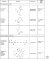
Structures of selected epigenetic drugs. Three nucleoside analogs are known that can inhibit DNA methylation after incorporation into DNA. 5-aza-CR (Vidaza) and 5-aza-CdR (decitabine) have been FDA approved for the treatment of the preleukemic disorder, myelodysplasia. Two HDAC inhibitors are also FDA approved for cutaneous T-cell lymphoma and several others are in clinical trials. Drugs targeting other epigenetic processes are in earlier stages of clinical development (see also Figs. 5 and 6 of Audia and Campbell 2014).

Similar articles

Cited by

References

    1. Ahuja N, Easwaran H, Baylin SB. 2014. Harnessing the potential of epigenetic therapy to target solid tumors. J Clin Invest 124: 56–63. - PMC - PubMed
    1. Allis CD, Jenuwein T, Reinberg D. 2014. Overview and concepts. Cold Spring Harb Perspect Biol 10.1101/cshperspect.a018739. - DOI
    1. Almouzni G, Cedar H. 2014. Maintenance of epigenetic information. Cold Spring Harb Perspect Biol 10.1101/cshperspect.a019372. - DOI - PMC - PubMed
    1. Amann JM, Nip J, Strom DK, Lutterbach B, Harada H, Lenny N, Downing JR, Meyers S, Hiebert SW. 2001. ETO, a target of t(8;21) in acute leukemia, makes distinct contacts with multiple histone deacetylases and binds mSin3A through its oligomerization domain. Mol Cell Biol 21: 6470–6483. - PMC - PubMed
    1. Araki S, Doi H, Sano Y, Tanaka S, Miyake Y. 2009. Preparation and CO(2) adsorption properties of aminopropyl-functionalized mesoporous silica microspheres. J Colloid Interface Sci 339: 382–389. - PubMed