Ageing and the pathogenesis of osteoarthritis
- PMID: 27192932
- PMCID: PMC4938009
- DOI: 10.1038/nrrheum.2016.65
Ageing and the pathogenesis of osteoarthritis
Abstract
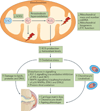
Ageing-associated changes that affect articular tissues promote the development of osteoarthritis (OA). Although ageing and OA are closely linked, they are independent processes. Several potential mechanisms by which ageing contributes to OA have been elucidated. This Review focuses on the contributions of the following factors: age-related inflammation (also referred to as 'inflammaging'); cellular senescence (including the senescence-associated secretory phenotype (SASP)); mitochondrial dysfunction and oxidative stress; dysfunction in energy metabolism due to reduced activity of 5'-AMP-activated protein kinase (AMPK), which is associated with reduced autophagy; and alterations in cell signalling due to age-related changes in the extracellular matrix. These various processes contribute to the development of OA by promoting a proinflammatory, catabolic state accompanied by increased susceptibility to cell death that together lead to increased joint tissue destruction and defective repair of damaged matrix. The majority of studies to date have focused on articular cartilage, and it will be important to determine whether similar mechanisms occur in other joint tissues. Improved understanding of ageing-related mechanisms that promote OA could lead to the discovery of new targets for therapies that aim to slow or stop the progression of this chronic and disabling condition.
Figures


Similar articles
-
Targeting of chondrocyte plasticity via connexin43 modulation attenuates cellular senescence and fosters a pro-regenerative environment in osteoarthritis.Cell Death Dis. 2018 Dec 5;9(12):1166. doi: 10.1038/s41419-018-1225-2. Cell Death Dis. 2018. PMID: 30518918 Free PMC article.
-
Cartilage regeneration and ageing: Targeting cellular plasticity in osteoarthritis.Ageing Res Rev. 2018 Mar;42:56-71. doi: 10.1016/j.arr.2017.12.006. Epub 2017 Dec 16. Ageing Res Rev. 2018. PMID: 29258883 Review.
-
Fibrates as drugs with senolytic and autophagic activity for osteoarthritis therapy.EBioMedicine. 2019 Jul;45:588-605. doi: 10.1016/j.ebiom.2019.06.049. Epub 2019 Jul 5. EBioMedicine. 2019. PMID: 31285188 Free PMC article.
-
Mechanisms and therapeutic implications of cellular senescence in osteoarthritis.Nat Rev Rheumatol. 2021 Jan;17(1):47-57. doi: 10.1038/s41584-020-00533-7. Epub 2020 Nov 18. Nat Rev Rheumatol. 2021. PMID: 33208917 Free PMC article. Review.
-
Aging-related inflammation in osteoarthritis.Osteoarthritis Cartilage. 2015 Nov;23(11):1966-71. doi: 10.1016/j.joca.2015.01.008. Osteoarthritis Cartilage. 2015. PMID: 26521742 Free PMC article. Review.
Cited by
-
Potential Methods of Targeting Cellular Aging Hallmarks to Reverse Osteoarthritic Phenotype of Chondrocytes.Biology (Basel). 2022 Jun 30;11(7):996. doi: 10.3390/biology11070996. Biology (Basel). 2022. PMID: 36101377 Free PMC article. Review.
-
Adherence to a Mediterranean diet is associated with lower prevalence of osteoarthritis: Data from the osteoarthritis initiative.Clin Nutr. 2017 Dec;36(6):1609-1614. doi: 10.1016/j.clnu.2016.09.035. Epub 2016 Oct 8. Clin Nutr. 2017. PMID: 27769781 Free PMC article.
-
Inflammatory signaling sensitizes Piezo1 mechanotransduction in articular chondrocytes as a pathogenic feed-forward mechanism in osteoarthritis.Proc Natl Acad Sci U S A. 2021 Mar 30;118(13):e2001611118. doi: 10.1073/pnas.2001611118. Proc Natl Acad Sci U S A. 2021. PMID: 33758095 Free PMC article.
-
Transcriptomic changes during the replicative senescence of human articular chondrocytes.bioRxiv [Preprint]. 2023 Nov 7:2023.11.07.565835. doi: 10.1101/2023.11.07.565835. bioRxiv. 2023. Update in: Int J Mol Sci. 2024 Nov 12;25(22):12130. doi: 10.3390/ijms252212130 PMID: 37986862 Free PMC article. Updated. Preprint.
-
Integrating transcriptome-wide association study and mRNA expression profile identified candidate genes related to hand osteoarthritis.Arthritis Res Ther. 2021 Mar 10;23(1):81. doi: 10.1186/s13075-021-02458-2. Arthritis Res Ther. 2021. PMID: 33691763 Free PMC article.
References
Publication types
MeSH terms
Substances
Grants and funding
LinkOut - more resources
Full Text Sources
Other Literature Sources
Medical

