Review
. 2015 Aug 27:1:15021.
doi: 10.1038/nrdp.2015.21.

Retinoblastoma

Affiliations
Review

Retinoblastoma

Helen Dimaras et al. Nat Rev Dis Primers. .

Abstract

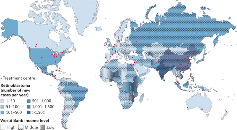
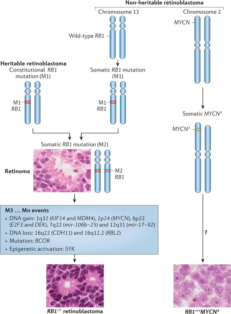
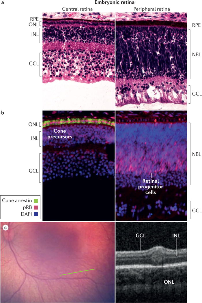
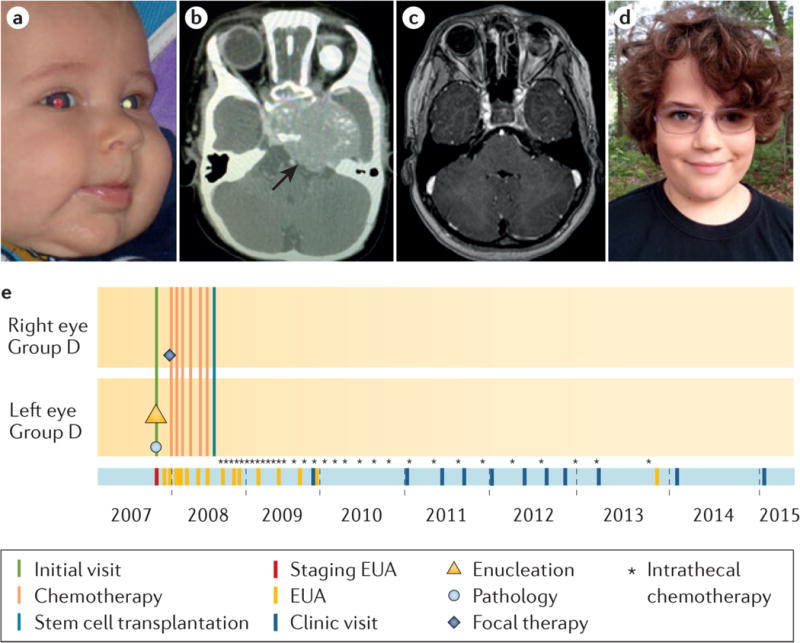
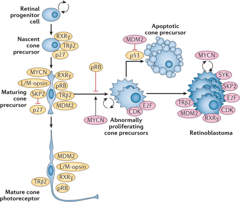
Retinoblastoma is a rare cancer of the infant retina that is diagnosed in approximately 8,000 children each year worldwide. It forms when both retinoblastoma gene (RB1) alleles are mutated in a susceptible retinal cell, probably a cone photoreceptor precursor. Loss of the tumour-suppressive functions of the retinoblastoma protein (pRB) leads to uncontrolled cell division and recurrent genomic changes during tumour progression. Although pRB is expressed in almost all tissues, cone precursors have biochemical and molecular features that may sensitize them to RB1 loss and enable tumorigenesis. Patient survival is >95% in high-income countries but <30% globally. However, outcomes are improving owing to increased disease awareness for earlier diagnosis, application of new guidelines and sharing of expertise. Intra-arterial and intravitreal chemotherapy have emerged as promising methods to salvage eyes that with conventional treatment might have been lost. Ongoing international collaborations will replace the multiple different classifications of eye involvement with standardized definitions to consistently assess the eligibility, efficacy and safety of treatment options. Life-long follow-up is warranted, as survivors of heritable retinoblastoma are at risk for developing second cancers. Defining the molecular consequences of RB1 loss in diverse tissues may open new avenues for treatment and prevention of retinoblastoma, as well as second cancers, in patients with germline RB1 mutations.


Conflict of interest statement

Competing interests

There is NO competing interest.

Figures

Figure 1
Figure 1. Progression of retinoblastoma
a) Anatomical features of a healthy eye. Genomic damage (orange lightning bolt) leads to mutation of RB1 resulting in biallelic functional loss of RB1 in a developing retinal cell (possibly a cone photoreceptor precursor cell that is dependent on pRB to stop proliferation). b) Genomic instability leads to a benign retinoma; only 5% of patients show retinoma without retinoblastoma. Inset shows a small retinoma/tumour not visible except by optical coherence tomography. c) Intraretinal retinoblastoma arises as additional genomic changes promote uncontrolled cell proliferation; the tumour grows and seeds become independent, floating under the retina and into vitreous. d) Retinoblastoma can invade adjacent tissues: into the optic nerve, uvea, or sclera to constitute high-risk pathologic features. e) Eventually, retinoblastoma can extend extraocularly into orbit and metastasize especially to the bone marrow, or into the brain (direct or via the cerebrospinal fluid).
Figure 2
Figure 2. Global retinoblastoma treatment centres and patient distribution relative to resources
One Retinoblastoma World (www.1rbw.org) shows Retinoblastoma Centres of Excellence, providing a resource for affected families to access expert care. The majority of patients reside in low and middle income countries, while the majority of Retinoblastoma Centres are in high income countries. Images from www.1rbw.org.
Figure 3
Figure 3. Genetic origins of retinoblastoma
Three genetic subtypes of retinoblastoma are known. Heritable retinoblastoma patients have a constitutive inactivating mutation (M1) in the RB1tumor suppressor gene in all cells of their body. A second, somatic mutation (M2) in a susceptible retinal cell can lead to benign retinoma. Further genetic and/or epigenetic events (M3…Mn) are required to transform to retinoblastoma. Non-heritable, RB1−/− retinoblastomas progress similarly, except both M1 and M2 occur in one susceptible retinal cell. RB1+/+MYCN-amplified (RB1+/+MYCNA) retinoblastoma is a rare, non-heritable retinoblastoma subtype driven by amplification of MYCN with normal RB1; other changes in these tumours remain uncharacterized. Retinoma histology shows distinct photoreceptor-like fleurettes, whereas RB1−/− retinoblastoma can show Flexner-Wintersteiner (insert) and Homer Wright rosettes (not shown). RB1+/+MYCNA retinoblastoma have a distinct morphology with rounded nuclei and prominent nucleoli related to the high MYCN protein.
Figure 4
Figure 4. Retinoblastomas originate in the retina
a, The retina has a complex structure and contains multiple cell types. Haematoxylin and eosin staining of post-fertilization week 19 retina shows three post-mitotic nuclear layers in the central retina near the fovea (left) and two nuclear layers in the less mature periphery of the same histologic section (right). Cell types in each layer are indicated. b, Cone precursors normally have high expression of the RB protein (pRB). Immunofluorescence staining of post-fertilization week 19 retina shows especially strong pRB signal (pink) in maturing cone precursors in the central retina (left, counterstained for cone arrestin, green) and in retinal progenitor cells in the peripheral retina of the same histologic section (right). pRB staining is less intense in DAPI-stained nuclei (blue) in other retinal cell types. c, Wide-angle retinal image (left image) shows no tumor. In the plane of the green line, optical coherence tomography (OCT; right image) shows a tiny intra-retinal tumor in a 2.5 month old infant, which seems to extend from the inner nuclear layer to the outer nuclear layer. The tumour has an uncertain epicentre making it difficult to infer the retinal layer from which the tumor arose. The layer-of-origin may be defined in the future with more images of higher resolution OCT of retina in very young children carrying an inherited germline RB1 mutation. Abbreviations: ONL, outer nuclear layer; INL, inner nuclear layer; GCL, ganglion cell layer; NBL, neuroblastic layer; RPE, retinal pigment epithelium.
Figure 5
Figure 5. Online diagnosis of retinoblastoma
a, Detection of photoleukocoria (white pupil) on this digital image led the parents to the diagnosis of retinoblastoma. The left eye was determined by examination under anaesthetic to have IIRC Group D retinoblastoma and was enucleated two days later; CT scan was scheduled to check for trilateral tumour. (Today, MRI would be recommended to reduce radiation exposure.) The pathologic examination of the eye showed no high-risk features. The other eye was normal at diagnosis but on the next examination a small tumour was detected and treated with only laser. b, One month after surgery, the child presented with high intracranial pressure. A large intracranial midline tumour (trilateral disease) was diagnosed on CT scan, which was treated with chemotherapy, high-dose chemotherapy with hematopoietic rescue by stem cell transplant, and intrathecal chemotherapy injections (directly into the cerebro-spinal fluid) through an implanted intraventricular catheter. c, Follow-up MRI at age 8 years shows no residual disease and d, the child is well with one good-looking artificial eye and one eye with normal vision; he wears poly-carbonate lenses to protect his only eye. e, The eCCRB timeline of treatments; not all intrathecal chemotherapy injections (*) are shown.
Figure 6
Figure 6. Different classification schemes for intraocular retinoblastoma confound comparison of outcomes
The features listed determine the overall classification, ranging from small tumours not threatening vision (“Group A”) to tumours clinically noted to have features suggesting potential spread outside the eye (“Group E”). Most importantly, size of tumour alone does not make an eye dangerous by Murphree, Children’s Oncology Group (COG), or TNM classification; but any eye with tumour >50% of eye volume is E (advanced-stage disease) by Shields classification. The consequence is widespread confusion in the literature undermining clinical research, since studies using the different classifications cannot be compared. The red boxes indicate the critical differences between the different classifications.
Figure 7
Figure 7. Primary treatment choices based on the Murphree IIRC
Treatment depends on the combined severity of each of the affected eyes (Eye 1 | Eye 2); the preferred option for each eye is depicted in the blue boxes. IIRC Group A eyes can be treated with only laser or cryotherapy (focal therapy consolidation). Group B and C eyes require several cycles of systemic intravenous chemotherapy (IVC) or intra-arterial chemotherapy (IAC) followed by focal therapy and intravitreal chemotherapy with melphalan and/or topotecan for residual or recurrent vitreous seeds. Isolated single tumours in Group B or C eyes may occasionally be appropriate for primary radioactive plaque therapy. Group D eyes require either IVC (with focal therapy consolidation) or IAC (with focal therapy consolidation). All eyes with features suggesting imminent extraocular extension (Group E) should be removed so that accurate pathological examination can be performed to determine risk of metastasis requiring adjuvant chemotherapy. IIRC, International Intraocular Retinoblastoma Classification.
Figure 8
Figure 8. Triplets with retinoblastoma
These triplets developed retinoblastomas in all six eyes illustrating the full expressivity and penetrance of RB1−/− germline mutations that completely abolish pRB expression. The pedigree indicates that the three children all have the same RB1 mutation, which is not detected in their parents or older brother. The eCCRB timelines reveal that all eyes had individualized therapy, with choices considering the overall impact on each child. Each child lost one eye, has a good-looking artificial eye and an eye with normal vision, and has disease control in less than one year. The number of eye exams under anaesthesia (EUA) was least for the child who had primary enucleation. OD, right eye; OS, left eye. Eye involvement at diagnosis is indicated in by the International Intraocular Retinoblastoma Classification; the eye labelled “Group 0” had no tumour at initial diagnosis but developed a tumour 6 weeks later.
Figure 9
Figure 9. Retinoblastoma treated with IVC
a, Detection of photoleukocoria in both eyes at age 8 months. b, Retinal photography at diagnosis showed total retinal detachment with underlying large tumours in the right (left image) and left eyes (right image). c, Both eyes have calcified tumor remnants and attached retinas 20 years after diagnosis, with 0.25 vision, right eye and 0.1 vision, left eye (Vision decimal system, 1 = normal, 0.1=legal blindness). d, eCCRB time line of shows treatment with IVC and focal therapy, then only follow-up visits.
Figure 10
Figure 10. Retinoblastoma treated with IAC
a, Before IAC for a massive exophytic (outward growing tumour) retinoblastoma with total retinal detachment; and b, after IAC, showing complete response and eye salvage, leaving a 6 mm calcified scar. c, Before IAC for an endophytic (inward growing tumour) retinoblastoma tumour with extensive vitreous seeding; and d, after IAC showing complete control with clinically intact fovea and visual acuity 20/25. e, Before IAC for an eye with macular retinoblastoma demonstrating excellent tumor regression away from the macula; and f, after IAC leaving intact fovea and hope for vision.
Figure 11
Figure 11. Child Life promotes effective coping through play, preparation, education, positive-touch and self-expression activities, based on natural child development
Child Life helps children and their families cope with challenging healthcare issues, hospitalization and therapeutic interventions. This child plays doctor and nurse, wearing gloves and stethoscopes to examine Puppet Kevin who had one eye removed for retinoblastoma. They also learn how to put in his artificial eye, to better understand their own artificial eye. Play (medical play and just general play) promotes a sense of control for children within their medical experience.
Figure 12
Figure 12. Proposed roles for cone precursor circuitry in retinoblastoma tumourigenesis and targeted therapy
Signalling circuitry specific to maturing L/M-cone precursors is uniquely conducive to oncogenic transformation in response to pRB loss.,, Specifically, these cone precursors express the transcription factors RXRγ and thyroid hormone receptor (TR) β2, and high levels of MYCN, MDM2, and pRB, and down regulate p27 via a SKP2-related mechanism. pRB depletion in maturing cone precursors blocks cone photoreceptor differentiation and enables proliferation and survival dependent upon MYCN, SKP2, and MDM2. Mouse models suggest that E2Fs and CDKs contribute to the proliferative response to pRB loss; these genes/proteins are also needed for proliferation and survival of human retinoblastoma cells., Identifying the molecular mechanism mediating the cone precursor proliferative response to pRB loss provides opportunities to prevent tumor initiation, at least in preclinical models. Potential drugs are inhibitors of MYCN expression (JQ1), inhibitors of MDM2-mediated p53 degradation (Nutlin 3a), inhibitors of SKP2-mediated p27 degradation (C25), and inhibitors of E2F and CDK activities (HLM006474 and R547, respectively). In addition, SYK was highly expressed in retinoblastoma but not normal retina. Additional targeted agents may be effective against growing tumours, such as improved versions of the SYK inhibitor R406.,

Similar articles

Cited by

References

    1. Knudson AG. Two genetic hits (more or less) to cancer. Nat Rev Cancer. 2001;1:157–162. - PubMed
    1. Dimaras H, et al. Loss of RB1 induces non-proliferative retinoma: increasing genomic instability correlates with progression to retinoblastoma. Hum Mol Genet. 2008;17:1363–1372. - PubMed
    1. Dimaras H, et al. Retinoblastoma. Lancet. 2012;379:1436–1446. doi: 10.1016/S0140-6736(11)61137-9. - DOI - PubMed
    1. Seregard S, Lundell G, Svedberg H, Kivela T. Incidence of retinoblastoma from 1958 to 1998 in Northern Europe: advantages of birth cohort analysis. Ophthalmology. 2004;111:1228–1232. doi: 10.1016/j.ophtha.2003.10.023. - DOI - PubMed
    1. Kivela T. The epidemiological challenge of the most frequent eye cancer: retinoblastoma, an issue of birth and death. Br J Ophthalmol. 2009;93:1129–1131. doi:93/9/1129 [pii]10.1136/bjo.2008.150292. - PubMed

Publication types

Substances

LinkOut - more resources