. 2016 May 23;213(4):415-24.
doi: 10.1083/jcb.201512034. Epub 2016 May 16.

Diaphanous formin mDia2 regulates CENP-A levels at centromeres

Affiliations

Diaphanous formin mDia2 regulates CENP-A levels at centromeres

Chenshu Liu et al. J Cell Biol. .

Abstract

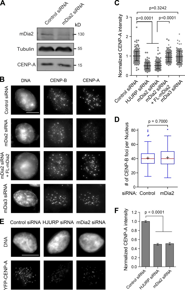
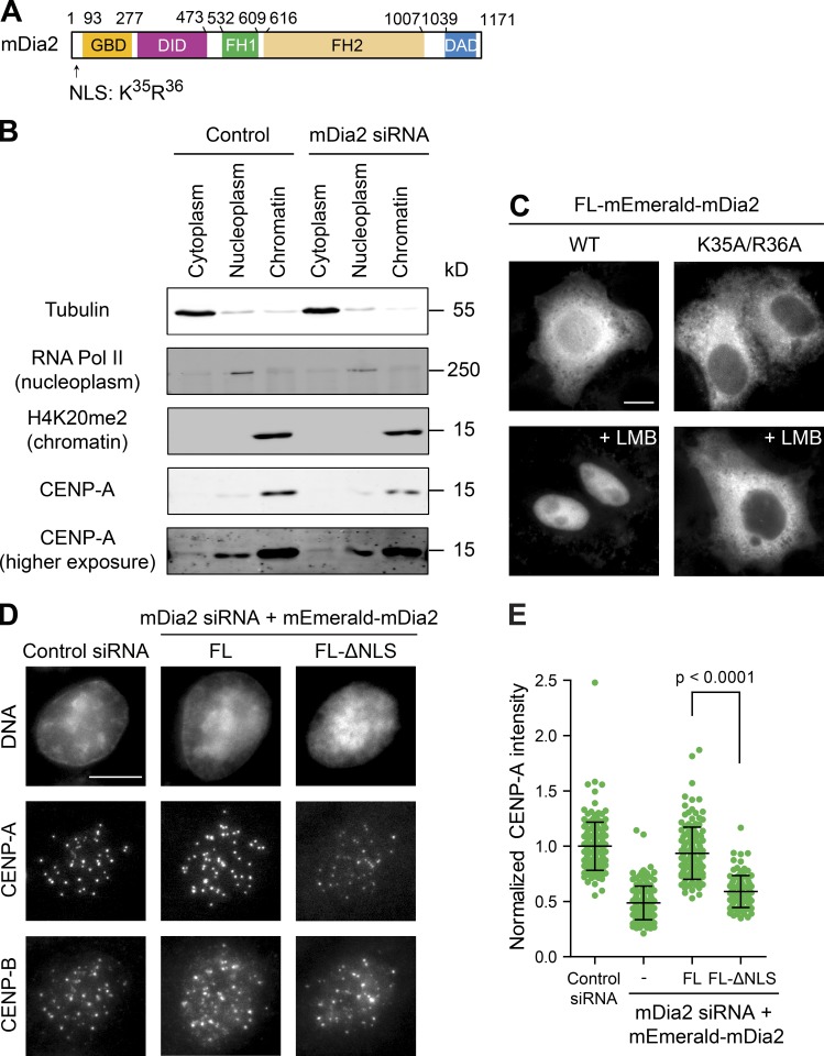
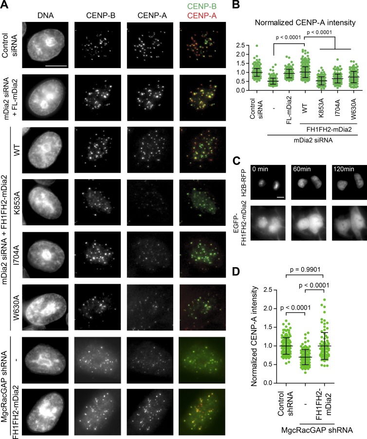
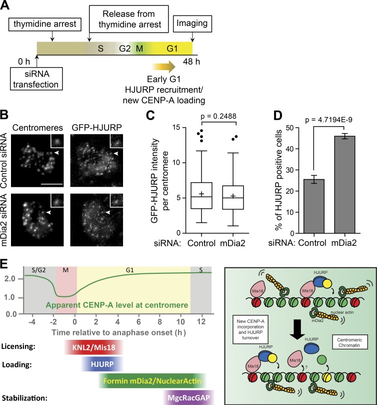
Centromeres of higher eukaryotes are epigenetically defined by centromere protein A (CENP-A), a centromere-specific histone H3 variant. The incorporation of new CENP-A into centromeres to maintain the epigenetic marker after genome replication in S phase occurs in G1 phase; however, how new CENP-A is loaded and stabilized remains poorly understood. Here, we identify the formin mDia2 as essential for stable replenishment of new CENP-A at centromeres. Quantitative imaging, pulse-chase analysis, and high-resolution ratiometric live-cell studies demonstrate that mDia2 and its nuclear localization are required to maintain CENP-A levels at centromeres. Depletion of mDia2 results in a prolonged centromere association of holiday junction recognition protein (HJURP), the chaperone required for CENP-A loading. A constitutively active form of mDia2 rescues the defect in new CENP-A loading caused by depletion of male germ cell Rac GTPase-activating protein (MgcRacGAP), a component of the small GTPase pathway essential for CENP-A maintenance. Thus, the formin mDia2 functions downstream of the MgcRacGAP-dependent pathway in regulating assembly of new CENP-A containing nucleosomes at centromeres.


Figures

Figure 1.
Figure 1.
The formin mDia2 is required to maintain CENP-A levels at centromeres. (A) Depletion of mDia2 does not affect CENP-A protein level. Immunoblotting analysis of HeLa cell lysates 48 h after transfection with control (GAPDH) and mDia2 siRNAs. (B) Immunofluorescence detection of CENP-A and CENP-B 48 h after transfection with the indicated siRNAs and the full-length mEmerald-tagged mDia2 (FL-mDia2) expression vector (DNA - DAPI). Transfected cells were identified by cotransfected fluorescence markers. Bar, 10 µm. (C) Scatterplot showing the distribution of normalized CENP-A integrated intensity per nucleus (mean ± SD overlaid with scatterplot) with the INCA method (Fig. S1). Control: n = 292; HJURP siRNA: n = 196; mDia2 siRNA: n = 260; mDia2 siRNA + FL-mDia2: n = 136; and mDia3 siRNA: n = 117 from three independent experiments. The p-value was computed using a two-tailed t test. (D) Depletion of mDia2 does not affect centromere numbers. Whisker-Tukey boxplots show the number of CENP-B foci. The boxes span the 25th to 75th percentile of the data, whereas the center bar denotes the median and + marks the mean. Control: n = 168, and mDia2 siRNA: n = 125, from three independent experiments. The p-value was computed using a two-tailed t test. (E) Depletion of mDia2 results in reduced levels of YFP-CENP-A at centromeres. Immunofluorescence images showing YFP-CENP-A in HeLa cells stably expressing YFP-CENP-A 48 h after transfection with indicated siRNAs (DNA - DAPI). Bar, 10 µm. (F) Quantification showing the mean levels of normalized integrated YFP-CENP-A intensity per nucleus (means ± 95% confidence intervals). Control (GAPDH siRNA): n = 456; HJURP siRNA: n = 250; and mDia2 siRNA: n = 158 cells from three independent experiments. The p-value was computed using a two-tailed t test.
Figure 2.
Figure 2.
The mDia2 protein is required for loading new CENP-A during G1. (A) High-resolution ratiometric live-cell imaging showing defective YFP-CENP-A loading upon mDia2 knockdown. Pseudocolored live imaging stills following cells through the 10-h time window after anaphase onset. Identical lookup table (LUT, linear and covering the full range of data) was used over time. Bar, 10 µm (insets are 3× magnified). (B) Quantification of centromeric YFP-CENP-A levels during G1 phase (plotted as mean ± SD). Control: n = 4,100 centromeres from 12 G1 pairs, and mDia2 siRNA: n = 4,310 centromeres from 13 G1-pairs were measured from three independent transfections (see Materials and methods and Fig. S2 for more details). (C) Scheme for the SNAP pulse chase labeling to distinguish existing centromeric CENP-A protein (old) from newly synthesized CENP-A loaded onto centromeres (new). (D) Immunofluorescence analysis showing old CENP-A labeled by TMR-Star and total CENP-A stained with anti-HA antibody. In merge: red, TMR-Star; green, HA-tag. Bars: (main) 10 µm; (insets) 1 µm. (E) Quantification of SNAP-tag-labeled CENP-A (old, red) and total CENP-A (total, green; means ± 95% confidence intervals). The p-value was computed using a two-tailed t test. (F) Quantification showing the normalized ratio between total CENP-A and old CENP-A (mean ± SD overlaid with scatterplot). Control: n = 215, and mDia2 siRNA: n = 217 cells from three independent experiments. The p-value was computed using a two-tailed t test.
Figure 3.
Figure 3.
Nuclear mDia2 is required for CENP-A levels at centromeres. (A) The mDia2 protein structure showing the relative positions of GBD, DID, FH1, FH2, and DAD domains as well as the position of the NLS. (B) Depletion of mDia2 does not affect CENP-A distribution in the cytoplasm and the nucleus. Cellular fractionation and immunoblotting analysis showing the distribution of CENP-A proteins in the cytoplasm, nucleoplasm, and insoluble materials associated with the chromatin marked by tubulin, RNA polymerase II, and H4K20me2, respectively. A higher exposure of CENP-A blot was shown for its cytoplasmic distribution. (C) Cells expressing wild-type mDia2 or the mDia2K35A/R36A mutant (with a defective NLS) fused with mEmerald were treated with or without LMB (20 nM for 60 min) before fixation and imaging mEmerald. Bar, 10 µm. (D) Immunofluorescence detection of CENP-A and CENP-B 48 h after transfection with the indicated siRNAs and the full-length mDia2 (FL mDia2) or mDia2 ΔNLS mutant expression vectors (DNA - DAPI). Bar, 10 µm. (E) Quantification showing the normalized CENP-A integrated intensity per nucleus (mean ± SD overlaid with scatterplot). The p-value was computed using a two-tailed t test. Control: n = 174, mDia2 siRNA: n = 149, mDia2 siRNA + FL-mDia2: n = 136, and mDia2 siRNA + FL-ΔNLS: n = 103.
Figure 4.
Figure 4.
The formin mDia2 is a downstream effector of the MgcRacGAP-dependent GTPase pathway to regulate epigenetic centromere maintenance. (A) HeLa cells 48 h after transfection of indicated siRNAs along with expression vectors were fixed and stained with DAPI (DNA), CENP-B, and CENP-A. Transfected cells were identified by fluorescence markers. Bar, 10 µm. (B and D) Quantifications of normalized CENP-A integrated intensity per nucleus plotted as means ± SD overlaid with scatterplot. The p-value was computed using a two-tailed t test. Control siRNA: n = 425, mDia2 siRNA: n = 260, mDia2 siRNA + FL-mDia2: n = 136, mDia2 siRNA + WT-FH1FH2-mDia2: n = 383, mDia2 siRNA + K853A-FH1FH2-mDia2: n = 151, mDia2 siRNA + I704A-FH1FH2-mDia2: n = 158, and mDia2 siRNA + W630A-FH1FH2-mDia2: n = 128. Control shRNA: n = 135, MgcRacGAP shRNA: n = 117, and MgcRacGAP shRNA + WT-FH1FH2-mDia2: n = 88 from at least three independent experiments. (C) Live-cell imaging stills showing EGFP-FH1FH2-mDia2 nuclear localization during G1 phase upon anaphase onset (0 min). Bar, 10 µm.
Figure 5.
Figure 5.
Depletion of mDia2 produces a prolonged HJURP localization at centromeres. (A) Scheme for the siRNA transfection and the thymidine arrest to examine centromere localization of HJURP in early G1 cells. (B) Immunofluorescence images of GFP-HJURP. Centromeres were identified using ACA antibodies. The arrowheads point to centromeres shown in insets. Bars, 5 µm (insets are 2× magnified). (C) Whisker-Tukey boxplots show the relative intensity of GFP-HJURP foci at centromeres. The boxes span the 25th to 75th percentile of the samples, whereas the center bar denotes the median and the + marks the mean. Control: n = 174 centromeres from 26 cells, and mDia2 siRNA: n = 183 centromeres from 20 cells. The p-value was computed using a two-tailed t test. (D) Mean percentage of GFP-HJURP centromere-positive cells with error bars showing standard deviations from three experiments (control: n = 594 cells, and mDia2 siRNA: n = 277 cells). The p-value was computed using a two-tailed z-test. (E) Model of mDia2 regulating CENP-A loading. (left) Timeline of the epigenetic inheritance of CENP-A over cell cycle. (right) Schematic model showing mDia2 to be important for HJURP-mediated CENP-A chromatin assembly and timely HJURP turnover. Green, histone H3; red, old CENP-A; yellow, new CENP-A.

Similar articles

Cited by

References

    1. Alberts A.S., Bouquin N., Johnston L.H., and Treisman R.. 1998. Analysis of RhoA-binding proteins reveals an interaction domain conserved in heterotrimeric G protein beta subunits and the yeast response regulator protein Skn7. J. Biol. Chem. 273:8616–8622. 10.1074/jbc.273.15.8616 - DOI - PubMed
    1. Baarlink C., Wang H., and Grosse R.. 2013. Nuclear actin network assembly by formins regulates the SRF coactivator MAL. Science. 340:864–867. 10.1126/science.1235038 - DOI - PubMed
    1. Barnhart M.C., Kuich P.H., Stellfox M.E., Ward J.A., Bassett E.A., Black B.E., and Foltz D.R.. 2011. HJURP is a CENP-A chromatin assembly factor sufficient to form a functional de novo kinetochore. J. Cell Biol. 194:229–243. 10.1083/jcb.201012017 - DOI - PMC - PubMed
    1. Bartolini F., Moseley J.B., Schmoranzer J., Cassimeris L., Goode B.L., and Gundersen G.G.. 2008. The formin mDia2 stabilizes microtubules independently of its actin nucleation activity. J. Cell Biol. 181:523–536. 10.1083/jcb.200709029 - DOI - PMC - PubMed
    1. Belin B.J., Cimini B.A., Blackburn E.H., and Mullins R.D.. 2013. Visualization of actin filaments and monomers in somatic cell nuclei. Mol. Biol. Cell. 24:982–994. 10.1091/mbc.E12-09-0685 - DOI - PMC - PubMed

Publication types

Associated data