Review
. 2016 Sep;7(5):637-60.
doi: 10.1002/wrna.1356. Epub 2016 May 16.

Anatomy of RISC: how do small RNAs and chaperones activate Argonaute proteins?

Affiliations
Review

Anatomy of RISC: how do small RNAs and chaperones activate Argonaute proteins?

Kotaro Nakanishi. Wiley Interdiscip Rev RNA. 2016 Sep.

Abstract

RNA silencing is a eukaryote-specific phenomenon in which microRNAs and small interfering RNAs degrade messenger RNAs containing a complementary sequence. To this end, these small RNAs need to be loaded onto an Argonaute protein (AGO protein) to form the effector complex referred to as RNA-induced silencing complex (RISC). RISC assembly undergoes multiple and sequential steps with the aid of Hsc70/Hsp90 chaperone machinery. The molecular mechanisms for this assembly process remain unclear, despite their significance for the development of gene silencing techniques and RNA interference-based therapeutics. This review dissects the currently available structures of AGO proteins and proposes models and hypotheses for RISC assembly, covering the conformation of unloaded AGO proteins, the chaperone-assisted duplex loading, and the slicer-dependent and slicer-independent duplex separation. The differences in the properties of RISC between prokaryotes and eukaryotes will also be clarified. WIREs RNA 2016, 7:637-660. doi: 10.1002/wrna.1356 For further resources related to this article, please visit the WIREs website.


Figures

Figure 1
Figure 1
Canonical biogenesis of miRNA in the human system. The gene of a miRNA is transcribed into primary miRNA (pri‐miRNA) and processed by the microprocessor, a complex of an RNase III enzyme Drosha, and its binding partner DGCR8. The product, pre‐miRNA, is transported to the cytoplasm and then cropped at its loop by Dicer. Eventually, the miRNA duplex is loaded onto an AGO protein to form the RISC. The N, PAZ, MID, and PIWI domains of the cartoon of AGO are colored in cyan, pink, wheat, and green, respectively. For clarity, two linker regions L1 and L2 are not shown. After binding to the target mRNAs, the RISC serves as a scaffold for GW182 and CCR4–NOT deadenylase complex that facilitates the mRNA degradation. The nucleotide region serving as the miRNA is colored in red. The generated 5′ monophosphate during the miRNA biogenesis is depicted as a yellow sphere. GW182 interacts with the RISC and poly‐(A) binding proteins (PABP) within its N‐terminal and C‐terminal regions, respectively. The process of the RISC assembly discussed in this review is highlighted with a dotted line.
Figure 2
Figure 2
Structures of the RISC of human Argonaute2 (a: PDB ID: 4W5N) and Argonaute1 (b: PDB ID: 4KXT). The transparent surface models of hAGOs are drawn with the same color codes as in Figure 1. The linkers, L1 and L2, are colored in yellow and gray, respectively. The nucleic acid‐binding channels are highlighted with dotted lines. The guide RNA (red) is depicted as a ribbon model. The disordered parts of the guide are shown as dotted lines.
Figure 3
Figure 3
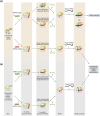
RISC assembly pathways and the downstream events in human system. (a) Catalytically active hAGO2 has two pathways dependent on the types of duplex loaded. The cleavage activity is depicted as scissors. This review proposes the existence of an intermediate state named ‘primary RISC (pri‐RISC)’ between the apo and the pre‐RISC. In this state, only the 5′ monophosphate is captured by the MID domain alone while the rest of the duplex is still exposed to solvent. (b) Catalytically inactive (slicer deficient) AGO proteins, hAGO1, hAGO3, and hAGO4, load siRNA and miRNA duplexes. Their RISC assembly is slicer independent regardless of the types of duplex.
Figure 4
Figure 4
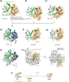
Structural evidence of an open conformation of guide‐free AGO proteins. (a–c) Crystal structures of Neurospora crassa QDE2 MID–PIWI domains (a: PDB ID: 2YHA), Pyrococcus furiosus AGO (b: PDB ID: 1U04), and Aquifex aeolicus AGO (c: PDB ID: 1YVU). For clarity, only the MID (wheat) and PIWI (light green) domains are shown in the ribbon models. The corresponding C‐terminal region that is disordered in TtAGO in complex with a guide of 10 nucleotide (nt) (see g) is colored in green. The MID domain is also drawn as a surface mode. The α‐helix on which the conserved tyrosine is stacked with the first nucleotide base of guide strand is colored in chocolate. (d) Superposed structures of (a)–(c) on their PIWI domain. The MID domains of NcQDE2, PfAGO, and AaAGO are colored in cyan, pink, and blue, respectively. Otherwise, the color code is the same as (a)–(c). (e) Superposed structures of TtAGO‐, KpAGO‐, hAGO1‐, and hAGO2‐RISCs (PDB IDs: 3DLH, 4F1N, 4KXT, and 4OLA, respectively) on their PIWI domain. The MID domains of TtAGO, KpAGO, hAGO1, and hAGO2 are colored in orange, cyan, blue, and pink, respectively. (f–g) Crystal structures of TtAGO in complex with a guide of 21 nt (f: PDB ID: 3DLH) and of 10 nt (g: PDB ID: 3DLB). The color code is the same to (a)–(c). (h) Positions of the phosphorylation sites, Y529 (cyan) and S798 (purple), on hAGO2. The structure of hAGO2‐RISC (PDB ID: 4OLA) is drawn as a surface model except for the MID domain that is shown as a cylinder model. The MID and PIWI domains are colored in wheat and light green, respectively. The bound guide RNA (red) is shown as a ribbon model. The guide is not shown on the right panel for clarity. (i) A hypothetical model of RISC assembly. Guide‐free AGO protein opens the hinged MID‐PIWI domains while the unstructured C‐terminal fragment (green) may be extended to solvent.
Figure 5
Figure 5
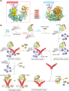
Model of chaperone‐mediated duplex loading. (a) Mapping of the chaperone‐binding sites on the structure of hAGO2 (PDB ID: 4W5N) based on the data of rat AGO256. The regions interacting strongly with Hsp90, p23, Hop, Hsp40, and Hsc70 (left) and weakly with Hsc70 and Hsp40 (right) are shown as a surface model. For clarity, the hAGO2‐bound guide RNA is not shown. (b) Duplex loading by co‐chaperone cycle of Hsp90 in humans. Nascent AGO peptide (aqua) is captured by a complex of Hsc70 (gray) and Hsp40 (pink). The PAZ domain is not shown in order to clarify the interaction between the L1 linker and chaperones. During folding of the AGO protein with the aid of the complex, Hop (slate) uses its TRP motif to recognize at the EEVD tetrapeptide of the Hsc70, which results in an AGO•Hsc70•Hop ternary complex. Hop uses another TRP motif to interact with Hsp90 (red) through the C‐terminal EEVD motif, forming an intermediate complex. Binding of ATP (yellow hexagon) to the N‐terminal domain of the Hsp90 drives the conformational changes, while a Hsp90 co‐chaperone, PPIase, containing a TRP domain (brown) binds to the remaining free EEVD motif of the Hsp90 to form an asymmetric complex. Further binding of PPIase to the C‐terminal EEVD motif of the Hsp90 results in the releases of Hop, which allows Hsp90 to capture the AGO protein. The resultant Hsp90•AGO complex in the closed conformation is stabilized by p23. The widely opened AGO protein accommodates a small RNA duplex. Probably, AGO sorts the 5′ nucleotide of the duplex using the MID domain until the bound duplex is completely accommodated into the channel (see Figure 6). ATP hydrolysis opens the structure of Hsp90 and releases ADP (blue hexagon), PPIase, and p23, along with the pre‐RISC. (Adapted from Ref 13)
Figure 6
Figure 6
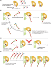
Models of stepwise duplex loading. (a) Interaction between the MID domain alone with nucleoside monophosphates, UMP (red), AMP (blue), CMP (magenta), and GMP (cyan). The MID domain preferentially binds U and A over C and G. The 5′ nucleotide‐binding pocket is divided into the 5′ monophosphate (red)‐ and the base (blue)‐binding sites by a conserved tyrosine residue (black) that is stacked with the 5′ base and its adjacent monophosphate (yellow). The base specificity loop is shown as a thick line (brown). (b) Model of sorting guide strand by the affinity of the MID domain to the 5′ base. Top: The MID domain alone binds to the uracil (or adenine) at the 5′ position of duplexes. The interaction is stable enough to endure until it forms the composite pocket with the PIWI domain. Bottom: The guanine (or cytosine) at the 5′ position of duplexes can bind to the 5′ base‐binding pocket. Owing to the aversion of the nucleotide specificity loop to cytosine and guanine, most of the complexes are dissociated before they form the composite pocket with PIWI domain. Therefore, the duplexes including guanine (or cytosine) at their 5′ position are easily released from the AGO protein. (c) Autoinhibition model. The MID and PIWI domains of unloaded AGO protein interact with each other such that the 5′ nucleotide‐binding pocket is not accessible. Chaperone machinery may pry open the autoinhibited conformation, and the resultant open pocket proceeds to (b). (d) Model of preorganized composite pocket. The MID and PIWI domains of unloaded AGO protein already complete the 5′ monophosphate‐binding site whose affinity is higher than that of the MID domain alone (a). The affinity to the 5′ monophosphate would overwhelm the aversion of the base‐binding site to guanine and cytosine. As a result, the preorganized composite pocket can bind any duplexes, regardless of the types of the 5′ base.
Figure 7
Figure 7
Two model mechanisms of passenger ejection. (a) Y‐shaped nucleic acid‐binding channel of AGO protein. Left: The crystal structure of hAGO2‐RISC (PDB ID: 4OLA) is cut with a section. The color codes of hAGO2 are the same as in Figure 1. Middle: The hAGO2 (light blue) and the bound guide RNA (red) are shown as a surface and a ball‐and‐stick model, respectively. The section area is colored in black. Right: The branched channels. The main channel and the branch are shown by orange and green arrows, respectively. The catalytic site is indicated by scissors. The α7 and α20 are drawn as spheres. (b) Model of the slicer‐dependent passenger ejection. The siRNA duplex composed of a guide (red) and a passenger (green) is loaded onto hAGO2 to form the pre‐RISC. The 5′ monophosphate is shown as a yellow sphere. The N and L1/L2 domains move outward (blue arrow) to expand the width of the nucleic acid‐binding channel. Only the guide RNA is anchored at its 5′ monophosphate and the sugar‐phosphate backbone in the seed region. The loaded siRNA duplex is pushed by the N and L1/L2 domains (cyan arrows) and squeezed out upon the passenger cleavage. The thermal dynamics of the PAZ domain shakes the 3′ end of the guide strand (pink arrows), which facilitates the ejection of the cleaved passenger strand. The territories of the PAZ domain at 25 and 37°C are drawn in a yellow and salmon circles, respectively. The guide 3′ end of siRNA duplex is positioned outside a territory in which the PAZ domain can reach at 25°C. Destabilized base pairs are depicted as dotted line. (c) Model of the slicer‐independent passenger ejection. The guide 3′ end is positioned within a territory that the PAZ domain can reach at 25°C. Because of its thermodynamic instability, the miRNA duplex is heavily distorted by inward pressure.
Figure 8
Figure 8
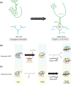
Difference between eukaryotic and prokaryotic AGO proteins. (a) Conformational change activates the AGO protein. In the catalytically inactive state (i.e., unplugged conformation), the region including the glutamate finger (green) folds into an α‐helix (left panel). In the active state (i.e., plugged‐in conformation), the α‐helix is partially unfolded and the glutamate finger is inserted into the DDX triad (aqua) to complete the catalytic DEDX tetrad (right panel). The residue numbers of KpAGO are indicated. (b and c) Requirements for the transition to the plugged‐in conformation. Eukaryotic AGO proteins transition to the plugged‐in state during the RISC assembly (b), whereas the prokaryotic counterparts need to incorporate a target strand to be plugged in (c). The source of guide strand for the prokaryotic AGO proteins remains unknown. The PIWI domain colored in green indicates the plugged‐in conformation.

Similar articles

Cited by

References

    1. Meister G. Argonaute proteins: functional insights and emerging roles. Nat Rev Genet 2013, 14:447–459. - PubMed
    1. Kawamata T, Tomari Y. Making RISC. Trends Biochem Sci 2010, 35:368–376. - PubMed
    1. Jinek M, Doudna JA. A three‐dimensional view of the molecular machinery of RNA interference. Nature 2009, 457:405–412. - PubMed
    1. Jonas S, Izaurralde E. Towards a molecular understanding of microRNA‐mediated gene silencing. Nat Rev Genet 2015, 16:421–433. - PubMed
    1. Huntzinger E, Izaurralde E. Gene silencing by microRNAs: contributions of translational repression and mRNA decay. Nat Rev Genet 2011, 12:99–110. - PubMed

Publication types

MeSH terms