. 2016 Jun 1;126(6):2077-92.
doi: 10.1172/JCI87037. Epub 2016 May 16.

Inhibition of SHP2 ameliorates the pathogenesis of systemic lupus erythematosus

Inhibition of SHP2 ameliorates the pathogenesis of systemic lupus erythematosus

Jianxun Wang et al. J Clin Invest. .

Abstract

Systemic lupus erythematosus (SLE) is a devastating multisystemic autoimmune disorder. However, the molecular mechanisms underlying its pathogenesis remain elusive. Some patients with Noonan syndrome, a congenital disorder predominantly caused by gain-of-function mutations in the protein tyrosine phosphatase SH2 domain-containing PTP (SHP2), have been shown to develop SLE, suggesting a functional correlation between phosphatase activity and systemic autoimmunity. To test this directly, we measured SHP2 activity in spleen lysates isolated from lupus-prone MRL/lpr mice and found it was markedly increased compared with that in control mice. Similar increases in SHP2 activity were seen in peripheral blood mononuclear cells isolated from lupus patients relative to healthy patients. To determine whether SHP2 alters autoimmunity and related immunopathology, we treated MRL/lpr mice with an SHP2 inhibitor and found increased life span, suppressed crescentic glomerulonephritis, reduced spleen size, and diminished skin lesions. SHP2 inhibition also reduced numbers of double-negative T cells, normalized ERK/MAPK signaling, and decreased production of IFN-γ and IL-17A/F, 2 cytokines involved in SLE-associated organ damage. Moreover, in cultured human lupus T cells, SHP2 inhibition reduced proliferation and decreased production of IFN-γ and IL-17A/F, further implicating SHP2 in lupus-associated immunopathology. Taken together, these data identify SHP2 as a critical regulator of SLE pathogenesis and suggest targeting of its activity as a potent treatment for lupus patients.


Figures
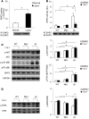
Figure 1
Figure 1. SHP2 activity is upregulated in both lupus patients and lupus-prone MRL/lpr mice, the normalization of which reverses aberrant ERK/MAPK signaling.
(A) SHP2 immune complex PTP assays were conducted using pNPP as a substrate on human PBMC lysates and (B) mouse splenic lysates generated from 18-week-old control C57BL/6 WT, strain-control MRL/MpJ (MpJ), and lupus-prone MRL/lpr (lpr) female mice that were either vehicle or SHP2 inhibitor (11a-1) treated (7.5 mg/kg/d) for 6 weeks, starting at 12 weeks of age. Immunoblotting controls for levels of immunoprecipitated SHP2, showing comparable recovery, are shown below each figure. n = 6 human samples/group and n = 3–8 mice/group, respectively. Splenic lysates isolated from 18-week-old WT, MpJ, and MRL/lpr mice subjected to either vehicle or 11a-1 were immunoblotted with (C) anti–phospho-AKT and anti–phospho-p70S6K or (D) anti–phospho-ERK, as indicated, followed by anti-AKT, anti-p70S6K, and anti-ERK, respectively, to control for loading. Parallel experimental samples were blotted with anti-SHP2 or anti-GAPDH to determine expression levels of these proteins. Quantification of data (n = 3 mice/group) is shown to the right of each representative Western blot figure. Data represent mean ± SEM; *P < 0.05, 1-way or 2-way ANOVA with Holm-Sidak post-test when ANOVA was significant.
Figure 2
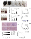
Figure 2. Inhibition of SHP2 activity reduces skin lesions, decreases splenomegaly, normalizes kidney structure and function, and prolongs MRL/lpr longevity.
Representative photographs of (A) skin lesions or (B) spleens from WT, MpJ, and MRL/lpr mice treated with vehicle or 11a-1 for 6 weeks. (C) Spleen weight to tibia length ratios (n = 8 mice/group) and (D) total splenocyte counts (n = 3–5 mice/group) from 18-week-old WT, MpJ, and MRL/lpr female mice treated for 6 weeks with either vehicle or 11a-1 (7.5 mg/kg/d). (E) Representative photograph of kidneys, (F) histograms showing kidney weight to tibia length (n = 8 mice/group), and (G) ratio of albumin to creatinine in urine (n = 7–8 mice per group), and (H) representative H&E-stained kidney sections and pathological scores (n = 4 mice/group) from 18-week-old WT, MpJ, and MRL/lpr female mice treated for 6 weeks with vehicle or 11a-1 (7.5 mg/kg/d). Scale bar: 500 μm. (I) Body weight measurements for 12- to 18-week-old WT, MpJ, and MRL/lpr mice subjected to either vehicle or 11a-1 (n = 8 mice/group). (J) Kaplan-Meier survival curve in 12- to 26-week-old WT, MpJ, and MRL/lpr mice subjected to either vehicle or 11a-1. n = 5 mice/group. Note: all vehicle-treated MRL/lpr mice died by 26 weeks, whereas only 20% of the 11a-1–treated mice died at that same time point. *P < 0.01; #P < 0.05, where P values were derived from 2-way ANOVA with Holm-Sidak post-test when ANOVA was significant.
Figure 3
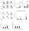
Figure 3. Inhibition of SHP2 reduces the number of circulating DN T cells in peripheral blood of MRL/lpr mice.
Total number of circulating (A) wbc and (B) lymphocytes in peripheral blood of 18-week-old WT, MpJ, and MRL/lpr female mice treated for 6 weeks with vehicle or 11a-1 (7.5 mg/kg/d). n = 7 mice/group. Peripheral blood from 18-week-old MRL/lpr female mice treated with vehicle or 11a-1 for 6 weeks was collected to determine percentage of (C) B and T cells and (D) various T cell subsets, including CD3+CD4+CD8TCRαβ+ (CD4+), CD3+CD4CD8+TCRαβ+ (CD8+), and CD3+CD4CD8TCRαβ+ (DN) T cells in CD45+ leukocytes. n = 4 mice/group. *P < 0.05, 2-way ANOVA with Holm-Sidak post-test when ANOVA was significant.
Figure 4
Figure 4. Normalization of SHP2 activity reduces T cell proliferation in MRL/lpr spleens.
(A) Representative flow cytometry of T cells (CD3+) and B cells (CD19+) isolated from spleens of 18-week-old WT, MpJ, and MRL/lpr female mice treated for 6 weeks with vehicle or 11a-1 (7.5 mg/kg/d). Quantification of the total number of (B) T cells and (C) B cells isolated from MRL/lpr spleens. n = 3–5 mice/group. (D) Representative flow cytometry of CD3+ gated T cells to identify T cell subsets: CD4+, CD8+, and CD4CD8 cells in spleens from 18-week-old WT, MpJ, and MRL/lpr female treated with vehicle or 11a-1 for 6 weeks. (E) Quantification of T cell subsets in WT, MpJ, and MRL/lpr mouse spleens. n = 3–5 mice/group. T cells isolated from spleens of 18-week-old WT, MpJ, and MRL/lpr mice were cultured (1 × 10⁵ cells/well) in 96-well plates for 48 hours in the presence of vehicle (DMSO) or 11a-1 (10 μg/ml) and either in the presence or absence of T cell–activating antibodies CD3 and CD28 to determine (F) proliferation (change in total number of cells) and (G) viability (measure of cell death). n = 3 independent experiments. *P < 0.01; #P < 0.05, 2-way ANOVA with Holm-Sidak post-test when ANOVA was significant.
Figure 5
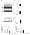
Figure 5. SHP2 activity specifically modulates DN T cell proliferation to promote aberrant ERK/MAPK, but not AKT/mTOR, signaling in this immune cell subset.
DN T cells isolated from lymph nodes of 18-week-old MRL/lpr mice were cultured (3 × 10⁶ cells/well) in 6-well plates for 48 hours in the presence of vehicle (DMSO) or 11a-1 (10 μg/ml) as well as T cell–activating antibodies anti-CD3 and anti-CD28. Cells were then harvested, lysed, and immunoblotted with (A) anti–phospho-AKT and anti–phospho-S6RP and (B) anti-phospho-ERK, as indicated, followed by anti-AKT, anti-S6RP, anti-SHP2, anti-GAPDH, and anti-ERK, respectively, to control for loading and protein expression levels. Quantification of Western blots from n = 3 independent experiments represents the mean ± SEM. DN T cells isolated from lymph nodes of 18-week-old MRL/lpr mice were cultured (1 × 10⁵ cells/well) in 96-well plates for 48 hours with either vehicle (DMSO) or 11a-1 (10 μg/ml) and in the presence or absence of T cell–activating antibodies anti-CD3 and anti-CD28 and were assessed for (C) proliferation or (D) viability. n = 3 independent experiments. *P < 0.01; #P < 0.05, 1-way or 2-way ANOVA with Holm-Sidak post-test when ANOVA was significant.
Figure 6
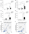
Figure 6. SHP2 activity mediates production of a specific subset of cytokines, IFN-γ, and IL-17A/F heterodimers in SLE.
Serum from 18-week-old WT, MpJ, and MRL/lpr female mice treated for 6 weeks with vehicle or 11a-1 (7.5 mg/kg/d) was collected and assessed for levels of (A) IL-17A/F or (B) IFN-γ. n = 3–8 mice/group. DN T cells were isolated from MRL/lpr mouse lymph nodes and cultured for 120 hours in the presence or absence of anti-CD3 and anti-CD28 antibodies and with either vehicle or 11a-1. These were assessed for levels of (C) IL-17A/F and (D) IFN-γ. n = 3 independent experiments. Pearson correlation analysis showing relationship of kidney dysfunction relative to the expression of serum cytokines (E) IL-17A/F and (F) IFN-γ from WT, MpJ, and MRL/lpr mice, treated with either vehicle or 11a-1 for 6 weeks. n = 30. *P < 0.01; #P < 0.05, 2-way ANOVA with Holm-Sidak post-test when ANOVA was significant.
Figure 7
Figure 7. SHP2 activity is not involved in autoantibody production.
(A) Serum IgG and (B) anti-dsDNA IgG were measured in 18-week-old WT, MpJ, and MRL/lpr female mice treated for 6 weeks with vehicle or 11a-1 (7.5 mg/kg/d). n = 7–8 mice/group. Representative flow cytometry and corresponding quantification of (C) extrafollicular T helper cells (Tefh) (TCRβ+CD4+PSGL-1CD62L) and (D) plasmoblasts (TCRβCD44hiB220+CD138+) and plasma cells (TCRβCD44hiB220CD138+) isolated from spleens of 18-week-old WT, MpJ, and MRL/lpr female mice treated for 6 weeks with vehicle or 11a-1 (7.5 mg/kg/d). n = 3–5 mice/group. *P < 0.01; #P < 0.05, 2-way ANOVA with Holm-Sidak post-test when ANOVA was significant.
Figure 8
Figure 8. SHP2 also modulates proliferation of T cells and expression of IFN-γ and IL-17A/F in human lupus.
Serum levels of (A) IL-17A/F and (B) IFN-γ were measured from either normal or lupus patients with SLEDAI scores higher than 7. n = 24–25/group. (C) Representative images of cultured normal or lupus patient T cells, isolated from PBMCs, following 48 hours of culture in the presence of vehicle (DMSO) or 11a-1 (10 μg/ml) and in the absence or presence of activating antibodies anti-CD3 and anti-CD28. Scale bar: 500 μm. T cells isolated from normal or lupus patients were assessed for (D) proliferation and (E) viability (n = 3 independent experiments) after 48 hours of culture. In addition, T cells isolated from normal or lups patients were measured for levels of (F) IL-17A/F or (G) IFN-γ following 120 hours of culture. n = 12–13/ group. *P < 0.05, 1-way or 2-way ANOVA with Holm-Sidak post-test when ANOVA was significant.

Comment in

Similar articles

Cited by

References

    1. Tsokos GC. Systemic lupus erythematosus. N Engl J Med. 2011;365(22):2110–2121. doi: 10.1056/NEJMra1100359. - DOI - PubMed
    1. Frostegard J. SLE, atherosclerosis and cardiovascular disease. J Intern Med. 2005;257(6):485–495. doi: 10.1111/j.1365-2796.2005.01502.x. - DOI - PubMed
    1. Kaplan MJ. Neutrophils in the pathogenesis and manifestations of SLE. Nat Rev Rheumatol. 2011;7(12):691–699. doi: 10.1038/nrrheum.2011.132. - DOI - PMC - PubMed
    1. Konya C, Paz Z, Tsokos GC. The role of T cells in systemic lupus erythematosus: an update. Curr Opin Rheumatol. 2014;26(5):493–501. doi: 10.1097/BOR.0000000000000082. - DOI - PubMed
    1. Mizui M, et al. IL-2 protects lupus-prone mice from multiple end-organ damage by limiting CD4-CD8- IL-17-producing T cells. J Immunol. 2014;193(5):2168–2177. doi: 10.4049/jimmunol.1400977. - DOI - PMC - PubMed

Publication types

MeSH terms