. 2016 Jul;30(7):693-708.
doi: 10.1210/me.2015-1299. Epub 2016 May 13.

Prolactin Signaling Stimulates Invasion via Na(+)/H(+) Exchanger NHE1 in T47D Human Breast Cancer Cells

Affiliations

Prolactin Signaling Stimulates Invasion via Na(+)/H(+) Exchanger NHE1 in T47D Human Breast Cancer Cells

Elena Pedraz-Cuesta et al. Mol Endocrinol. 2016 Jul.

Abstract

Prolactin (PRL) and its receptor (PRLR) are implicated in breast cancer invasiveness, although their exact roles remain controversial. The Na(+)/H(+) exchanger (NHE1) plays essential roles in cancer cell motility and invasiveness, but the PRLR and NHE1 have not previously been linked. Here we show that in T47D human breast cancer cells, which express high levels of PRLR and NHE1, exposure to PRL led to the activation of Janus kinase-2 (JAK2)/signal transducer and activator of transcription-5 (STAT5), Akt, and ERK1/2 signaling and the rapid formation of peripheral membrane ruffles, known to be associated with cell motility. NHE1 was present in small ruffles prior to PRL treatment and was further recruited to the larger, more dynamic ruffles induced by PRL exposure. In PRL-induced ruffles, NHE1 colocalized with activated Akt, ERK1/2, and the ERK effector p90Ribosomal S kinase (p90RSK), known regulators of NHE1 activity. Stimulation of T47D cells with PRL augmented p90RSK activation, Ser703-phosphorylation of NHE1, NHE1-dependent intracellular pH recovery, pericellular acidification, and NHE1-dependent invasiveness. NHE1 activity and localization to ruffles were attenuated by the inhibition of Akt and/or ERK1/2. In contrast, noncancerous MCF10A breast epithelial cells expressed NHE1 and PRLR at lower levels than T47D cells, and their stimulation with PRL induced neither NHE1 activation nor NHE1-dependent invasiveness. In conclusion, we show for the first time that PRLR activation stimulates breast cancer cell invasiveness via the activation of NHE1. We propose that PRL-induced NHE1 activation and the resulting NHE1-dependent invasiveness may contribute to the metastatic behavior of human breast cancer cells.


Figures
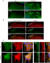
Figure 1.
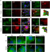
Figure 1.. Stimulation with PRL induces peripheral membrane ruffling, cytoskeletal reorganization, and NHE1-PRLR colocalization to ruffles in T47D cancer cells.
A, Detail of PRL-induced ruffling in T47D cells. Cells were treated or not with PRL (10 nM, 5 min) and stained for vinculin (green) as a marker of focal adhesions and ruffles. Nuclei are labeled with DAPI (blue), the dotted line indicates the lamellipodial edge, and the arrows the localization of ruffles. Ai, iii, At the cell-coverslip interface, vinculin is predominantly found in FAs; Aii, iv, 1–2 μm above the site of adhesion to the coverslip, vinculin is found in large membrane ruffles induced by PRL. Scale bar, 5 μm. B, T47D cells were treated with PRL (10 nM, 5 min), labeled for pTyr576/577-FAK (red) and NHE1 (green), and imaged at the cell-coverslip interface (FAs) and a few micrometers above (ruffles). The dotted line indicates the lamellipodial edge, and the arrowheads mark the colocalization of NHE1 and p-FAK in ruffles. Note that NHE1 does not appear to localize to FAs but localizes prominently to membrane ruffles. Scale bar, 10 μm. C, Immunofluorescence analysis of NHE1 (green) and the PRLR (red) localization in T47D cells in the absence and presence of PRL (10 nM, 5 min). Where indicated, images are merged with DAPI nuclear staining and a higher-magnification view (×2 zoom) of the indicated square is shown. Arrows mark regions of prominent colocalization. Scale bars (full images), 10 μm; (detail panel), 5 μm. All images are representative of three to four independent experiments per condition. Ctrl, control.
Figure 2.
Figure 2.. NHE1 colocalization with pAkt and pERK in PRL-induced ruffles.
Immunofluorescence analysis of T47D cells treated or not with PRL (10 nM, 5 min) and stained for NHE1, pSer473-Akt, pThr202/Tyr204-ERK1/2, and DAPI (nuclear stain, blue), as indicated. Arrows mark regions of prominent colocalization (A–D) or NHE1 localization to ruffles (E). A, NHE1 (green) and pSer473-Akt (red) colocalize in PRL-induced ruffles. Scale bar, 10 μm. B, z-stack imaging, maximal intensity projection, and isosurface projection show details of NHE1-p-Akt colocalization in ruffles. Note the strong p-Akt enrichment in ruffles seen in both panels A and B. Scale bar, 5 μm. C, NHE1 (green) and pThr202/Tyr204-ERK1/2 (red). D, z-stack imaging, maximal intensity projection and isosurface projection show details of NHE1-p-ERK colocalization in ruffles. Scale bar, 5 μm. Scale bar, 10 μm. E, T47D cells were treated with Akti-1/2 or U0126 10 μM for 1 hour, followed by treatment or not with PRL, as indicated, and stained for NHE1 (green) with DAPI nuclear counterstain. Scale bar, 10 μm. All images are representative of at least three independent experiments per condition. Ctrl, control.
Figure 3.
Figure 3.. PRL treatment increases ruffling and NHE-GFP recruitment in MDCK cells.
A, MDCK cells stably expressing NHE1-GFP. The box indicates the region shown in panels B and C, Scale bar, 10 μm. B, Montages of NHE1-GFP in ruffle dynamics before addition of PRL. C, Montages of NHE1-GFP in ruffle dynamics of the same region as in panel B after addition of PRL. Panels B and C are shown as inverted-contrast images (upper panels) and with a gradient pseudocolor of GFP intensity (lower panels). The intensity scale is shown in arbitrary units on the right-hand side. The montages are chosen from movies with a total length of 5 minutes (Supplemental Videos 1 and 2, respectively) each, with 10 seconds between each frame. Arrows point to NHE1-GFP in the ruffle edge. Scale bar, 3 μm. Data shown are representative of eight independent experiments. Ctrl, control.
Figure 4.
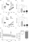
Figure 4.. PRL stimulates NHE1 activity and acid extrusion in T47D cells.
A, pHi recovery over time after an NH4Cl prepulse-induced acid load in MCF10A cells ± 10 nM PRL and/or 5 μM EIPA, as indicated. Error bars represent SD from measurements on at least six cells in a single experiment. B, Relative pHi recovery rates in MCF10A cells were calculated from the initial, linear phase of the recovery curve and are shown as mean with SEM error bars of four independent experiments. ***, P ≤ .001, one-way ANOVA with Tukey multiple comparisons posttest. C, As in panel A, except in T47D cells. D, Relative pH recovery rates in T47D cells, calculated from the initial, linear phase of the recovery curve, and are shown as mean with SEM error bars of five independent experiments. *, P ≤ .05, ***, P ≤ .001, ****, P ≤ .0001, one-way ANOVA with Tukey multiple comparisons test. E and F, pHe was measured in T47D cells using Fluorescein-DHPE. E, Sample trace. The arrow shows the time of addition of PRL (10 nM). Error bars are SD from measurements on seven cells in a single experiment. F, Data as in panel E summarized as the change in pHe per minute the last minute before PRL addition and at time 5 minutes after PRL addition. Error bars represent SEM for the four independent experiments. *, Paired, two-way Student's t test. Ctrl, control.
Figure 5.
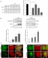
Figure 5.. PRL elicits NHE1 phosphorylation on S703, and active p90RSK colocalizes with NHE1 in PRL-induced ruffles.
A, Left panel, Representative immunoblots of Ser703-phosphorylated and total NHE1, with p150 as a loading control. Right panel, Quantification of pSer703-NHE1 relative to total NHE1. Data are based on three independent experiments. **, P ≤ .01, one-way ANOVA with Tukey posttest. B, Top panel, Representative immunoblots of p380-p90RSK, p90RSK1, pThr202/Tyr204-ERK, ERK, and p150 (loading control). Bottom panel, Quantification of pERK relative to total ERK and p380p90RSK relative to total p90RSK. Data are based on three independent experiments. *, P ≤ .05, **, P ≤ .01, one-way ANOVA with Tukey posttest. C and D, IF analysis of T47D cells treated or not with PRL (10 nM, 5 min), as indicated. Arrows mark regions of prominent colocalization. C, NHE1 (green) and p90RSK1 (red). D, NHE1 (green) and pSer380-RSK (red). Images are merged and DNA (blue) is stained by DAPI. Scale bar, 10 μm. data are representative of three independent experiments per condition. Ctrl, control.
Figure 6.
Figure 6.. PRL induces invasiveness of T47D cells in a NHE1-dependent manner.
Invasiveness of MCF10A (A) and T47D (A and B) cells was assessed in matrigel-coated Boyden chambers. PRL (10 nM) was applied as a chemoattractant to the lower chamber, as indicated, and where indicated, EIPA (5 μM) (A) or cariporide (10 μM) (B) was applied to both chambers to inhibit NHE1. Data are shown as relative cell invasion (invasion relative to migration) in the presence or absence of PRL and/or EIPA or cariporide as indicated. Error bars represent SEM of four (MCF10; A), five (T47D; A), or three (T47D; B) independent experiments. *, P ≤ .05, **, P ≤ .01, ***, P ≤ .001, one-way ANOVA with Tukey posttest. Ctrl, control.

Similar articles

Cited by

References

    1. Teilum K, Hoch JC, Goffin V, Kinet S, Martial JA, Kragelund BB. Solution structure of human prolactin. J Mol Biol. 2005;351:810–823. - PubMed
    1. Marano RJ, Ben-Jonathan N. Minireview: extrapituitary prolactin: an update on the distribution, regulation, and functions. Mol Endocrinol. 2014;28:622–633. - PMC - PubMed
    1. Clevenger CV, Furth PA, Hankinson SE, Schuler LA. The role of prolactin in mammary carcinoma. Endocr Rev. 2003;24:1–27. - PMC - PubMed
    1. Clevenger CV, Gadd SL, Zheng J. New mechanisms for PRLr action in breast cancer. Trends Endocrinol Metab. 2009;20:223–229. - PubMed
    1. Brooks CL. Molecular mechanisms of prolactin and its receptor. Endocr Rev. 2012;33:504–525. - PMC - PubMed

MeSH terms

Grants and funding

This work was supported by the Danish Council for Independent Research Grants 12–127290 (to S.F.P.) and 12–125862 (to B.B.K.) and support from the Hartmann Foundation (to S.F.P.), the Novo Nordisk Foundation (to B.B.K.), the Harboe Foundation (to S.F.P.); the Lundbeck Foundation (to L.N.N.), the Carlsberg Foundation (to L.N.N.), and MEMBRANES (to L.N.N.) and the Graduate School of Science and Technology (to H.H.J.).