. 2016 May 13:6:25838.
doi: 10.1038/srep25838.

Derivation of Porcine Embryonic Stem-Like Cells from In Vitro-Produced Blastocyst-Stage Embryos

Affiliations

Derivation of Porcine Embryonic Stem-Like Cells from In Vitro-Produced Blastocyst-Stage Embryos

Dao-Rong Hou et al. Sci Rep. .

Abstract

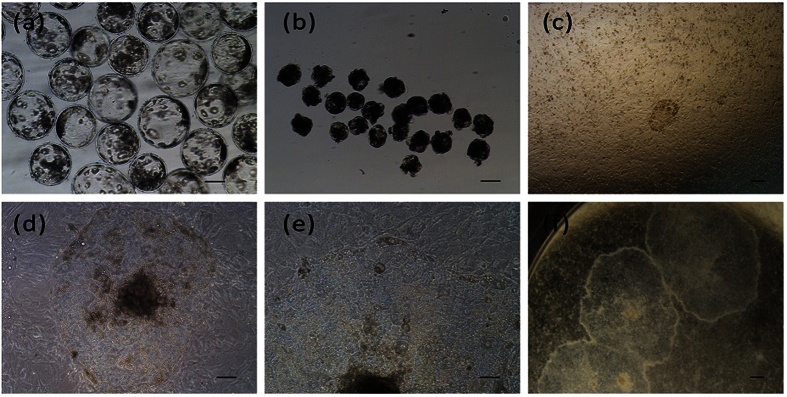
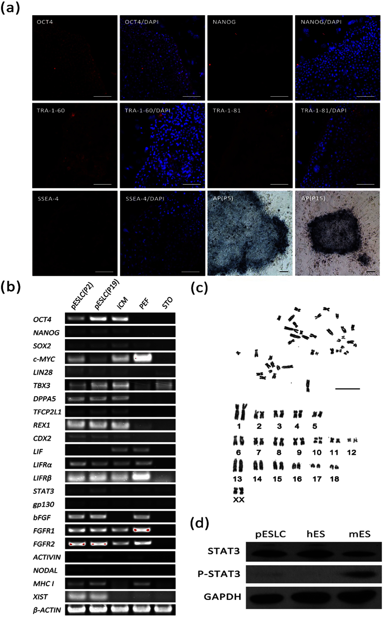
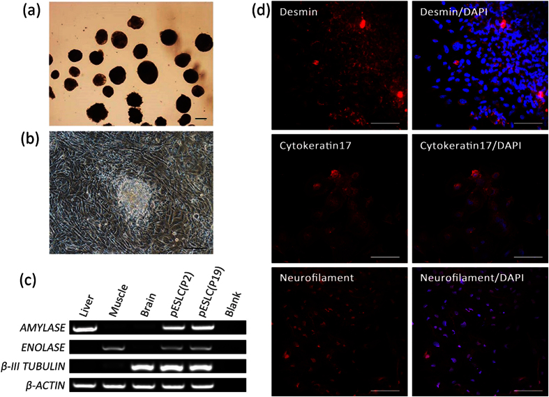
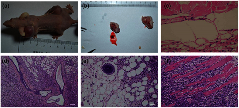
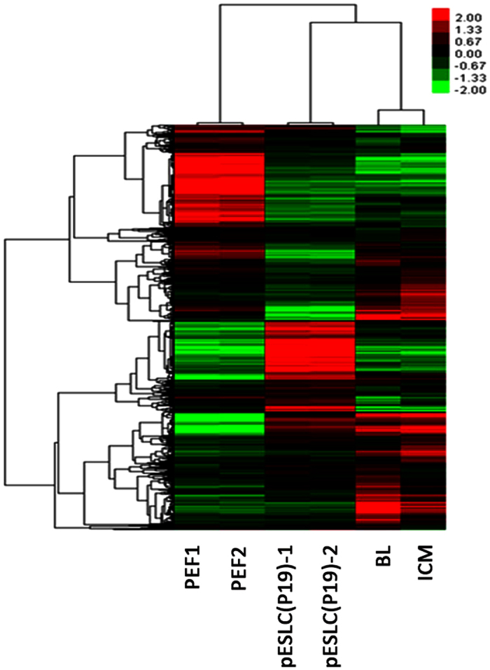
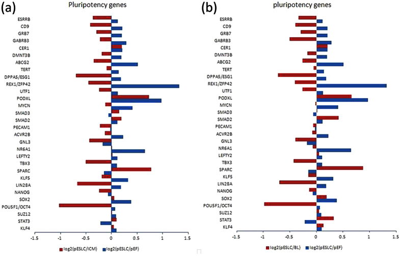
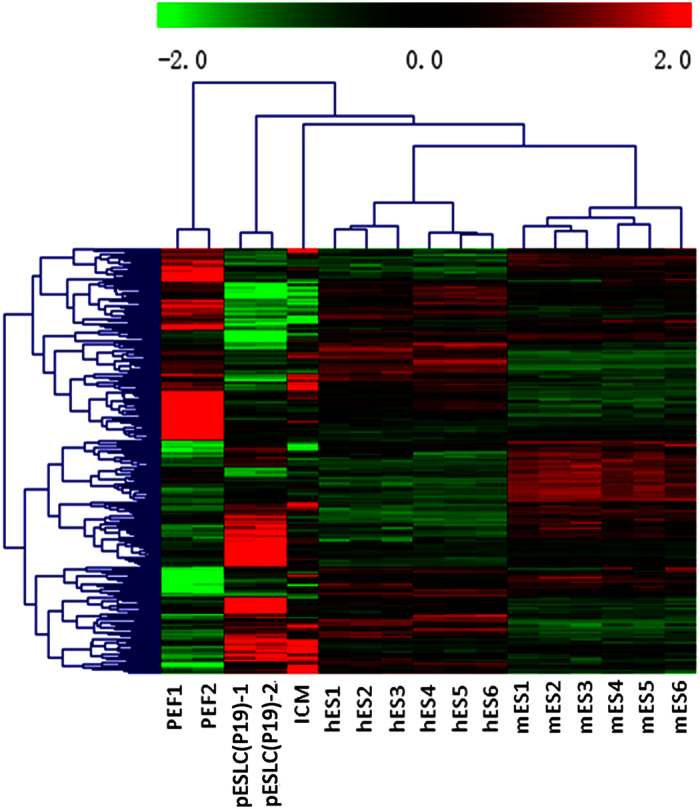
Efficient isolation of embryonic stem (ES) cells from pre-implantation porcine embryos has remained a challenge. Here, we describe the derivation of porcine embryonic stem-like cells (pESLCs) by seeding the isolated inner cell mass (ICM) from in vitro-produced porcine blastocyst into α-MEM with basic fibroblast growth factor (bFGF). The pESL cells kept the normal karyotype and displayed flatten clones, similar in phenotype to human embryonic stem cells (hES cells) and rodent epiblast stem cells. These cells exhibited alkaline phosphatase (AP) activity and expressed pluripotency markers such as OCT4, NANOG, SOX2, SSEA-4, TRA-1-60, and TRA-1-81 as determined by both immunofluorescence and RT-PCR. Additionally, these cells formed embryoid body (EB), teratomas and also differentiated into 3 germ layers in vitro and in vivo. Microarray analysis showed the expression of the pluripotency markers, PODXL, REX1, SOX2, KLF5 and NR6A1, was significantly higher compared with porcine embryonic fibroblasts (PEF), but expression of OCT4, TBX3, REX1, LIN28A and DPPA5, was lower compared to the whole blastocysts or ICM of blastocyst. Our results showed that porcine embryonic stem-like cells can be established from in vitro-produced blastocyst-stage embryos, which promote porcine naive ES cells to be established.


Figures

Figure 1
Figure 1. Derivation and morphology of pESL cells.
(a) In vitro fertilized blastocysts at day 7. (b) Blastocysts after immunosurgery treatment. (c) Day 3 attached ICM. (d,e) The morphology of pESL cells colony at passage 2. (f) The morphology of pESL cells colony at passage 15. Scale bars = 100 um.
Figure 2
Figure 2. Characteristics of pESL cells.
(a) The immunofluorescence staining of pluripotency markers OCT4, NANOG, SSEA-4, TRA-1-60, TRA-1-81 and alkaline phosphatase staining in pESLC colonies cultured on STO cells are shown. Scale bars = 100 um. (b) RT-PCR analysis of relative transcript concentrations of pluripotent and lineage-specific genes in pESL cells at passage 2 and passage 19, ICM, PEF, and STO. (c) Karyotype analysis of pESL cells at passage 7. Scale bars = 100 um. (d) Phosphorylation status of STAT3 in the pESLCs at passage 18. mES cells and hES cells were used as control. ICM: inner cell mass; PEF: porcine embryonic fibroblast cell; STO: feeder layer cell, SIM mouse thioguanne and ouabain resistant fibroblast cells.
Figure 3
Figure 3. In vitro differentiation potential of pESL cells.
(a) EB derived from pESLCs at passage 19 by hanging drop culture on no adhesive culture dishes for 10 days. (b) EB spread on dishes coated with gelatin and displayed distinct signs of differentiation. (c) Expression of the β-III TUBULIN (endoderm), ENOLASE (mesoderm), and AMYLASE (ectoderm) were detected in EB resulted from pESLCs at passage 2 and passage 19. Brain, liver and muscle tissues of Landrace pig were used as positive control. (d) Expression of differentiation marker Cytokeratin 17 (endoderm), Desmin (mesoderm) and Neurofilament (ectoderm) from differentiated pESL cells were confirmed by the immunocytochemistry analysis. Scale bar = 100 um.
Figure 4
Figure 4. Teratoma formation from injected pESL cells.
(a) Gross image of the mouse showing subcutaneous teratomas in the neck and dorsal flank regions. (b) Tumors removed from mouse (a) after 90 days injection. (cf) Three germ layers present in teratomas derived from the pESL cell line at passage 21. The images of H&E-stained sections showing representative endoderm (d), branched glands and duct, mesoderm (c,f), striated muscle and smooth muscle and ectoderm lineage (e), neural rosette. Scale bar = 100 um.
Figure 5
Figure 5. Gene expression profiling of pESL cells using Affymetrix microarrays data.
Two-color heat map representation of globe gene expression data that derived from Affymetrix gene chip analysis of PEF1, PEF2, pESLC (P19), BL and ICM. Each row represents the expression of a single gene and columns indicate samples. PEF1, 2: Porcine embryonic fibroblast cell line PEF1 and PEF2 at passage 3; pESLC (P19)-1, pESLC (P19)-2: pESL cells at passage 19; BL: blastocysts; ICM: inner cell mass.
Figure 6
Figure 6. Pluripotency gene expression analysis of pESL cells was performed using Affymetrix microarrays data.
(a) Log2-fold change in pluripotency genes expression in pESLCs compared with ICMs (red bars) and PEFs (blue bars). (b) Log2-fold change in pluripotency gene expression in pESLCs compared with in vitro fertilized d7 blastocysts (red bars) and PEFs (blue bars).
Figure 7
Figure 7. Hierarchical cluster analysis of the orthologous genes expression profile of pESLCs.
Two-color heat map representation of the orthologous genes expression data of PEF, pESLCs, hES cells and mES cells. Each row represents the expression of a single gene and columns indicate samples; PEF1, 2: Porcine embryonic fibroblast cell line PEF1 and PEF2 at passage 3; pESLC(P19)-1, pESLC(P19)-2: pESL cells at passage 19; hES 1, 2 and 3: hES cell line HUES6; hES 4, 5 and 6: hES cell line H9; mES 1, 2 and 3: mES cell line R1; mES 4, 5 and 6: mES cell line J1.

Similar articles

Cited by

  • Tetraploid embryo aggregation produces high-quality blastocysts with an increased trophectoderm in pigs.
    Lee J, Cai L, Kim M, Choi H, Oh D, Jawad A, Lee E, Hyun SH. Lee J, et al. Front Cell Dev Biol. 2023 Nov 16;11:1239448. doi: 10.3389/fcell.2023.1239448. eCollection 2023. Front Cell Dev Biol. 2023. PMID: 38033873 Free PMC article.
  • Derivation of Porcine Extra-Embryonic Endoderm Cell Lines Reveals Distinct Signaling Pathway and Multipotency States.
    Zhang ML, Jin Y, Zhao LH, Zhang J, Zhou M, Li MS, Yin ZB, Wang ZX, Zhao LX, Li XH, Li RF. Zhang ML, et al. Int J Mol Sci. 2021 Nov 29;22(23):12918. doi: 10.3390/ijms222312918. Int J Mol Sci. 2021. PMID: 34884722 Free PMC article.
  • Modeling lethal X-linked genetic disorders in pigs with ensured fertility.
    Matsunari H, Watanabe M, Nakano K, Enosawa S, Umeyama K, Uchikura A, Yashima S, Fukuda T, Klymiuk N, Kurome M, Kessler B, Wuensch A, Zakhartchenko V, Wolf E, Hanazono Y, Nagaya M, Umezawa A, Nakauchi H, Nagashima H. Matsunari H, et al. Proc Natl Acad Sci U S A. 2018 Jan 23;115(4):708-713. doi: 10.1073/pnas.1715940115. Epub 2018 Jan 8. Proc Natl Acad Sci U S A. 2018. PMID: 29311328 Free PMC article.
  • Generation and characterization of stable pig pregastrulation epiblast stem cell lines.
    Zhi M, Zhang J, Tang Q, Yu D, Gao S, Gao D, Liu P, Guo J, Hai T, Gao J, Cao S, Zhao Z, Li C, Weng X, He M, Chen T, Wang Y, Long K, Jiao D, Li G, Zhang J, Liu Y, Lin Y, Pang D, Zhu Q, Chen N, Huang J, Chen X, Yao Y, Yang J, Xie Z, Huang X, Liu M, Zhang R, Li Q, Miao Y, Tian J, Huang X, Ouyang H, Liu B, Xie W, Zhou Q, Wei H, Liu Z, Zheng C, Li M, Han J. Zhi M, et al. Cell Res. 2022 Apr;32(4):383-400. doi: 10.1038/s41422-021-00592-9. Epub 2021 Nov 30. Cell Res. 2022. PMID: 34848870 Free PMC article.
  • Establishment of porcine and human expanded potential stem cells.
    Gao X, Nowak-Imialek M, Chen X, Chen D, Herrmann D, Ruan D, Chen ACH, Eckersley-Maslin MA, Ahmad S, Lee YL, Kobayashi T, Ryan D, Zhong J, Zhu J, Wu J, Lan G, Petkov S, Yang J, Antunes L, Campos LS, Fu B, Wang S, Yong Y, Wang X, Xue SG, Ge L, Liu Z, Huang Y, Nie T, Li P, Wu D, Pei D, Zhang Y, Lu L, Yang F, Kimber SJ, Reik W, Zou X, Shang Z, Lai L, Surani A, Tam PPL, Ahmed A, Yeung WSB, Teichmann SA, Niemann H, Liu P. Gao X, et al. Nat Cell Biol. 2019 Jun;21(6):687-699. doi: 10.1038/s41556-019-0333-2. Epub 2019 Jun 3. Nat Cell Biol. 2019. PMID: 31160711 Free PMC article.

References

    1. Nichols J. & Smith A. The origin and identity of embryonic stem cells. Development 138, 3–8, doi: 10.1242/dev.050831 (2011). - DOI - PubMed
    1. Evans M. J. a. Kaufman, M. Establishment in culture of pluripotential cells from mouse embryos. Nature 292, 154–156 (1981). - PubMed
    1. Martin G. R. Isolation of a pluripotent cell line from early mouse embryos cultured in medium conditioned by teratocarcinoma stem cells. Proc. Natl. Acad. Sci. USA 78, 7634–7638 (1981). - PMC - PubMed
    1. Thomson J. et al.. Isolation of a Primate Embryonic Stem Cell Line. PNAS. 92, 7844–7848 (1995). - PMC - PubMed
    1. Thomson J. A. et al.. M. Embryonic Stem Cell Lines Derived from Human Blastocysts. Science 282, 1145–1147 (1998). - PubMed

Publication types