. 2016 Apr 21;1(5):e85923.
doi: 10.1172/jci.insight.85923.

Central role for GSK3β in the pathogenesis of arrhythmogenic cardiomyopathy

Affiliations

Central role for GSK3β in the pathogenesis of arrhythmogenic cardiomyopathy

Stephen P Chelko et al. JCI Insight. .

Abstract

Arrhythmogenic cardiomyopathy (ACM) is characterized by redistribution of junctional proteins, arrhythmias, and progressive myocardial injury. We previously reported that SB216763 (SB2), annotated as a GSK3β inhibitor, reverses disease phenotypes in a zebrafish model of ACM. Here, we show that SB2 prevents myocyte injury and cardiac dysfunction in vivo in two murine models of ACM at baseline and in response to exercise. SB2-treated mice with desmosome mutations showed improvements in ventricular ectopy and myocardial fibrosis/inflammation as compared with vehicle-treated (Veh-treated) mice. GSK3β inhibition improved left ventricle function and survival in sedentary and exercised Dsg2mut/mut mice compared with Veh-treated Dsg2mut/mut mice and normalized intercalated disc (ID) protein distribution in both mutant mice. GSK3β showed diffuse cytoplasmic localization in control myocytes but ID redistribution in ACM mice. Identical GSK3β redistribution is present in ACM patient myocardium but not in normal hearts or other cardiomyopathies. SB2 reduced total GSK3β protein levels but not phosphorylated Ser 9-GSK3β in ACM mice. Constitutively active GSK3β worsens ACM in mutant mice, while GSK3β shRNA silencing in ACM cardiomyocytes prevents abnormal ID protein distribution. These results highlight a central role for GSKβ in the complex phenotype of ACM and provide further evidence that pharmacologic GSKβ inhibition improves cardiomyopathies due to desmosome mutations.


Figures
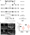
Figure 1
Figure 1. Targeting of murine Dsg2, encoding desmoglein-2, recapitulates ACM.
(A) Structure of targeting vector for Dsg2mut/mut mutant mice and Dsg2mut allele after Flp- and Cre-mediated recombination. Note that Dsg2mut cDNA has two stop codons (TGA) in exon 6. (B) Representative short-axis m-mode and left ventricle (LV) long-axis echocardiography from WT and Dsg2mut/mut mice at 8 weeks of age. Echocardiographic images are representative of n = 7 and n = 6 for WT and Dsg2mut/mut mice, respectively. (C) Decreased fractional shortening and ejection fraction in Dsg2mut/mut mice at 8 weeks of age. Horizontal bars indicate the medians, boxes indicate 25th to 75th percentiles, and whiskers indicate 10th and 90th percentiles. Mean ± SEM, n ≥ 6/genotype, *P < 0.05 for WT vs. Dsg2mut/mut using 2-way ANOVA with Tukey’s post-hoc analysis.
Figure 2
Figure 2. Biventricular fibrosis and abnormal distribution of junctional proteins in Dsg2mut/mut mice at 16 weeks of age.
(A) Western immunoblots and immunolabeled myocardium probed for desmoglein-2 (DSG2) showed a complete absence of protein and junctional distribution of DSG2 in Dsg2mut/mut mice compared with WT mice. Scale bar: 10 μm. (B) Representative images from myocardium immunolabeled for plakoglobin (PLK), connexin43 (Cx43), and N-cadherin. Scale bar: 20 μm. (C and D) Gross pathology shows right ventricle (white arrows) and left ventricle (white arrowhead) epicardial and endocardial scars. (E) Masson’s trichrome–immunostained (MTC-immunostained) myocardium from Dsg2mut/mut mice shows extensive epicardial and endocardial scarring compared with WT controls. Scale bar: 1 mm. Images are representative of n = 7 and n = 6 for WT and Dsg2mut/mut mice, respectively. (F) Percentage fibrosis presented as mean ± SEM. P < 0.05 for WT vs. Dsg2mut/mut using 2-tailed t test with equal variance.
Figure 3
Figure 3. Transgenic myocyte-specific expression of human 2157del2 plakoglobin recapitulates ACM.
(A) Representative Western immunoblots from WT and JUP2157del2 mouse hearts. Antibodies directed against the plakoglobin N-terminus (PLK, Nt) detect both the endogenous PLK (black arrowhead) and truncated mutant transgene (black asterisk). Antibodies directed against the C-terminus (PLK, Ct) do not detect the mutant transgenic protein and confirm that endogenous Jup expression is not reduced in transgenic mice compared with WT. n = 3/genotype. (B) JUP2157del2 hearts display focal areas of inflammation (scale bar: 100 μm) and increased TUNEL-positive nuclei (white arrowheads; scale bar: 20 μm). H&E: n = 6 for WT and n = 10 for JUP2157del2; TUNEL: n = 5/genotype. The percentage of scarring and the percentage of TUNEL-positive nuclei are presented as mean ± SEM. *P < 0.05 for WT vs. JUP2157del2 using 2-tailed t test with equal variance. (C) Representative images from JUP2157del2 and WT control hearts immunolabeled for N-cadherin, plakoglobin, connexin43 (Cx43), and synapse-associated protein 97 (SAP97). Images are representative of n ≥ 5/genotype. Scale bar: 20 μm.
Figure 4
Figure 4. SB216763 treatment improves myocardial injury and cardiac function in JUP2157del2 and Dsg2mut/mut mice.
(A) Representative images of ventricular myocardia from JUP2157del2 and Dsg2mut/mut mice treated with vehicle (Veh) or SB216763 (SB2). Myocardia were immunostained with H&E, Masson’s trichrome (MTC), and TUNEL. Scale bar: 50 μm. Images are representative of n ≥ 4/genotype (white arrows, TUNEL-positive nuclei). (B) Percentage fibrosis and TUNEL-positive nuclei in hearts from SB2- and Veh-treated mutant mice. Fibrosis: n = 4/genotype; TUNEL: n = 4 for WT and Dsg2mut/mut mice and n = 6 for WT and JUP2157del2 mice. Mean ± SEM. P < 0.05 for WT vs. ACM mice using 2-tailed paired t test. (C) Quantitative ECG telemetry analysis of SB2-treated JUP2157del2 mice exhibited decreased bouts of single and >2 premature ventricular complexes. Representative ECG telemetry tracing from a vehicle-treated JUP2157del2 mouse. Mean ± SEM. P < 0.05 for SB2-treated JUP2157del2 (n = 16) vs. Veh-treated JUP2157del2 (n = 6) mice using 2-tailed t test with equal variance. (D) Echocardiography and ECG telemetry analysis of Veh- and SB2-treated Dsg2mut/mut mice. In box-and-whisker plots, horizontal bars indicate the medians, boxes indicate 25th to 75th percentiles, and whiskers indicate 10th and 90th percentiles. Mean ± SEM, n = 4/genotype/treatment. *P < 0.05 for SB2-treated Dsg2mut/mut vs. Veh-treated Dsg2mut/mut mice using 2-way ANOVA with Tukey’s post-hoc analysis.
Figure 5
Figure 5. In vivo reversal of protein distribution in SB216763-treated ACM mice.
(A and B) Representative myocardium immunolabeled with plakoglobin (PLK) and connexin43 (Cx43) from vehicle- (Veh-) and SB216763-treated (SB2-treated) WT, Dsg2mut/mut, and JUP2157del2 mice. Scale bar: 20 μm. Images are representative of n = 4/genotype/treatment. (C and D) Western analysis of ventricular lysates probed for PLK, desmoplakin (DSP), and Cx43, normalized to GAPDH. Mean ± SEM, n = 4/genotype/treatment. P < 0.05 for SB2-treated mice vs. Veh-treated mice using 2-tailed paired t test.
Figure 6
Figure 6. GSK3β localization is uniquely abnormal in ACM, and SB216763 normalizes it.
(A) Representative images of formalin-fixed, paraffin-embedded ventricular myocardia (FFPE-VM) (scale bar: 20 μm) from JUP2157del2 and Dsg2mut/mut mice and neonatal rat ventricular myocytes (NRVM) (scale bar: 10 μm) expressing JUP2157del2 and PKP21851del123 transgenes immunolabeled for anti-GSK3β. Images are representative of n = 4/genotype/treatment (DAPI, blue; GSK3β, red; white arrows, ID localization of GSK3β; yellow arrows, absence of ID localization of GSK3β). (B) Western blots probed for GSK3α, GSK3β, and phosphorylated GSK3β (pGSK3β-S9) from WT, Dsg2mut/mut, and JUP2157del2 mice. (C) Quantitative GSK3β and GSK3α protein levels from WT, Dsg2mut/mut, and JUP2157del2 mice, normalized to GAPDH. Mean ± SEM, n = 4/genotype/treatment. P < 0.05 for SB2-treated mice vs. Veh-treated mice using 2-tailed paired t test. *Dsg2mut/mut vs. WT; JUP2157del2 vs. WT. (D) GSK3β-immunolabeled patient biopsies. Representative images taken from endomyocardial biopsy samples show GSK3β distribution at IDs (white arrows) in all patients with ACM (20 of 20) differ from control hearts (n = 10) and patients diagnosed with sarcoidosis (n = 15), giant cell myocarditis (GCM, n = 5), and end-stage ischemic, dilated cardiomyopathy (DCM), and hypertrophic cardiomyopathy (HCM, n = 5 for each) (yellow asterisks, punctate cytosolic pools of GSK3β; yellow arrows, absence of GSK3β signal at IDs).
Figure 7
Figure 7. SB216763 treatment improves response to exercise in Dsg2mut/mut mice.
(A) Percentage survival of drug-treated exercised Dsg2mut/mut and WT mice. Mean ± SEM, n values are shown. (B) Percentage ejection fraction and fractional shortening from exercised vehicle- (Veh-) and SB216763-treated (SB2-treated) Dsg2mut/mut mice at 16 weeks of age. Mean ± SEM, n = 4/genotype/treatment. *P < 0.05 using 1-way ANOVA. (C) Myocardia immunolabeled with TUNEL from exercised Dsg2 mutant and WT mice treated with Veh or SB2. Scale bar: 20 μm. Images are representative of n = 4/genotype/treatment. (D) GSK3β immunolabeled myocardium. Images are representative of n = 4/genotype/treatment (white arrows, junctional signal for GSK3β; yellow arrowheads, absence of junctional signal for GSK3β). Scale bar: 20 μm. (E) Representative images from exercised drug-treated Dsg2mut/mut and Dsg2mut/+ mice immunostained for plakoglobin (PLK) and connexin43 (Cx43). Images are representative of n = 4/genotype/treatment. Scale bar: 20 μm.
Figure 8
Figure 8. GSK3β knockdown prevents loss of junctional signal of ID proteins in ACM neonatal rat ventricular myocytes.
(A) GSK3β knockdown was confirmed via Western immunoblots in GSK3β shRNA–transfected neonatal rat ventricular myocytes (NRVMs) compared with untransfected controls. Cotransfected NRVM lysates probed for changes in plakoglobin (PLK), connexin43 (Cx43), and GSK3α/β, normalized to GAPDH. Immunoblots are representative of n = 3/cohort. (B) Representative images of untransfected controls and GSK3β shRNA–transfected NRVMs. Note that GSK3β shRNA–transfected NRVMs display control-like PLK and Cx43 distribution. Images are representative of n = 3/cohort. (C) Mutant ACM expressing NRVMs displayed increased nuclear PLK localization and near absent Cx43 signal. Cotransfected NRVMs (ACM expressing construct and GSK3β shRNA transgene) immunostained for changes in PLK and Cx43 distribution. Note that abnormal PLK and Cx43 localization was prevented in cotransfected NRVMs, and Cx43 signal was increased compared with ACM myocytes without GSK3β shRNA construct (white arrows, junctional signal; yellow arrowheads, nuclear localization). Scale bar: 50 μm. Images are representative of n = 3/cohort.
Figure 9
Figure 9. Dsg2 mutant mice with constitutively active GSK3β demonstrate increased myocardial fibrosis and cardiac dysfunction.
(A) Representative short-axis, m-mode echocardiography from WT and heterozygous and homozygous Dsg2 mutant mice with 1 or 2 copies of mutant GSK3β-S9A alleles. Images are representative of n ≥ 4/genotype. (B) Quantitative echocardiography analysis of WT and heterozygous and homozygous Dsg2 mutant mice expressing 1 or 2 copies of mutant GSK3β-S9A at 4, 8, and 16 weeks of age. Mean ± SEM, n ≥ 4/genotype/time point. *P < 0.05 for Dsg2mut/mut; GSK3βS9A/+ and Dsg2mut/mut; GSK3βS9A/S9A vs. WT using 2-way ANOVA with Tukey’s post-hoc analysis. (C) Representative images of ventricular myocardia from WT, heterozygous, and homozygous Dsg2 mutant mice with either 1 or 2 copies of mutant GSK3β-S9A stained with Masson’s trichrome (MTC). Images are representative of n = 4/genotype. Scale bar: 1 mm.

Similar articles

Cited by

References

    1. Sen-Chowdhry S, McKenna WJ. Sudden death from genetic and acquired cardiomyopathies. Circulation. 2012;125(12):1563–1576. doi: 10.1161/CIRCULATIONAHA.111.025528. - DOI - PubMed
    1. Garcia-Gras E, et al. Suppression of canonical wnt/beta-catenin signaling by nuclear plakoglobin recapitulates phenotype of arrhythmogenic right ventricular cardiomyopathy. J Clin Invest. 2006;116(7):2012–2021. doi: 10.1172/JCI27751. - DOI - PMC - PubMed
    1. Asimaki A, et al. Identification of a new modulator of the intercalated disc in a zebrafish model of arrhythmogenic cardiomyopathy. Sci Transl Med. 2014;6(240):e85923. doi: 10.1126/scitranslmed.3008008. - DOI - PMC - PubMed
    1. Eldar-Finkelman H, Martinez A. GSK-3 inhibitors: Preclinical and clinical focus on CNS. Front Mol Neurosci. 2011;4:e85923. - PMC - PubMed
    1. Coghlan MP, et al. Selective small molecule inhibitors of glycogen synthase kinase-3 modulate glycogen metabolism and gene transcription. Chem Biol. 2000;7(10):793–803. doi: 10.1016/S1074-5521(00)00025-9. - DOI - PubMed