. 2016 Oct 1;594(19):5593-609.
doi: 10.1113/JP272205. Epub 2016 Jul 18.

Functional and structural properties of ion channels at the nerve terminal depends on compact myelin

Affiliations

Functional and structural properties of ion channels at the nerve terminal depends on compact myelin

Emmanuelle Berret et al. J Physiol. .

Abstract

Key points: In the present study, we document the role of compact myelin in regulating the structural and functional properties of ion channels at the nerve terminals, using electrophysiology, dynamic Na(+) imaging and immunohistochemistry. The subcellular segregation of Na(+) channel expression and intracellular Na(+) dynamics at the heminode and terminal was lost in the dysmyelinated axon from Long-Evans shaker rats, which lack compact myelin. In Long-Evans shaker rats, loss of the Nav β4 subunit specifically at the heminode reduced resurgent and persistent Na(+) currents, whereas K(+) channel expression and currents were increased. The results of the present study suggest that there is a specific role for compact myelin in dictating protein expression and function at the axon heminode and in regulating excitability of the nerve terminal.

Abstract: Axon myelination increases the conduction velocity and precision of action potential propagation. Although the negative effects of demyelination are generally attributed to conduction failure, accumulating evidence suggests that myelination also regulates the structural properties and molecular composition of the axonal membrane. In the present study, we investigated how myelination affects ion channel expression and function, particularly at the last axon heminode before the nerve terminal, which regulates the presynaptic excitability of the nerve terminal. We compared the structure and physiology of normal axons and those of the Long-Evans shaker (LES) rat, which lacks compact myelin. The normal segregation of Na(+) channel expression and dynamics at the heminode and terminal was lost in the LES rat. Specifically, NaV -α subunits were dispersed and NaV β4 subunit was absent, whereas the density of K(+) channels was increased at the heminode. Correspondingly, resurgent and persistent Na(+) currents were reduced and K(+) current was increased. Taken together, these data suggest a specific role for compact myelin in the orchestration of ion channel expression and function at the axon heminode and in regulating excitability of the nerve terminal.

Keywords: Calyx of held; Kv channels; Myelin; Nav channels; Presynaptic terminal.


Figures

Figure 1
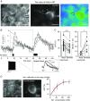
Figure 1. Na+ transients during action potentials in the calyx of Held nerve terminal
A, differential interference contrast and pseudocolour fluorescence images of the calyx of Held axon and terminal in the MNTB of the WT rat (P15) loaded with the Na+ indicator, CoroNa‐green (100 μm), during whole‐cell recording. Ovals indicate subcellular regions of interest [axon heminode (A, black) and terminal (T, grey)] from which fluorescence measurements were obtained. B, intracellular Na+ transients, measured as relative changes in CoroNa‐green fluorescence (ΔF/F), elicited by AP trains (180 Hz, 1.5 s and 3 s) recorded at the axon heminode (A, black trace) and presynaptic terminal (T1 and T2, grey traces). Note the rapid rise in Na+ at the axon heminode (A) and the slower and smaller rise at the presynaptic terminal (T1 and T2). Inset, representative trace of AP train at 180 Hz (1.5 s) and a scaled trace of the presynaptic terminal (grey) to the peak of signal from the axon heminode (black) to compare the decay time course of Na+ transient at the heminode (black, τ = 12 s) and terminal (grey, τ = 30 s). C, summary of the intensity (ΔF/F) and decay time constant (τ) of Na+ transients elicited by AP trains for (3 s, 180 Hz) at heminodes and terminals in the individual myelinated axon. Broken lines join paired data points obtained from the same axon. Data were analysed using a paired t test: *** P < 0.0001 and * P < 0.05, respectively. D, differential interference contrast and fluorescence images of the calyx of Held terminal, loaded with a Na+ indicator, CoroNa‐green (100 μm), using whole‐cell recordings. The black circle indicates the measuring area at presynaptic terminal and the white circle indicates the background area. Na+ calibration curve relating the change in fluorescence intensity as function of the applied Na+ concentrations ([Na+]): Fitting curve (red) was obtained using the Hill equation. [Colour figure can be viewed at wileyonlinelibrary.com]
Figure 2
Figure 2. Lack of myelin alters the kinetics of Na+ transients at axon heminodes and terminals from LES rats
A, differential interference contrast and fluorescence images of the LES calyx axon and terminal (P16), which was loaded with CoroNa‐green (100 μm) during whole‐cell recordings. Ovals indicate heminode and terminal regions of interest in the dysmyelinated calyx axon from which fluorescence measurements were obtained. B, representative trace of Na+ transient (ΔF/F) in response to AP trains (180 Hz, 3 s) at the axon heminode (black) and presynaptic terminal (grey) in the LES rat. C, summary of changes in the intensity (ΔF/F) and decay time constant (τ) of Na+ transients at heminode and terminal in the same dysmyelinated axon from LES rats. Broken lines link data from the axon heminode and terminal of the same identified cell. Paired t test: * P < 0.05 and non‐significant (NS), respectively. D, comparison of Na+ transient (ΔF/F) in response to AP trains (180 Hz, 3 s) at axon heminodes and terminals from WT and LES rats. E, comparison of decay time constant (τ) of Na+ transients elicited by AP trains at axon heminodes and terminals from WT and LES rats. *** P < 0.0001, ** P < 0.001 and * P < 0.05, respectively. [Colour figure can be viewed at wileyonlinelibrary.com]
Figure 3
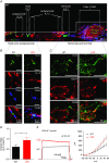
Figure 3. Myelin loss specifically disrupts Na+ channels at the axon terminal heminode
A, expression of NavPan (green), Caspr (red) and calretinin (CR, blue) immunoreactivity along the calyx of Held axon and terminal in the MNTB in the WT rat. The dashed lines delimit nodal and heminodal Nav cluster, as well as the calyx of Held. Blue horizontal arrows indicate the distance between the heminode and the next node, and the internodal distance, respectively. B, expression of Caspr (red) and NavPan (green) at axon nodes in the MNTB from WT and LES rats. Arrows indicate NavPan at nodes. C, expression of NavPan (green) and vGluT1 (red) at axon heminodes and calyx terminals. Arrows indicate the end of NavPan signals for measuring the length of Na+ channel cluster. D and E, summary of the length of Na+ channel cluster at nodes of Ranvier and axon heminodes. F and G, summary of the distance between the axon heminode and the next node, and between two nodes (internodal distance). *** P < 0.0001.
Figure 4
Figure 4. Dispersed Nav1.6 channel expression extends into the presynaptic terminal
A, heminodal staining at calyx of Held presynaptic terminal with Nav1.6 (green) and vGluT (red) in WT and LES rats (left). Nodal and paranodal staining with Nav1.6 (green, yellow arrows) and Caspr (red) in WT and LES rats (right). B, representative traces of the outside‐out patch recordings from the calyx terminals in WT and LES rats. Voltage‐activated Na+ and K+ currents were evoked by step depolarization (from −100 mV to 50 mV; in the presence of TEA 10 mm and 4‐AP 2 mm) in the outside‐out patches. Inset, expanded scale of Na+ currents in the boxed area from the LES recording. C, summary of the proportion of the presynaptic terminals displaying detectable I Na in WT and LES rats in the outside‐out patch recordings.
Figure 5
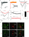
Figure 5. Reduced resurgent and persistent Na+ currents at axon terminal in the LES rat
A and B, representative traces of resurgent Na+ currents evoked by step repolarization (from +20 mV to −70 mV) after a brief depolarization (+30 mV, 5 ms) to generate transient Na+ currents (INaT) at the calyx of Held terminals in WT (A) and LES rats (B). C, current–voltage (IV) relationship of resurgent Na+ currents form WT (black) and LES rats (red). D, persistent Na+ currents were recorded by ramp depolarization from −90 mV to +40 mV (100 mV/s) at the calyx of Held terminals in WT (black) and LES rats (red). Representative traces of persistent Na+ currents after TTX subtraction. E, in current clamp recordings, TTX (1 μm) hyperpolarized the calyx terminal in the MNTB from WT but not LES rats. F, summary of changes in calyx membrane resting potential (ΔV m) by the application of TTX (1 μm) in the MNTB from WT and LES rats. G, Navβ4 (green) expression at the axon heminode with calretinin (CR, red) in WT and LES rats. Circles indicate the heminodal region and arrows indicate the Navβ4 cluster. Unpaired t test, * P < 0.05. [Colour figure can be viewed at wileyonlinelibrary.com]
Figure 6
Figure 6. Lack of compact myelin disrupts the cluster of Kv1.2 at juxtaparanodes and Kv3.1 at heminodes
A, the calyx of Held axon and terminal were immunolabelled for Kv3.1 (green), Kv1.2 (red) and calretinin (CR, blue) in the WT rat brainstem. Dashed arrows indicate heminodes, nodes and juxtaparanodes, as well as the calyx terminal. Note, Kv3.1 was expressed at the node and the heminode and Kv1.2 was expressed at the juxtaparanode. B, expression of Kv3.1 (green), Caspr (blue) and Kv1.2 (red) at nodes, paranodes and juxtaparanodes along the axon in WT and LES rats. Arrows indicate Kv3.1 at nodes. C, at the axon heminode and terminal, expression of Kv3.1 (green) and vGluT1 (red) in WT and LES rats. Yellow arrows indicate the extent of Kv3.1 expression, which invades the presynaptic terminal in the LES. D, summary of the intensity ratio of Kv3.1 signal by vGluT1 at presynaptic terminals in WT and LES rats obtained in (C). E, representative trace of TEA‐sensitive HVA‐K+ (at +30 mV; sensitive to TEA 1 mm) at holding −70 mV recorded in the calyx terminal from WT and LES rats. Traces were obtained after TEA subtraction. The current–voltage (IV) relationships of TEA‐sensitive HVA‐K+) currents in WT and LES rats obtained after the TEA subtraction. Unpaired t test, * < 0.05. [Colour figure can be viewed at wileyonlinelibrary.com]
Figure 7
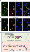
Figure 7. Lack of condensed myelin affects the distribution of Nav and Kv clusters at the heminode and calyx of Held terminal in the MNTB
A, expression of Kv3.1 (green) and Nav1.6 (red) at the axon heminode and terminal marked with vGluT1 (blue) in WT and LES rats. Kv3.1 (yellow arrow) and Nav1.6 clusters (white arrow) were well co‐localized at the axon heminode in WT rats, whereas their clusters completely disrupted at heminode and dispersed to the terminal in LES rats (yellow arrows). B, nodal and juxtaparanodal staining with Kv3.1 (green), Nav1.6 (red) and Kv1.2 (blue) in WT and LES rats. Kv3.1 and Nav1.6 clusters are well preserved at the node, although juxtaparanodal structures stained with Kv1.2 are disrupted in LES axons. C, summary diagram of effects of myelin loss on the distribution of voltage‐gate activated ion channels (Nav1.6, Navβ, Kv1 and Kv3) at the last heminode. [Colour figure can be viewed at wileyonlinelibrary.com]

Similar articles

Cited by

References

    1. Alle H & Geiger JR (2006). Combined analog and action potential coding in hippocampal mossy fibers. Science 311, 1290–1293. - PubMed
    1. Aman TK, Grieco‐Calub TM, Chen C, Rusconi R, Slat EA, Isom LL & Raman IM (2009). Regulation of persistent Na current by interactions between beta subunits of voltage‐gated Na channels. J Neurosci 29, 2027–2042. - PMC - PubMed
    1. Arroyo EJ, Xu T, Grinspan J, Lambert S, Levinson SR, Brophy PJ, Peles E & Scherer SS (2002). Genetic dysmyelination alters the molecular architecture of the nodal region. J Neurosci 22, 1726–1737. - PMC - PubMed
    1. Bant JS & Raman IM (2010). Control of transient, resurgent, and persistent current by open‐channel block by Na channel beta4 in cultured cerebellar granule neurons. Proc Natl Acad Sci USA 107, 12357–12362. - PMC - PubMed
    1. Boiko T, Rasband MN, Levinson SR, Caldwell JH, Mandel G, Trimmer JS & Matthews G (2001). Compact myelin dictates the differential targeting of two sodium channel isoforms in the same axon. Neuron 30, 91–104. - PubMed

Publication types

LinkOut - more resources