. 2016 Dec;76(12):1387-1416.
doi: 10.1002/dneu.22400. Epub 2016 Jun 6.

dHb9 expressing larval motor neurons persist through metamorphosis to innervate adult-specific muscle targets and function in Drosophila eclosion

Affiliations

dHb9 expressing larval motor neurons persist through metamorphosis to innervate adult-specific muscle targets and function in Drosophila eclosion

Soumya Banerjee et al. Dev Neurobiol. 2016 Dec.

Abstract

The Drosophila larval nervous system is radically restructured during metamorphosis to produce adult specific neural circuits and behaviors. Genesis of new neurons, death of larval neurons and remodeling of those neurons that persistent collectively act to shape the adult nervous system. Here, we examine the fate of a subset of larval motor neurons during this restructuring process. We used a dHb9 reporter, in combination with the FLP/FRT system to individually identify abdominal motor neurons in the larval to adult transition using a combination of relative cell body location, axonal position, and muscle targets. We found that segment specific cell death of some dHb9 expressing motor neurons occurs throughout the metamorphosis period and continues into the post-eclosion period. Many dHb9 > GFP expressing neurons however persist in the two anterior hemisegments, A1 and A2, which have segment specific muscles required for eclosion while a smaller proportion also persist in A2-A5. Consistent with a functional requirement for these neurons, ablating them during the pupal period produces defects in adult eclosion. In adults, subsequent to the execution of eclosion behaviors, the NMJs of some of these neurons were found to be dismantled and their muscle targets degenerate. Our studies demonstrate a critical continuity of some larval motor neurons into adults and reveal that multiple aspects of motor neuron remodeling and plasticity that are essential for adult motor behaviors. © 2016 Wiley Periodicals, Inc. Develop Neurobiol 76: 1387-1416, 2016.

Keywords: Drosophila; dHb9; eclosion; metamorphosis; motor neuron remodeling.


Figures

Figure 1
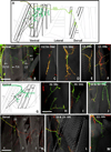
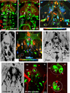
Figure 1. Expression pattern of dHb9>GFP in the third instar larva
(A) A subset of NMJs in the body wall is labeled with GFP. A1n= A1 nerve; Arrowhead indicates cleft innervation of MF 15/16. (*)= Muscle fibers 13 and 12 in A2. (B) The number of dHb9 axons in each nerve tract is indicated. (C) Three clusters of dHb-9> GFP expressing cell bodies are observed: dorsal (red), Intermediate (green and yellow) and ventral (blue). Dashed line indicates the dorsal midline of the CNS. (D–F) Of the five dorsal neurons (green) revealed in OK371>mCD8GFP, three are dHb9-positive neurons (E, red; D, green & red) and two are even-skipped positive (F, red). (G) Number of dHb9-positive neuronal cells closely matches with the number of NMJs. Bars represent 200 µm in (A), 40 µm in (B), 20 µm in (D) and 35µm in (E and F).
Figure 2
Figure 2. Morphological attributes of NMJs made by dHb9>GFP expressing motor neurons
(A) Schematic representation of the NMJs made by dHb9 expressing neurons which populate four of six known nerve branches: SNb, SNc, SNd and ISN. Muscle fibers innervated by the corresponding motor neurons are indicated in bold. Double labeling with HRP indicates that GFP reveals only a subset of boutons at each of the muscle fibers. Muscles closer to the cuticle are indicated in dashed lines. (B) A group of four ventral muscles that are innervated by dHb9>GFP expressing motor neurons. (C–F) Higher magnification images showing the NMJs at each of the 6 muscle fibers shown in (B). (G) Schematic depicting a group of ventral muscles (dashed lines) that lie beneath the ones shown in B. These include (H) MF 26 (SNc), (I) MF 28 (SNb), (J) MFs 30, 14 (SNb). (K) Three dorsal muscles are innervated by dHb9>GFP expressing motor neurons, all of which receive axons through the ISN branch. These include MFs 20, 19 (L), and MF 11 (M). Scale bars represent 50 µm for (C–F), 65 µm for (H, I, J, L), 45 µm for (M) and 110 µm for (B and K),
Figure 3
Figure 3. Adult innervation revealed by dHb9>GFP in the third instar larva
(A–C) The mature pattern of dHb9>GFP innervation is present at 72h APF. White asterisks: Persistent larval muscle fibers 12 and 13; White dotted line: a large triangular muscle arrangement in A1 (MFs 80, 81; Crossley, 1980). In a 2-day old adult, MFs 12 and 13 are absent in A1 and A2. (D) Virgin Adult: Three NMJs can be observed in the A2 ventral muscle group (D’) Close-up of the A2 segmental nerve, which carries a total of 5 axons, 3 to the ventral muscle group and 2 to the dorsal muscles (D”). (E) In a 2-day old adult, a single motor axon in A2 innervates the ventral muscles, and two motor axons project to the dorsal muscles. (F) The number of motor axons to ventral muscles decreases from 3 to 1 in A2; in A3, there is no change. Dorsal muscle targets of dHb9 expressing neurons at virgin (G) and 2-day old adult (H) stages. (G’ and H’): Schematic representation of G and H respectively. Filled gray muscles indicate targets innervated by dHb9>GFP neurons. Black dotted lines indicate the larger persistent larval muscle fibers, which are eliminated between virgin and 2-day old adult stages. Scale bar in A represents 80 µm for (A and B) and 95 µm for (C). Scale bars in (D and E) represent 50 µm, and 90 µm in (G and H).
Figure 4
Figure 4. Matching pre and post synaptic partners by FLP-induced GFP expressing single motor neurons
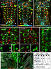
(A–D) 9 dHb9>GFP expressing motor neurons are represented in the four examples of larval CNS containing FLP induced GFP labeling in subsets of dHb9 neurons. (E–G) Axons of MN6/7-Ib and MN14-Ib exit the CNS contralaterally to innervate MFs 6/7 and 14 respectively. (H, I) Axon of MN12-Ib exits the CNS contralaterally to innervate MF 12, while the axon of MN13-Ib exits the CNS ipsilaterally to innervate MF 13. (J, K) MN28-Ib exits the CNS ipsilaterally through the SNb nerve to innervate MF 28. (L, M) Axon of MN15/16-I exits the CNS ipsilaterally through the SNd nerve to innervate MF 15/16 (N, O) Axon of MN26-Ib exits the CNS ipsilaterally through the SNc nerve to innervate MF 26. (P, R) Cell body positions and motor axonal projections of MN20-Ib and MN11-Ib in segments A3 and A2 respectively. Axons of MN20-Ib and MN11-Ib exit the CNS ipsilaterally through the ISN nerve to innervate MF 20 (Q) and MF 11 (S) respectively. (T, U) dHb9>GFP flip out preps labeled with anti-Hb9 antibody (red) and anti-GFP antibody (green). The CNS midline is shown with a white dotted line. (T) Relative positions of MN6/7-Ib, MN14-Ib, MN15/16-I, MN20-Ib, MN12-Ib and MN13-Ib in A2 and A3 segments of the VNC. (U) Relative positions of MN11-Ib and MN13-Ib in A2 and A4 segments of the VNC. Scale bars represent 25 µm in (E, H, J, L, P, R, T and U), 30 µm in (N), 55 µm in (F, G and I), 60 µm in (K, Q and S), 70 µm in (M), 100 µm in (G) 115 µm in (0).
Figure 5
Figure 5. Relative positions of dHb9-positive motor neurons in the third instar larva
(A) Schematic summarizing the relative positions of dHb9-positive motor neurons in the A1 and A2 segments of the larval CNS (based on an examination of 165 clones) A color spectrum, from red to blue has been used to depict their distance from the dorsal surface of the VNC. (B) Muscle targets and characteristic neuromuscular junctions of these motor neurons are shown. Neuromuscular junctions have been color coded to match with corresponding cell bodies.
Figure 6
Figure 6. Persistence of dHb9>GFP positive motor neurons during early metamorphosis (0–20h APF)
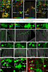
A–C: Pattern of cell bodies from 8h–20h APF. The lateral cluster in A1 and A2 (Arrows), and the dorsal cluster in A1 and A2 (ovals) are indicated in the ovals. Smaller dashed circular outlines (C) indicate cells that persist through early metamorphosis in A3–A6, and collectively form an outer row of cells. These were identified to be MN13-Ib. Red arrowheads (C) indicate MN12-Ib in A3–A6, which collectively form an inner row. D–H: Programmed Cell death (PCD) detected in a subset of dHb9>mCD8GFP expressing neurons at 10h APF, using TUNEL assay. Circles indicate the dorsal clusters of dHb9-positive neurons in A1 (D) and A2 (E). Arrows indicate apoptotic dHb9-positive neurons in A1 (D and D’), A2 (E and E’) and A3 (D and D”) hemisegments. The boxed area in (F) shows a pair of MN6/7-Ib in A2 hemisegments which also label with TUNEL (red) at 10h APF. The boxed area is shown in (G, H) at low magnification, allowing the labeled NMJs in the periphery to be seen. The projections of MN6/7-Ib into the periphery reveals the typical cleft innervation at MFs 6/7. (I) Schematic diagram summarizing the early metamorphic (0–12h APF) fate of dorsally located motor neurons (MN6/7-Ib, MN14-Ib and MN30-Ib). Scale bars represent 40 µm in (D, E, F, G and H), 15 µm in (D’, D’’, E’ and F’), 20 µm in (G’).
Figure 7
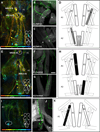
Figure 7. Establishment of the adult pattern of dHb9>GFP expressing neurons (30h-Adult)
(A) 30h APF: The pattern of cell bodies resembles what is seen at 20h APF. (B) 48h: Due to expansion of the thoracic neuromeres, the cell bodies of MN12-Ib move closer to the midline (white arrowheads). (C, C’) MN12-Ib in A1 and A2 (arrowheads) send anteriorly projecting trajectories that then cross the midline. (D, E) Virgin Adults: Persistent cells include the dorsal clusters in A1 and A2, MN12-Ib in A1 and A2 segments (white arrowheads), lateral clusters in A1 and A2 (arrows), and MN13-Ib in posterior abdominal segments (dashed white circles). (F) 2-day adult: Fewer cell bodies are observed as compared with virgin adults. 2 cells remain in the A1 dorsal cluster. MN12-Ib is absent in A1 and A2. Several cells in the lateral clusters in A1 and A2 persist. Fewer cells express dHb9 in A3 onwards. (G, G’) 4h after eclosion: A pair of dorsally located dHb9>GFP-positive neurons in A1 and A2 undergoes programmed cell death (PCD) as detected by TUNEL labeling. Scale bars represent 70 µm in (A: larva), 85 µm in (A: 30h APF), 90 µm in (A: 20h APF and 48h APF), 95 µm in (A: adult), 50 µm in (B, C and F), 45 µm in (E), 10µm in (G’).
Figure 8
Figure 8. Remodeling of innervation to persistent larval muscles 12 and 13 during the larval- adult transition
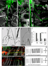
(A–C) Retraction of larval NMJs and histolysis of larval muscles occurs during the 0–12 h APF period, and by 20h, most of the larval muscles have histolyzed, and persistent larval muscles 12 and 13 can be seen only in A1 and A2 (*). (D–G) Remodeling of NMJs on MFs 12 and 13 during 0–72h APF. Scale Bar in C= 90µm in A–C; 50µm in D–G
Figure 9
Figure 9. Longitudinal imaging of MN13-Ib and MN12-Ib from larva to the adult stage reveals that MN13-Ib switches its muscle target
(A) The NMJs made by MN13-Ib and MN12-Ib in the A1 hemi-segment of a second instar larva are imaged through the cuticle. (B) The same animal imaged as a virgin adult, shows GFP labeled NMJs on the persistent MF 12, and on MF 80 but not on persistent MF 13. (C) Schematic representation of an adult A1 hemisegment indicating the innervated muscles seen in (B): MF 13 (gray) and MF 80 (black). (D) In a different animal, NMJs made by MN12-Ib and MN14-Ib are visualized through the cuticle. This animal imaged at the virgin adult stage (E) shows NMJs on persistent larval MFs 12 and 13. (F) Schematic representation of an adult A1 hemisegment indicating the innervated muscles [MF 12, 13 (gray], seen in E. (G–H) The terminals of MN14-Ib visualized at 30h APF are seen on MF 13 (*). (I–J) The terminals of MN13-Ib visualized at 30h APF are seen on MF 13 (*). (K–M) The terminals of MN14-Ib visualized at 60h APF are seen on MF 13 (*). (O–P) The terminals of MN13-Ib visualized at 60h APF are seen on MF 80. Scale bar in A represents 50 µm in (B, E) and 70 µm in (A, D). Scale bar in G represents 35 µm in (G), 40 µm in (O), 45 µm in (H, I, K, and O), 70 µm in (L), 100 µm in (J).
Figure 10
Figure 10. Adult muscle targets of dHb9-expressing neurons in A1 and A2 as revealed by FLP-induced labeling
For each pre-synaptic cell (s) labeled with GFP, the postsynaptic muscle target shown with the NMJs. Schematics identify the muscle targets in the context of the other surrounding muscle fibers in that hemisegment. (A) MN14-Ib in A1 and A2 (dorsal cluster) innervate MF13 in corresponding bodywall segments (B, C). (E) MN30-Ib and MN13-Ib which are cells of the dorsal cluster, persist to innervate MF 80 and 81 respectively (F, G). (I–N) Subsets of neurons of the lateral cluster in A1 are labeled in (I) and (L); their muscle targets include MFs 80 and 81 (J, K, M, N). (O–P”) Two distinct neurons in the lateral cluster of A2 are labeled. These innervate non-overlapping sets of dorsal muscles in the virgin adult. (Q) MN12-Ib is labeled in A1 and A2, which contralaterally innervate the persistent larval muscle 12 in the bodywall (R, S). Scale bars represent 25 µm in (B, F and J), 40 µm in (C), 35 µm in (G), 30 µm in (M and R), 45 µm in (S) and 40 µm in (O’ and P’).
Figure 11
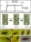
Figure 11. Effects of DT-1 expression on dHb9 neurons
(A) Temperature regimens to manipulate dHb9-expressing neurons during metamorphosis.(B) Monitoring eclosion behavior defects with video recordings. White dotted lines indicate control flies. Video frames of control (C and C’) and experimental flies (D and D’). (E and E’) Successive video frames of an experimental fly showing a gap between the head and the anterior end of the pupal casing even after 115hr-equivalent age, indicating a lack of head expansion (F) A ‘half-eclosed’ experimental fly showing emergence of the head and thorax, but the abdomen is still within the case.
Figure 12
Figure 12. dHb9>GFP expressing cell bodies and NMJs in controls and DT-1 induced experimental flies
A–F: The ventral ganglion of control and experimental animals. The dorsal cluster of cells in A1 and A2 is indicated in the ovals. (A) Control (dHb9-Gal4/UASmCD8GFP/W1118): Four neurons are present in A1 and two neurons are present in A2. (B) Un-eclosed: Three neurons are present in A1, and no dorsally located cell is seen in A2. (C) Half-eclosed: Four neurons are present in A1, and none in A2. (D) Eclosed: Four neurons are present in A1, and one A2. Scale bar in A represents 50 µm in (A), 55 µm in (B–D). E–H. Persistent larval muscle fibers 12 and 13 of the animals shown in A–F (E and F) Control (dHb9/GFP/w1118): A2 Ventral muscles (including PLMFs 13 and 12) receive inputs from both dHb9>GFP positive and non-dHb9 expressing neurons, indicating two distinct classes of motor neurons innervate this muscle. White dotted lines show the region of PLM 13. NMJs that do not express dHb9 are distinguished by visualizing boutons which are only HRP-positive (white arrows). (G) Un-eclosed: White dotted lines show the area of PLM13. Non-dHb9-positive NMJs (HRP-positive; white arrows) are mainly seen on PLMF 13 in A2. (H) Measurement of relative dHb9>GFP expression levels in cell bodies of control and experimental (treatment 1) flies. Error bars on the graph represent S.E.M. Asterisks represents significant differences (p< 0.0005). Scale bar in E represents 30µm for (E and F), and 35µm for (G).
Figure 13
Figure 13. Schematic representation of the muscle targets of four prominent dHb9>GFP expressing motor neurons in A1–A3 during the larval and adult stages
MN14-lb and MN30-lb are in the dorsal cluster and project contra-laterally to their muscle targets. In A1 and A2, MN14-Ib innervates persistent muscle fiber 13, and is absent in A3. MN30-Ib is only present in A1 and innervates a segment specific adult muscle, MF 81. MN13-lb is in the intermediate cluster and projects ipsilaterally. In A1 it innervates a segment specific adult muscle (MF 80), whereas in A2 and A3 it innervates an adult muscle, MF 111. MN12-Ib represents the ventral cluster and innervates persistent muscle fiber 12 in A1 and A2.
Figure 14
Figure 14. Schematic representation of the location of dHb9>GFP expressing motor neurons and their muscle targets at adult stage
Segment specific patterns (A1–A3) of dHb9 expressing MNs are depicted in the newly eclosed adult. The colors red, green/yellow, blue, represent the dorsal, intermediate and ventral clusters respectively. Neuromuscular junctions have been color coded to match the corresponding cell bodies.

Similar articles

Cited by

References

    1. Arber S, Han B, Mendelsohn M, Smith M, Jessell TM, Sockanathan S. Requirement for the homeobox gene Hb9 in the consolidation of motor neuron identity. Neuron. 1999;23:659–674. - PubMed
    1. Baek M, Mann RS. Lineage and birth date specify motor neuron targeting and dendritic architecture in adult Drosophila. J Neurosci. 2009;29:6904–6916. - PMC - PubMed
    1. Bellen HJ, Develyn D, Harvey M, Elledge SJ. Isolation of Temperature-Sensitive Diphtheria Toxins in Yeast and Their Effects on Drosophila Cells. Development. 1992;114:787–796. - PubMed
    1. Broihier HT, Skeath JB. Drosophila homeodomain protein dHb9 directs neuronal fate via crossrepressive and cell-nonautonomous mechanisms. Neuron. 2002;35:39–50. - PubMed
    1. Budnik V, Koh YH, Guan B, Hartmann B, Hough C, Woods D, Gorczyca M. Regulation of synapse structure and function by the Drosophila tumor suppressor gene dlg. Neuron. 1996;17:627–640. - PMC - PubMed

Publication types

LinkOut - more resources