. 2016 May 10;23(5):811-20.
doi: 10.1016/j.cmet.2016.04.006.

Parabrachial CGRP Neurons Control Meal Termination

Affiliations

Parabrachial CGRP Neurons Control Meal Termination

Carlos A Campos et al. Cell Metab. .

Abstract

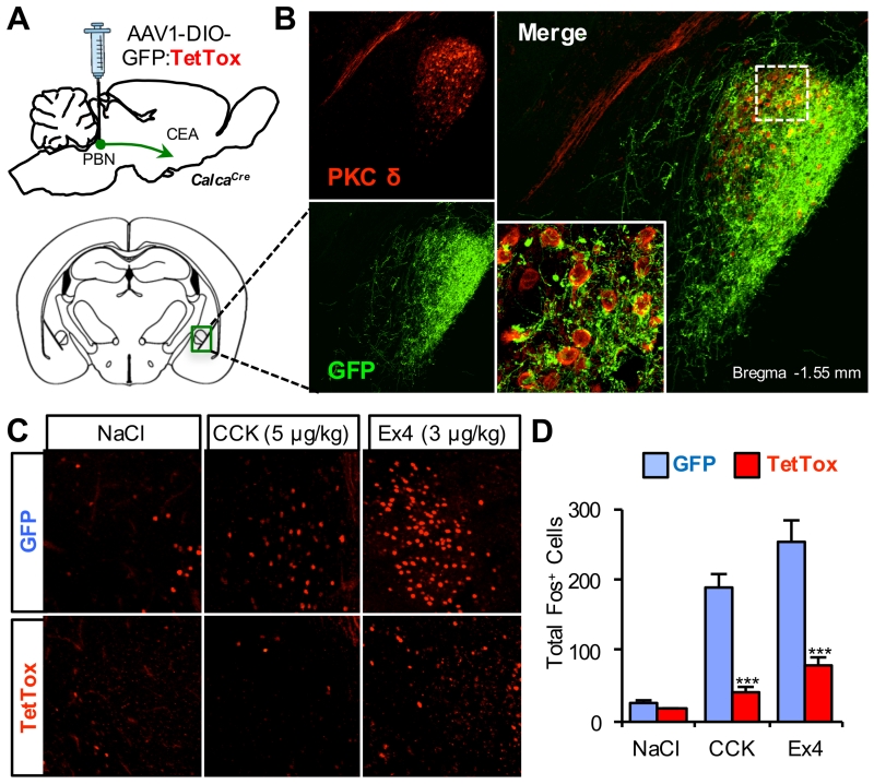
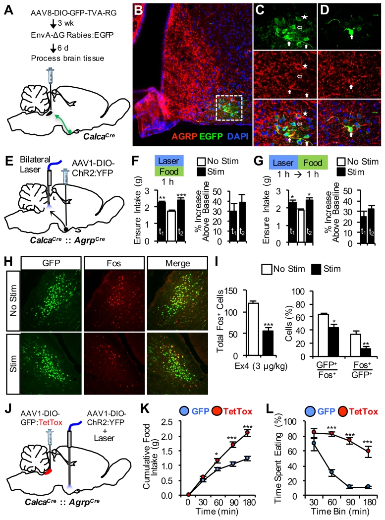
The lateral parabrachial nucleus is a conduit for visceral signals that cause anorexia. We previously identified a subset of neurons located in the external lateral parabrachial nucleus (PBel) that express calcitonin gene-related peptide (CGRP) and inhibit feeding when activated by illness mimetics. We report here that in otherwise normal mice, functional inactivation of CGRP neurons markedly increases meal size, with meal frequency being reduced in a compensatory manner, and renders mice insensitive to the anorexic effects of meal-related satiety peptides. Furthermore, CGRP neurons are directly innervated by orexigenic hypothalamic AgRP neurons, and photostimulation of AgRP fibers supplying the PBel delays satiation by inhibiting CGRP neurons, thereby contributing to AgRP-driven hyperphagia. By establishing a role for CGRP neurons in the control of meal termination and as a downstream mediator of feeding elicited by AgRP neurons, these findings identify a node in which hunger and satiety circuits interact to control feeding behavior.


Figures

Figure 1
Figure 1. CGRPPBel Neurons are Activated by a Meal and Control Meal Termination
(A) Representative histological images showing Fos immunoreactivity (red) in GFP-labeled CGRPPBel neurons (green) after fast-refeeding. (B) Quantification of Fos immunoreactivity in the PBel after fast-refeeding (n = 6 per group). (C) Unilateral delivery of AAV carrying Cre-dependent hM3Dq:mCherry into the PBel of CalcaCre mice. (D and E) Cumulative food intake (D) and meal pattern analysis (E) following saline or CNO (1 mg/kg, i.p.) administration immediately prior to onset of dark cycle in non-food-deprived mice (n = 8). All data shown are means ± s.e.m. * P < 0.05; ** P < 0.01; *** P < 0.001. Statistical analysis was performed with an unpaired student’s t-test (B) and two-way ANOVA followed by Bonferroni’s post-hoc test (D-E). See also Figure S1.
Figure 2
Figure 2. Functional Inactivation of CGRPPBel Neurons Disrupts Control of Meal Size
(A) Bilateral delivery of AAV carrying Cre-dependent GFP:TetTox into the PBel of CalcaCre mice. (B) Representative bout-size recordings from individual TetTox and GFP control mice during a 24-h recording period. (C-E) Group-wide, meal-pattern analysis (n = 9 per group) was obtained from bout size recordings using set meal parameters. TetTox inactivation of CGRPPBel neurons increased the size and duration of meals (C) but was accompanied by a decrease in meal frequency (D) such that overall food intake was unaltered (E). (F) Food intake measured following a 24-h food deprivation (n = 11 per group). (G) Intake of a palatable liquid diet (Ensure) from non-food-deprived mice given 2 h ad libitum access during the light cycle (n = 11 per group). All data shown are means ± s.e.m. * P < 0.05; ** P < 0.01; *** P < 0.001. Statistical analysis was performed with an unpaired student’s t-test (C-D and G) and two-way ANOVA followed by Bonferroni’s post-hoc test (F). See also Figure S2.
Figure 3
Figure 3. CGRPPBel Neuronal Activation is Required for CCK and Leptin-induced Anorexia
(A) Bilateral delivery of AAV carrying Cre-dependent GFP:TetTox into the PBel of CalcaCre mice. (B) CCK administration (i.p.) decreases 2-h cumulative food intake in non-food-deprived GFP control mice (n = 8) injected immediately before the dark cycle. (C) TetTox inactivation of CGRPPBel neurons abolishes CCK-induced anorexia (n = 10). (D) Leptin administration (i.p.) decreases 12-h cumulative food intake by decreasing meal size in non-food-deprived GFP control mice (n = 6) injected immediately before the dark cycle. (E) Leptin-induced decrease of 12-h cumulative food intake and reduction of meal size is absent in TetTox mice (n = 9). All data shown are means ± s.e.m. * P < 0.05; ** P < 0.01; *** P < 0.001. Statistical analysis was performed with a one-way ANOVA followed by Tukey’s post-hoc test (B-C) and paired student’s t-test (D-E).
Figure 4
Figure 4. CGRPPBel Neuronal Activation Contributes to Anorexia Induced by a GLP-1 Receptor Agonist
(A) Representative histological images showing Fos immunoreactivity (red) in GFP-labeled CGRPPBel neurons (green) following saline or Ex4 administration. (B) Quantification of Fos immunoreactivity in the PBel after saline (n = 3) or Ex4 (n = 4) administration. (C) Ex4 injection (i.p.) decreases 12-h cumulative food intake by decreasing meal size in non-food-deprived GFP control mice (n = 8) administered immediately before the dark cycle. (D) Ex-induced decrease of 12-h cumulative food intake and reduction of meal size is absent in TetTox mice (n = 8). (E and F) Twice daily i.p. administration of Ex4 to diet-induced obese mice with access to high-fat diet (TetTox, n = 6; GFP, n = 8). Ex4-induced anorexia (E) and body weight-loss (F) was attenuated in TetTox mice. All data shown are means ± s.e.m. * P < 0.05; ** P < 0.01; *** P < 0.001. Statistical analysis was performed with an unpaired student’s t-test (B), paired student’s t-test (C-D), and two-way ANOVA followed by Bonferroni’s post-hoc test (E-F). See also Figures S3.
Figure 5
Figure 5. Activation of CeA Following Gut Peptide Administration Requires CGRPPBel Neuronal Activation
(A) Bilateral delivery of AAV carrying Cre-dependent GFP:TetTox into the PBel of CalcaCre mice. (B) Representative histological images showing overlap of PKC-δ immunoreactivity (red) and GFP-labeled CGRPPBel axon projections (green) in the CeA. (C and D) Representative histological images (C) and quantification of Fos immunoreactivity (D) in the CeA following saline, CCK, or Ex4 administration to GFP and TetTox mice (n = 4-6 per treatment). All data shown are means ± s.e.m. *** P < 0.001. Statistical analysis was performed with a one-way ANOVA followed by Tukey’s post-hoc test (D). See also Figure S4.
Figure 6
Figure 6. CGRPPBel Neurons Limit Hyperphagic Response to AgRP Neuron Activation
(A) Procedure for unilateral delivery of viruses into the PBel of CalcaCre mice allowing for retrograde monosynaptic rabies tracing from CGRPPBel neurons. (B) Representative confocal image of the arcuate hypothalamus showing overlap of upstream EGFP-labeled neurons (green) and AgRP immunoreactivity (red). (C and D) High-magnification optical sections of (B) demonstrating examples of EGFP and AgRP double-labeling of soma and fibers (filled arrows), single-labeled EGFP neurons (hollow arrows), and single-labeled EGFP neurons in close apposition to AgRP labeling (star). (E) Bilateral delivery of AAV carrying Cre-dependent ChR2:YFP into the arcuate hypothalamus of CalcaCre::AgrpCre mice with fiber-optic cannulae bilaterally implanted over the PBel. (F and G) Concurrent palatable food access and AgRP→PBel photostimulation (F) or AgRP→PBel photostimulation for 1 h immediately prior to palatable food access (G) increases food intake compared to experimental trials without photostimulation (n = 5). Photostimulation trial 1, t1; trial 2, t2. (H and I) Representative histological images (H) and quantification (I) of Ex4-induced Fos immunoreactivity in GFP-labeled CGRPPBel neurons ipsilateral and contralateral to unilateral AgRP→PBel photostimulation. (J) Bilateral delivery of AAV carrying Cre-dependent GFP:TetTox into the PBel and bilateral delivery of AAV carrying Cre-dependent ChR2:YFP into the arcuate hypothalamus of CalcaCre::AgrpCre mice with a fiber-optic cannula implanted over the arcuate nucleus. (K and L) Cumulative food intake (K) and time spent eating (L) following photostimulation of AgRP soma for 2 h in TetTox mice compared to GFP control mice (n = 4 per group). All data shown are means ± s.e.m. * P < 0.05; ** P < 0.01; *** P < 0.001. Statistical analysis was performed with a one-way ANOVA followed by Dunnett’s post-hoc test (F-G), unpaired student’s t-test (I), and two-way ANOVA followed by Bonferroni’s post-hoc test (K-L). See also Figure S5, S6, and Video S1.

Similar articles

Cited by

References

    1. Abbott CR, Monteiro M, Small CJ, Sajedi A, Smith KL, Parkinson JR, Ghatei MA, Bloom SR. The inhibitory effects of peripheral administration of peptide YY(3-36) and glucagon-like peptide-1 on food intake are attenuated by ablation of the vagal-brainstem-hypothalamic pathway. Brain Res. 2005;1044:127–131. - PubMed
    1. Alhadeff AL, Baird JP, Swick JC, Hayes MR, Grill HJ. Glucagon-like Peptide-1 receptor signaling in the lateral parabrachial nucleus contributes to the control of food intake and motivation to feed. Neuropsychopharmacology. 2014a;39:2233–2243. - PMC - PubMed
    1. Alhadeff AL, Golub D, Hayes MR, Grill HJ. Peptide YY signaling in the lateral parabrachial nucleus increases food intake through the Y1 receptor. Am. J. Physiol.-Endoc. M. 2015;309:E759–E766. - PMC - PubMed
    1. Alhadeff AL, Hayes MR, Grill HJ. Leptin receptor signaling in the lateral parabrachial nucleus contributes to the control of food intake. Am. J. Physiol. Regul. Integr. Comp. Physiol. 2014b;307:R1338–1344. - PMC - PubMed
    1. Aponte Y, Atasoy D, Sternson SM. AGRP neurons are sufficient to orchestrate feeding behavior rapidly and without training. Nat. Neurosci. 2011;14:351–355. - PMC - PubMed