

Kaposi's sarcoma herpesvirus (KSHV) microRNA K12-1 functions as an oncogene by activating NF-κB/IL-6/STAT3 signaling
- PMID: 27166260
- PMCID: PMC5078101
- DOI: 10.18632/oncotarget.9221
Kaposi's sarcoma herpesvirus (KSHV) microRNA K12-1 functions as an oncogene by activating NF-κB/IL-6/STAT3 signaling
Abstract
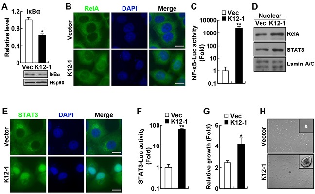
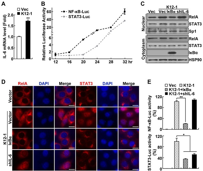
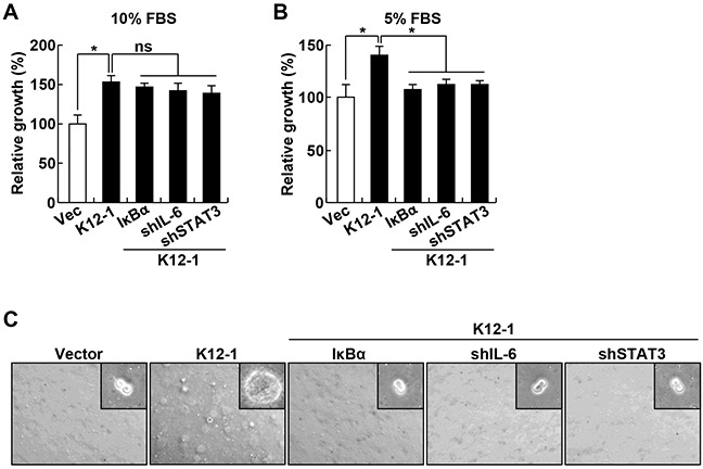
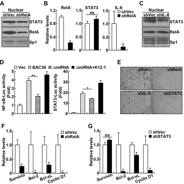
The human oncogenic virus Kaposi's sarcoma herpesvirus (KSHV) is the most common cause of malignancies among AIDS patients. KSHV possesses over hundred genes, including 25 microRNAs (miRNAs). The roles of these miRNAs and many other viral genes in KSHV biology and pathogenesis remain largely unknown. Accordingly, the molecular mechanisms by which KSHV induces tumorigenesis are still poorly defined. Here, we identify KSHV miRNA K12-1 (miR-K12-1) as a novel viral oncogene by activating two important transcription factors, nuclear factor-κb (NF-κB) and signal transducer and activator of transcription 3 (STAT3). Interestingly, miR-K12-1 activates STAT3 indirectly through inducing NF-κB activation and NF-κB-dependent expression of the cytokine interleukin-6 (IL-6) by repressing the expression of the NF-κB inhibitor IκBα. Accordingly, expression of ectopic IκBα or knockdown of NF-κB RelA, IL-6 or STAT3 prevents expression of cell growth genes and suppresses the oncogenicities of both miR-K12-1 and KSHV. These data identify miR-K12-1/NF-κB/IL-6/STAT3 as a novel oncogenic signaling underlying KSHV tumorigenesis. These data also provide the first evidence showing that IL-6/STAT3 signaling acts as an essential mediator of NF-κB oncogenic actions. These findings significantly improve our understanding of KSHV pathogenesis and oncogenic interaction between NF-κB and STAT3.
Keywords: IL-6; KSHV/HHV8; NF-κB; STAT3; miR-K12-1.
Conflict of interest statement
The authors declare no conflict of interest.
Figures
Similar articles
-
HIV-1 Vpr Inhibits Kaposi's Sarcoma-Associated Herpesvirus Lytic Replication by Inducing MicroRNA miR-942-5p and Activating NF-κB Signaling.J Virol. 2016 Sep 12;90(19):8739-53. doi: 10.1128/JVI.00797-16. Print 2016 Oct 1. J Virol. 2016. PMID: 27440900 Free PMC article.
-
Upregulation of MicroRNA 711 Mediates HIV-1 Vpr Promotion of Kaposi's Sarcoma-Associated Herpesvirus Latency and Induction of Pro-proliferation and Pro-survival Cytokines by Targeting the Notch/NF-κB-Signaling Axis.J Virol. 2018 Aug 29;92(18):e00580-18. doi: 10.1128/JVI.00580-18. Print 2018 Sep 15. J Virol. 2018. PMID: 29976660 Free PMC article.
-
Modified Cross-Linking, Ligation, and Sequencing of Hybrids (qCLASH) Identifies Kaposi's Sarcoma-Associated Herpesvirus MicroRNA Targets in Endothelial Cells.J Virol. 2018 Mar 28;92(8):e02138-17. doi: 10.1128/JVI.02138-17. Print 2018 Apr 15. J Virol. 2018. PMID: 29386283 Free PMC article.
-
Regulation of the MIR155 host gene in physiological and pathological processes.Gene. 2013 Dec 10;532(1):1-12. doi: 10.1016/j.gene.2012.12.009. Epub 2012 Dec 14. Gene. 2013. PMID: 23246696 Review.
-
KSHV miRNAs decrease expression of lytic genes in latently infected PEL and endothelial cells by targeting host transcription factors.Viruses. 2014 Oct 23;6(10):4005-23. doi: 10.3390/v6104005. Viruses. 2014. PMID: 25341664 Free PMC article. Review.
Cited by
-
The regulatory role of protein phosphorylation in human gammaherpesvirus associated cancers.Virol Sin. 2017 Oct;32(5):357-368. doi: 10.1007/s12250-017-4081-9. Epub 2017 Oct 30. Virol Sin. 2017. PMID: 29116588 Free PMC article. Review.
-
The interplay between EBV and KSHV viral products and NF-κB pathway in oncogenesis.Infect Agent Cancer. 2020 Oct 15;15:62. doi: 10.1186/s13027-020-00317-4. eCollection 2020. Infect Agent Cancer. 2020. PMID: 33072180 Free PMC article. Review.
-
Dynamic Expression of EpCAM in Primary and Metastatic Lung Cancer Is Controlled by Both Genetic and Epigenetic Mechanisms.Cancers (Basel). 2022 Aug 25;14(17):4121. doi: 10.3390/cancers14174121. Cancers (Basel). 2022. PMID: 36077658 Free PMC article.
-
Reactivation and Lytic Replication of Kaposi's Sarcoma-Associated Herpesvirus: An Update.Front Microbiol. 2017 Apr 20;8:613. doi: 10.3389/fmicb.2017.00613. eCollection 2017. Front Microbiol. 2017. PMID: 28473805 Free PMC article. Review.
-
Viral MicroRNAs in Herpes Simplex Virus 1 Pathobiology.Curr Pharm Des. 2024;30(9):649-665. doi: 10.2174/0113816128286469240129100313. Curr Pharm Des. 2024. PMID: 38347772 Review.
References
-
- Chang Y, Cesarman E, Pessin MS, Lee F, Culpepper J, Knowles DM, Moore PS. Identification of herpesvirus-like DNA sequences in AIDS-associated Kaposi's sarcoma. Science. 1994;265:1865–1869. - PubMed
-
- Cesarman E, Chang Y, Moore PS, Said JW, Knowles DM. Kaposi's sarcoma-associated herpesvirus-like DNA sequences in AIDS-related body-cavity-based lymphomas. N Engl J Med. 1995;332:1186–1191. - PubMed
-
- Soulier J, Grollet L, Oksenhendler E, Cacoub P, Cazals-Hatem D, Babinet P, d'Agay MF, Clauvel JP, Raphael M, Degos L, et al. Kaposi's sarcoma-associated herpesvirus-like DNA sequences in multicentric Castleman's disease. Blood. 1995;86:1276–1280. - PubMed
MeSH terms
Substances
Grants and funding
LinkOut - more resources
Full Text Sources
Other Literature Sources
Medical
Miscellaneous
